
Research Article
Austin J Orthopade & Rheumatol. 2017; 4(2): 1051.
Open Reduction and Fixation of Capitellum Fractures of the Elbow
Acosta-Olivo C*, Blanco-Rivera J, Villarreal- Villarreal G, Galván-Esquivel A, Vilchez-Cavazos F and Peña-Martinez V
Department of Orthopaedics and Traumatology, Universidad Autónoma de Nuevo León, Mexico
*Corresponding author: Carlos Acosta-Olivo, Department of Orthopaedics and Traumatology, Universidad Autónoma de Nuevo León, Mexico
Received: February 17, 2017; Accepted: March 16, 2017; Published: March 23, 2017
Abstract
Background: Elbow capitellum fractures are rare and represent an incidence of 1.5 per 100,000 distal humerus fractures. The objective of this workis to present a functional evaluation of patients treated with open reduction and internal fixation of isolated capitellum fractures.
Material and Methods: Retrospective study of four patients with capitellum fractures, with one year of follow-up. The patients were evaluated with functional scales: the Mayo Elbow Score and the Disability of Arm, Shoulder and Hand scale, and measured the range of motion of the elbow in flexion, extension, pronation and supination.
Results: The range of age was 14 to 71 years old. In the Mayo Elbow Score, at the end of follow-up, all the patients obtained a result of 100 points; and in the Disability of Arm, Shoulder and Hand scale, the result was 0. The mobility of the elbow was 130° of flexion, -5° of extension, pronation and supination of 80°.
Conclusion: The functional results of isolated capitellum fractures treated with open reduction and internal fixation was excellent in this case series. We need to know and recognize this fracture to make and adequate treatment and obtain a good clinical result.
Level of Evidence: IV Case series.
Keywords: Capitellum fracture; Internal fixation; Mayo elbow score
Introduction
The isolated fractures of the capitellum are rare, with an incidence of 1.5 per 100,000 population; these fractures have a bimodal distribution with one peak less than 19 years of age and other above the 80 years of age, with a female predominance. This type of fracture is usually associated with high energy forces in the younger population, and with osteoporosis in the older patients [1]. The diagnosis is usually done with an elbow anteroposterior and lateral radiographs, and could be complemented with a CT scan, to determinate the extension of the lesion, and the presence of comminution. Bryan and Morrey1-3 classified this fractures as: type I involves the capitellar articular surface along with the subcondral bone, type II consists of a capitellar articular surface along with a thin shell of subcondral bone, type III are the comminuted capitellar fractures, and type IV (described by Mckee) consists of a type I with medial extensión to include the lateral half of the trochlea [3,4]. The AO classification place the articular humerus fractures as type B3, where the B3-1 are the capitellum fractures, B3-2 trochlea fractures and B3-3 a combined fracture [1,3].
The management of the capitellar fractures could be nonoperative or operative [1]. However, the nonoperative management that includes a closed reduction and casting, and is only recommended in younger patients, has a high failure index [2]. Therefore, the treatment of choice is open reduction and internal rigid fixation using headless screws [1-3]. The most common complication after the surgical management is elbow stiffness [5]. Nowadays, there are few studies that report long or mid-term functional results of isolated capitellar fractures [3,6,7]. The purpose of our revision is to measure the functional outcome of four patients with isolated fractures of the capitellum treated with an open reduction and fixation with headless screws with one year follow up.
Material and Methods
In a one year revision, we received four patients with the diagnosis of isolated capitellum fractures treated with open surgery and internal fixation with headless screws. A mean follow up of 12 months, all treated by the same surgeon. All patients were initially evaluated with simple X-rays, an anteroposterior and lateral view of the elbow (Figure 1). A CT scan was taken to determine the fracture pattern. Fractures were classified with the Brayan and Morrey and AO classifications. All patients were classified as Brayan and Morrey type 1, and B3 according to the AO classification, and were initially treated with a back splint a 90° of flexion of the elbow.

    Figure 1:  Antero-posterior and lateral view of the elbow. Coronal displaced
fracture of the capitellum.
The patients were treated with open surgery, using the Kocher lateral approach [1]. Headless screws were used for fixation, with a disposition anterior to posterior and lateral to medial (Figure 2). Patient’s follow-up consisted in radiographic evaluation, Mayo Elbow Scorethat evaluates four aspects: pain, stability, range of motion and functional capacity; scores >90 points is excellent, 75-89 good, 60-74 fair and <60 bad [8]. Other scale used was quick-DASH (The Disabilities of the Arm, Shoulder and Hand) that consists of 11 questions; scoring without difficulty, mild, moderate, severe and unable [9]. Besides the range of motion in flexion, extension, pronation and supination were evaluated. All evaluation was made at two and four weeks, and two, six and twelve months. All the data are expressed as the means ± standard deviations, considering a value of p < 0.05 significant. Statistical tests were performed using SPSS 23.0 software for Windows 7.0.
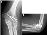
    Figure 2:  Anteroposterior y lateral view of the elbow. Three headless screw
were used to perform an anatomical reduction.
Results
Of our four patients, three were male and one female, the age range was from 14-71 years. Clinical and functional improvement was observed in all patients. The average of initial Mayo elbow score was poor result and the final average score was excellent. The initial mean of quick-DASH was 71.3 points with a final score of 0 points (P=<0.05). At 2 months of the follow up patients had recover 80% of their capacities. Range of motion improved throughout the one year of follow up. (Table 1) Only 1 patient presented implant failure and was re-operated with another headless Herbert screw. All patients showed radiographic consolidation on average of 4.2 months of evolution.
![]()
    Mayo    score
    quick-DASH
    Flexion
    Extension
    Pronation
    Supination


    Two    weeks
    46.6 ±    33.2
    71.3 ±    7.4
    90° ± 0
    -73.3° ± 2.8
    11.6° ± 2.8
    13.3° ± 5.7


    Four    weeks
    78.3 ±    15.2
    56.4 ±    11.6
    90° ± 0
    -51.6° ± 22.5
    20° ± 10
    21.6° ± 7.6


    Two    month
    91.3 ±    14.4
    32 ±    26.8
    109° ± 9.0
    -50° ± 18.0
    51.6° ± 29.2
    58.6° ± 28.2


    Sixth    month
    100 ± 0
    15.6    ±16.8
    121.6° ± 7.6
    -15° ± 5
    75° ± 13.2
    78.3° ± 7.6


    One year
    100 ± 0
    0 ± 0
    130° ± 0
    -5° ± 5
    83.3° ± 2.8
    83.3° ± 2.8
Table 1: Average of evaluation of patients.
Discussion and Conclusion
We report a short case series of isolated capitellum fractures treated with open reduction and internal fixation with headless screw that we evaluated with clinical scales and radiographic studies. The best clinical results for patients with these injuries, are obtained with an open reduction and internal fixation with headless screws (Lopiz 2016, Bilsel 2013) [2-5,10,11]. Mighell et al. [2] treated 18 patients using Herbert screws with a 26 month mean of follow up, they found a mean of 128° of flexion-extension with 176° of prono-supination; andan average of 93.3 in the Broberg Morey scale. In our study, the patients achieved similar outcomes, besides the fact that our sample was smaller, we only did a 1 year follow up and the range of ages was from 14 to 71 years.
Other studies have shown similar good results in patients treated with internal fixation, the average of Mayo Elbow Score was 91-93 points, however, in these studies the fixation where from posterior to anterior (Dubberley, Mahirogullari)., whether we performed an anterior to posterior fixation to avoid any disruption in the blood flow [4], and because it allows a better compression to the site of fracture minimizing damage to the articular surface [2]. In a retrospective review of 23 patients, followed for 48 months, with an average age of 71 years (66-79), and 65% of cases were women, the fractures were mostly non-dominant side, average of union fracture was observed at a 4.8 month. The limitations of the study, is that it is a retrospective review, also by the age of the patients they do not have a measure of bone density that could influence the increase in the frequency of fractures [6]. Other retrospective review of 30 patients with humeral trochlea and capitelum fractures, compared 12 simple fractures, against 18 comminuted fractures, and observed a higher rate of nonunion in patients with comminution (8 cases of 18), while the simple fractures all consolidated [12]. In our patients, we don´t observed any comminution. Retrospective of 18 patients, 12 were women, with an average age of 45.3 years (16-70), it is concluded that internal fixation with screws, cannulated, to maintain anatomical reduction, provides the best functional outcomes of patients [7].
In comparison to other studies, we only had one complication, which was the lost of reduction in one patient who went back to the operating room for a new ORIF. We did not find avascular necrosis or post-traumatic arthritis, but there is more time follow up to needed rule it out. The reported index of avascular necrosis is between 10- 30% [8]. There are other types of fixation which had shown good to excellent outcomes, as the use of bioabsorbable pins. Kraan et al. used an absorbable polyglycolide pin in a 24 year old male, reporting a full recovery with normal range of motion in a period of 6 months [13]. Also, Hirvensalo et al. reported the use of absorbable pins in type I capitellum fractures with a stable reduction and a good long term functional outcome, but with one case of asceptic synovitis as a complication [14]. One of the limitations of our study was the small number of patients as well as a short follow-up period, however we could found excellent functional and radiographic results. We can conclude that in an isolated capitellum fracture the best option of treatment is an anatomical reduction and fixation with cannulated headless screws.
References
- Bucholz RW, Heckman J.D, Court-Brown C.M, Tornetta P. Distal Humerus Fractures. In: Bucholz R.W, Court-Brown CM, editors. Rockwood and Greenâs Fractures in Adults. Texas: Lippincott Williams and Wilkins, 2010; 989- 993
- Mahirogullari M, Kiral A, Solakoglu C, Pehlivan O, Akmaz I, Rodop O. Treatment of Fractures of the Capitellum Using Herbert Screws. J Hand Surg Br. 2006; 31: 320-325.
- Ashwood N, Verma M, Hamlet M, Garlapati A, Fogg Q. Transarticular Shear Fractures of the Distal Humerus. J Shoulder Elbow Surg. 2010; 19: 46-52
- Mighell M, Virani N, Shannon R, Echols E, Badman B, Keating C. Large Coronal Shear Fractures of the Capitellum and Trochlea treated with Headless Compression Screws. J Shoulder Elbow Surg. 2010; 19: 38-45.
- Dubberley J, Faber K, MacDermid J, Patterson S, King G. Outcome After Open Reduction and Internal Fixation of Capitellar and Trochlear Fractures. J Bone Joint Surg. 2006; 88: 46-54.
- Lopiz Y, Rodríguez-González A, García-Fernández C, Marco F. Open reduction and internal fixation of coronal fractures of the capitellum in patients older than 65 years. J Shoulder Elbow Surg. 2016; 25: 369-375.
- Bilsel K, Atalar KC, Erdil M, Elmadag M, Sen C, Demirhan M. Coronal plane fractures of the distal humerus involving the capitellum and trochlea treated with open reduction internal fixation. Arch Orthop Trauma Surg. 2013; 133: 797-804.
- Dawson J, Fitzpatrick R, Carr A, Murray D. Questionnaire on the perceptions of patients about total hip replacement. J Bone Joint Surg Br. 1996; 78: 185- 190.
- Hudak PL, Amadio PC, Bombardier C. Development of an upper extremity outcome measure: the DASH (disabilities of thearm, shoulder and hand) [corrected]. The Upper Extremity Collaborative Group (UECG) Am J Ind Med. 1996; 29: 602-608.
- Lambert P. Fractures of humeral capitellum: Herbert Screw fixation. J R Coll Surg Edinb. 1994; 39: 321-323.
- Sabo MT, Fay K, McDonald CP, Ferreira L, Johnson J, King G. Effects of Coronal Shear Fractures of the Distal Humerus on Elbow Kinematics and Stability. J Shoulder Elbow Surg. 2010; 19: 670-680.
- Brouwer KM, Jupiter JB, Ring D. Nonunion of operatively treated capitellum and trochlear fractures. J Hand Surg. 2011; 36: 804-807.
- Kraan G, Krijnen M, Eerenberg J. Internal Fixation for Coronal Shear Fracture of Capitellum with Polylactide Resorbable Fixation. BMJ Case Rep. 2013.
- Hirvensalo E, Bostman O, Partio E, Tormala P, Rokkanen P. Fracture of the Humeral Capitellum Fixed with Absorbable Polyglycoide Pins. Acta Ortho Scand. 1993; 64: 85-86.