
Research Article
Austin J Otolaryngol. 2016; 3(3): 1082.
Comparative Quantitative Analysis of Outcome of Radical Muscle Dissection on Velopharyngeal Closure: Randomised Prospective Study
Singh D, Rajnikanth K, Borle R, Bhola N, Yadav A and Pushkar Gadre*
Department of Oral and Maxillofacial Surgery, Sharad Pawar Dental College Sawangi Meghe, Maharashtra, India
*Corresponding author: Pushkar Gadre, Department of Oral and Maxillofacial Surgery, Sharad Pawar Dental College Sawangi Meghe, India
Received: October 04, 2016; Accepted: November 15, 2016; Published: November 17, 2016
Abstract
Background: Cleft palate deformity hampers not only the soft and skeletal tissue growth but also has a distressing effect on patient’s speech. Palatoplasty techniques have evolved since their inception and presently the emphasis is on regaining the functional benefits by improvement in speech. Perceptual speech evaluation along with anatomic imaging of the velopharyngeal port is the universally accepted protocol.
Aims: The aim of the present study was to compare the outcome of palatoplasty with radical muscle dissection on perceptual speech evaluation along with lateral videofluroscopic assessment of the velopharyngeal port.
Settings and Design: This was a randomized prospective single blinded clinical trial on cleft palate patients operated with and without radical muscle dissection at Acharya Vinobha Bhave Rural Hospital, Sawangi, Wardha.
Methods and Materials: 30 patients, aging between 5 and 25 years, with either unilateral or bilateral complete cleft palate, bilateral incomplete cleft palate or submucous cleft palate deformity were included and randomly divided into two groups of 15 patients each. Pre and post operatively perceptual speech evaluation was done to assess hypernasality and speech understandability. Along with this linear and angular measurements were done on lateral videofluroscopy to assess the velopharyngeal anatomy.
Statistical Analysis Used: Study variables were assessed with tests like chi-square test, student’s paired t test and student’s unpaired t test.
Results and Conclusions: Radical muscle dissection greatly improved the levator function, evident from significant improvement in linear and angular measurement of velar excursion. It did not have a significant improvement in palatal lengthening. Lateral videofluroscopy is an important diagnostic tool for velopharyngeal incompetence.
Keywords: Cleft palate; Videofluroscopy; Pharyngeal incompetence
Introduction
Cleft is a three-dimensional anomaly involving soft and skeletal tissues that changes in the fourth dimension with growth and function. The Indian sub-continent is one of the most populous areas of the world with an estimated population of 1.22 billion in India alone. This yields an estimated 24.5 million births per year with prevalence of clefts, somewhere between 27,000 and 33,000 per year. This means around 78 affected infants every day, or 3 infants with clefts every hour are born [1].
A plethora of problems have been associated with cleft palate. These include feeding problems, failure to thrive [2], reduction in weight gain [3], hearing impairment due to recurrent otitis media with or without effusion leading to sensori-neural losses [4,5] and the most distressing of all, impaired speech.
Peterson-Falzone SJ et al defined velopharyngeal insufficiency as closure problems related to deficiencies of tissue or ‘‘space’’ inadequacies, such as when the velum is too short or the nasopharynx too deep. Velopharyngeal insufficiency results in the characteristic speech problems of hypernasality, audible/visible nasal emission, and weak pressure consonants [6].
Correction of VPI involves perceptual speech evaluation along with image assessment to identify the role that structural anatomy plays in speech disorder. Imaging modalities for VPI have evolved from primitive aids like lateral cephalometrics to videofluoroscopy, nasopharyngeal endoscopy, and more recently, Magnetic Resonance Imaging (MRI). Out of these, videofluoroscopy has distinct advantages of recording and replaying immediately, assessment of velopharyngeal function in sagittal, coronal, and transverse planes and a dynamic view during connected speech from beginning to end. It is non invasive with better patient compliance in younger age group who form a large part of cleft population.
The purpose of present study was to quantitatively evaluate the velopharyngeal competence in cleft palate patients operated by two flap palatoplasty with and without radical muscle dissection using lateral videofluroscopy.
Aim and Objective
The aim of the present study was to compare the outcome of palatoplasty with radical muscle dissection on perceptual speech evaluation along with lateral videofluroscopic assessment of the velopharyngeal port.
Materials and Methods
30 cleft palate patients were randomly selected for the present prospective randomized single blinded clinical trial, independent of the sex of the patient. 15 patients were assigned to each group A and B by simple randomization process. The groups differentiated on the basis of palatoplasty with or without radical muscle dissection. The investigator was blinded to the type of procedure being performed for each patient.
The patients included in the study were non syndromic belonging to age group of 5-25 years, ASA I and II category and diagnosed with complete unilateral and bilateral cleft palate or bilateral incomplete cleft palate or cleft of soft palate uvula or submucous cleft palate. ASA III and ASA IV patients, syndromic patients, orofacial cleft patients and patients with defective speech due to poor understanding or neurologic dysfunction or after primary palatoplasty requiring a secondary repair or hearing impairment were excluded from the study.
Pre and post operative speech evaluation consisted of repeating the syllables comprising of labial stops like pa pa pa.., ba ba ba and velar stops like ka ka ka…, ga ga ga. Conversational speech samples included asking patient to speak their name, father’s name, school name, counting from sixty to seventy and counting from one to
ten in the regional language so as to have alternate vowel and consonant speech. From this, speech pathologist did a perceptual speech evaluation to assess hypernasality and overall speech understandability according to 4- point scale given by Henningsson et al in 2008 [7] (Table 1).
![]()
    Hypernasality-Single Words
    Speech Understandability-Conversational Speech


    0 = within normal limits
    0 = within normal limits: speech is always easy to understand.


    1 = mild
    1 = mild: speech is occasionally hard to understand.


    2 = moderate
    2 = moderate: speech is often hard to understand


    3 = severe
    3 = severe: speech is hard to understand most or all of the time.


    X = missing data
    X = missing data.
Table 1: Universal perceptual speech assessment parameters.
With the same speech protocol, lateral videofluroscopy was done for each patient pre and post operatively. Post operative evaluations were done after 3 months period. The equipment used was PHILLIPS ALLURA XCELLRA FD 20. Midline of dorsum of tongue was coated with Microbar HD High Density Low viscosity fruit flavoured barium sulphate powder mixed with 2-3 drops of saline. . Simultaneous speech recording was done using a 16.1 mega pixel W series 5x optical zoom cyber shot Sony camera Videofluroscopic parameters included both linear and angular variables, listed as follows [8,9]:
- Presence or absence of velopharyngeal closure.
- Linear measurements:
- Angular measurements:
- Reddy SG, Reddy RR, Bronkhorst EM, Prasad R, Ettema AM, Sailer HF, et al. Incidence of Cleft Lip and Palate in the state of Andhra Pradesh, South India. Indian J Plast Surg. 2010; 43: 184-189.
- Pandya AN, Boorman JG. Failure to thrive in the Babies with cleft lip and palate. Br J Plast Surg. 2001; 54: 471-475.
- Jones WB. Weight Gain and Feeding in the neonate with cleft: a three centre study. Cleft Palate J. 1988; 25: 379-384.
- Ramesh Sharma, Vipul Nanda. Problems of Middle Ear and Hearing in Cleft Children. Indian J Plast Surg. 2009; 42: 144-148.
- Paradise JL. Middle ear problems associated with cleft palate. Cleft Palate J. 1975; 12: 17-22.
- Peterson-Falzone SJ, Hardin-Jones MA, Karnell MP. Cleft palate speech. 3rd ed. St. Louis, MO: Mosby. 2002.
- Golding-Kushner KJ, Argamaso RV, Cotton RT, Grames LM, Henningsson G, Jones DL. Standardization for the Reporting of Nasopharyngoscopy and Multiview Videofluroscopy: A Report from an International Working Group. Cleft Palate J. 1990; 27: 337-348.
- Coffey JP, Hamilton D, Fitzsimons M, Freyne PJ. Image processing of videofluroscopy of patients with velopharyngeal insufficiency and hypernasal speech. Clin Radiol. 1993; 48: 260-263.
- Lipira AB, Grames LM, Molter D, Govier D, Kane AA, Woo AS. Videofluoroscopic and Nasendoscopic Correlates of Speech in Velopharyngeal Dysfunction. Cleft Palate Craniofac J. 2011; 48: 550-560.
- Arosarena OA. Cleft lip and palate. Otolaryngol Clin North Am. 2007; 40: 27-60.
- Kriens OB. An anatomical approach to veloplasty. Plast Reconstr Surg. 1969; 43: 29-41.
- Sommerlad BC. A technique for cleft palate repair. Plast Reconstr Surg. 2003; 112: 1542-1548.
- Sommerlad BC, Henley M, Birch M, Harland K, Moiemen N, Boorman JG. Cleft palate re-repairs- a clinical and radiographic study of 32 consecutive Cases. Br J Plast Surg. 1994; 47: 406-410.
- Birch MJ, Sommerlad BC, Fenn C, Butterworth M. A Study of the Measurement Errors Associated with the Analysis of velar Movements Assessed from Lateral Videofluroscopic Investigations. Cleft Palate Craniofac J. 1999; 36: 499-507.
- Sinclair SW, Davies DM, Bracka A. Comparative reliability of nasal pharyngoscopy and videofluorography in the assessment of velopharyngeal incompetence. Br J Plast Surg. 1982; 35: 113-117.
- Peter Randall, Don LaRossa, Betty Jane McWilliams, Marilyn Cohen, Cynthia Solot, Jawad AF. Palatal Length in Cleft Palate as a Predictor of Speech Outcome. Plast Reconstr Surg. 2000; 106: 1254-1261.
- Bae YC, Kim JH, Lee J, Hwang SM, Kim SS. Comparative study of the extent of palatal lengthening by different methods. Ann Plast Surg. 2002; 48: 359-364.
- Marsh JL. The evaluation and management of velopharyngeal dysfunction. Clin Plast Surg. 2004; 31: 261-269.
- Shi B. Consideration and treatment of clinical problems of lip cleft and palate cleft. J Oral Maxillofac Surg. 2005; 15: 125-127.
- Lu Y, Shi B, Zheng Q, Wang ZY, Hu QG. Logistic regression analysis on factors associated with velopharyngeal competence after primary repair of cleft palate. Chinese Journal of Aesthetic Medicine. 2006; 15: 1279-1281.
- David Kuehn, Lisa Henne. Speech evaluation and treatment for patients with cleft palate. Am J Speech Lang Pathol. 2003; 12: 103-109.
- Andrades P, Espinosa-de-los- Monteros A, Shell DH, Thurston TE, Fowler JS, Xavier ST, et al. The Importance of Radical Intravelar Veloplasty during Two-Flap Palatoplasty. Plast Reconstr Surg. 2008; 122: 1121-1130.
- Nyberg J, Westberg LR, Neovius E, Larson O, Henningsson G. Speech Results after One-Stage Palatoplasty With or Without Muscle Reconstruction for Isolated Cleft palate. Cleft Palate Craniofac J. 2010; 47: 92-103.
Distance of velar tip from posterior end of hard palate (Figure 1).
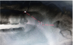
    Figure 1:  Linear measurements -distance of velar tip from posterior end of
hard palate.
b) Maximum excursion of velum during phonation towards the anterior arch of atlas during phonation (Figure 2).
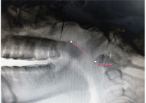
    Figure 2:  Linear measurements - Maximum excursion of velum during
phonation towards the anterior arch of atlas during phonation.
c) Maximum velar lift by estimation of distance between posterior tip of the hard palate and highest point of velar lift during phonation (Figure 3).
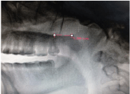
    Figure 3:  Linear measurements -Maximum velar lift by estimation of distance
between posterior tip of the hard palate and highest point of velar lift during
phonation.
d) The maximum distance of the velar midpoint, at rest from the anterior arch of the atlas (Figure 4).
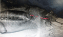
    Figure 4:  Linear measurements -The maximum distance of the velar
midpoint, at rest from the anterior arch of the atlas.
a) Velum- hard palate angle (Figure 5).
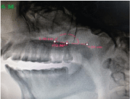
Figure 5: Angular measurements- Velum- hard palate angle.
b) Genu angle (Figure 6).
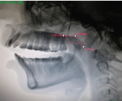
Figure 6: Angular measurements -Genu angle.
Operative technique
The patients were operated by a team of senior consultants. To reduce the surgical bias, a standard protocol of surgical procedure was followed. Standard two flap palatoplasty was performed. Group A underwent radical muscle dissection whereby levator muscle sling was reconstructed followed by three layer closure. Group B did not have radical muscle dissection performed and so closure was done in two layers only. Patients were discharged on 6th postoperative day after speech therapy by a professional speech pathologist. Patients were trained to perform exercises like touching the tip of the tongue at anterior region of hard palate, blowing candles and bits of paper. They were also counselled and encouraged to practice speaking slowly, with an effort to improve their pronunciation.
Ethical approval
The present clinical trial on human beings was approved by the local ethical committee.
Results
Demographic comparison revealed that, Group A had a mean age of 15 years with a maximum of six patients, falling in the age group of 5-10 years. Patients in Group B had mean age of patients was 11 years with a maximum of 8 patients falling in the age group of 5-10 years.
In Group A velopharyngeal closure was seen in all cleft palate types except the cleft of soft palate and uvula. Group B on the other hand had only one patient showing the velopharyngeal closure, who belonged to bilateral incomplete cleft palate type.
Maximum distance of velar midpoint at rest assessed the velopharyngeal gap size at rest. It indicated elongation of the velum due to reduction in gap size post operatively. Group A and Group B both had a significant change post operatively, though the change in the latter group was less significant (Group A, P= 0.000 and Group B, P = 0.006 <0.05). Comparison of mean difference between Group A and Group B gave P = 0.81(>0.05) implying that palatal lengthening occurred in both the groups and was not solely a contribution of radical muscle dissection.
Distance of velar tip from posterior end of hard palate is a direct measure of palatal length and its comparison indicated elongation of the palate. Group A and Group B both showed post operative palatal elongation but comparison of mean difference gave the P value=0.088 (p>0.05) indicating that pushback in both the groups led to an improvement in palatal lengthening.
Maximum velar excursion was taken as a function of both palatal lengthening and levator reconstruction. Group A showed improvement in maximum velar excursion (P=0.027) while Group B did not (P=0.313). This could be attributed to non functional levator sling in Group B.
Maximum velar excursion was taken as a function of both palatal lengthening and levator reconstruction. Group A showed improvement in maximum velar excursion (P=0.027) while Group B did not (P=0.313). This could be attributed to non functional levator sling in Group B.
Maximum velar lift assessed both palatal lengthening and postero superior movement of velum due to excursion of the reconstructed levator sling. Group A (P=0.000) showed a better post operative change in velar lift than Group B (P=0.017). Comparison of mean maximum velar lift gave P value as 0.93 (<0.05). This could be attributed to increase in post operative palatal length in both the groups.
Velar hard palate angle was a direct function of the reconstructed levator muscle sling. It reflected the angular arc through which the palate moves during closure, reflecting the mobility of the soft palate [11]. Group A showed an improvement in this parameter (P=0.001) while Group B did not show an improvement (P=0.22). Comparison of mean velar hard palate angle gave P=0.029 (Table 2). It indicated that radical muscle dissection contributes significantly to improvement of velar function by enhancing its posterosuperior movement (Table 3).
![]()
Group
N
Mean
Std. Deviation
Std. Error Mean
t-value
p-value
Group A
15
10.81
8.04
2.07
0.46
0.64
NS, p>0.05Group B
15
13.01
16.71
4.31
Table 2: Comparison of mean Genu angle in both the groups.
![]()
    Group
    N
    Mean
    Std.    Deviation
    Std.    Error Mean
    t-value
    p-value


    Group    A
    15
    5.53
    5.13
    1.32
    2.29
    0.029
      S, p<0.05


    Group    B
    15
    1.48
    4.50
    1.16
Table 3: Comparison of mean velum hard palate angle in both the groups.
Post operative improvement in genu angle was more in Group A (P=0.000) than that in Group B (P=0.009). Comparison of mean genu angle gave P= 0.64 (Table 4). Genu angle is not only a function of intrinsic activity of levator muscle but also depends on pharyngeal wall contact. False contact of velum with adenoids may be related to enhanced genu angle in Group B patients also and thus yielding a non significant outcome from mean comparison (Table 3).
![]()
    Groups
    Mild
    %
    Moderate
    %
    Severe


    Group    A
    10
    66.67
    5
    33.33
    0


    Group    B
    3
    20
    12
    80
    0
Table 4: Comparison of post operative hypernasality in both the groups.
66.67% patients in Group A had post operative change in hypernasality to mild and 33.33 % patients to moderate hypernasality. Group B, had 20 % patients with post operative change to mild hypernasality while 80% had moderate hypernasality (Table 4). Thus greater number of patients showed improvement in hypernasality in Group A. None of the patients in both the groups had severe hypernasality in the post operative phase.
Comparison of speech understandability revealed that in Group A, 66.67 % patients had mild discrepancies. 26.67 % patients had moderate discrepancies and their speech was often hard to understand (Table 5). One patient in Group A showed no improvement and had severe discrepancies in speech. Group B, 60% patients had moderate discrepancies in their speech and 40 % showed no improvement in their speech post operatively.
![]()
    Groups
    Mild
    %
    Moderate
    %
    Severe
    %


    Group    A
    10
    66.67
    4
    26.67
    1
    6.67


    Group    B
    0
    0
    9
    60
    6
    40
Table 5:  Comparison of post operative speech understand ability in both the
groups.
Discussion
The management of the CP has evolved from obturation (1700s); to simple repairs of the cleft soft palate in early 1800s; to two-flap palatoplasties, such as von Langenbeck’s palatoplasty; to palatal lengthening repairs like Veau-Wardill- Kilner V-to-Y advancement technique; to repairs that along with palatal lengthening, correctly align the palatal musculature. Recreation of the levator sling during CP repair has been associated with a higher probability of successful speech development [10].
Kriens intravelar veloplasty technique (1969) elaborated on reconstruction of the levator sling [11]. Over the time this technique was modified by surgeons like Sommerlad (2003) who advocated the use of microscope for muscle repair [12].
Sommerlad (1994) in his study evaluated degree of closure, changes in rate of closure, changes in velar extensibility and velar lift using lateral videofluroscopy as the evaluating tool [13]. In the present study, these parameters were evaluated as described by Coffey et al (1993) [8]. The present clinical trial included the distance of the velar tip from the posterior end of the hard palate, maximum distance of the velar midpoint, at rest from the anterior arch of the atlas, maximum excursion of the velum and maximum velar lift. Birch et al (1999) in their study concluded that measurements of velopharyngeal distance, extension of soft palate at maximum closure and the angular lift of soft palate above the plane of the hard palate can be reliably and accurately assessed from lateral videofluroscopic images [14]. Two angular measurements co relating with speech as given by Angelo Lipira et al (2011), the velar hard palate angle and the genu angle were also evaluated [9]. The use of lateral videofluroscopy was supported by Sinclair et al (1982) when a comparison of nasal pharyngoscopy, basal and lateral fluorography was done and the conclusion derived was that lateral fluorography provides a high proportion of material with good definition of the velopharyngeal isthmus (80%) [15]. Sommerlad studied 32 cleft palate patients whereby he carried out clinical assessment of nasal resonance and nasal escape and radiographic assessment on lateral videofluroscopy for degree of closure, changes in rate of closure, changes in velar extensibility and velar lift. Lateral videofluroscopy, was concluded to be the most accurate method of measuring velar movement [13]. The finding that greater number of patients operated with radical muscle dissection had post operative velopharyngeal closure was given by Sommerlad (1994).
In the present study, distance of velar tip from posterior end of hard palate evaluated the palatal length which is a predictor of speech outcome as supported by LaRossa et al (2000) [16] in their study. Bae (2002) [17] demonstrated that retro positioning of the velar muscles itself allows some lengthening of the soft palate. In Group A, owing to the additive effect of radical muscle dissection, the post operative increase in palatal length was more than that in Group B. However, on comparing the two groups the difference was no significant with P= 0.088. Thus it may be concluded that the retropositioning of levator muscle doesn’t have significant influence on palatal lengthening but can certainly improves the velar function.
Marsh (2004), stated the angular mobility of the velum in the posterior and superior directions is a reflection of function of levator palatine muscles [18]. In the present study, significant change occurred in velar hard palate angle post operatively in Group A with a P=0.029.
Shi proposed that the optimal time for primary repair of cleft palate was 12 to 18 months old, according to the relationship between the operative age and the growth of the maxilla and velopharyngeal function [19]. Many clinical reports indicated that younger patients with cleft palate had a higher postoperative Velopharyngeal Closure (VPC) rate than older patients, the rate decreased obviously when operative age was older than 2 years. It suggested that with increased operative age, patients will suffer a higher rate of Velopharyngeal Insufficiency (VPI) after primary repair [20]. In Group A, where radical muscle dissection was done, the closure occurred in 30% patients (n=5). Out of these five patients, four belonged to the age groups between 8- 12 years and one adult patient with age of 25 years had velopharyngeal closure. On the other hand, patients who did not have the closure belonged to the age groups varying from 9-25 years of age. Group B, where palatoplasty was done without radical muscle dissection, only 1 patient had the velopharyngeal closure.
Henningsson et al (2008) [7] developed a system of universal parameters for reporting speech outcomes in cleft palate patients. Perceptual parameters were devised that characterized speech production behaviour regardless of the language or languages spoken. Five parameters had chosen included hypernasality, hyponasality audible nasal air emission and /or nasal turbulence, consonant production errors and voice disorders. Universal parameters included were speech understandability and speech acceptability. The position of soft palate lowers during rest and during nasal speech sounds. On the other hand, velar lift/ elevation occurs for non- nasal speech. For vowels, the positioning of the velum depends on adjacent speech sounds also. If vowels are surrounded by non- nasal consonants, the velum tends to elevate, felicitating velopharyngeal closure throughout the word. In contrast, if vowels are surrounded by nasal consonants, the velum will normally tend to be lowered leading to velopharyngeal opening throughout the word [21].
Speech samples in the present study were consistent with the fact that non nasal vowel ‘a’ was surrounded by non nasal consonants like ‘p’, ‘b’ and ‘k’. Also the single word speech followed the structure of consonant-vowel-consonant, which was consistent with the one as described as universal speech sample. There was velar lift throughout the speech which made it easy to assess the velar morphology on videofluroscopy. Conversational speech samples helped in assessing general speech understandability.
Andrades et al (2008) [22] retrospectively assessed the importance of radical muscle dissection during two- flap palatoplasty. They evaluated the outcome measures in terms of perceptual speech evaluation by assessing change in hypernasality, nasal emission, articulation errors, speech intelligibility and velopharyngeal competence. Significant improvement in hypernasality, articulation and velopharyngeal competence was observed but no statistically significant change in speech intelligibility was seen in the group where radical muscle dissection was performed. Nyberg et al (2010) [23] also assessed retrospectively speech results after one stage palatoplasty with and without muscle reconstruction. It was concluded that the technique of muscle reconstruction did not improve articulation or velopharyngeal function. They stated that muscle reconstruction being more extensive left a large raw wound area which resulted in a scar tissue, reducing the effect of muscle reconstruction.
Considering the speech understandability, results though in favour of radical muscle dissection, could not be generalised in view of limited sample size and the fact that conversational speech did not have structured sentences to be spoken. Single word speech assessment showed an improvement of speech but the results could vary with sentence speech samples. All the patients in the present study had crossed the speech formative age and thus were habituated to the abnormal speech. Single word samples when asked to speak in presence of speech pathologist showed improvement but patients reverted back to their habitual speech when made to have a conversational speech.
Summery and Conclusion
It can be concluded that radical muscle dissection improved the action of levator muscle owing to its reconstruction and thus improved the perceptual speech evaluating symptoms also. Two main limitations of the present study were the age group of patients included, well beyond the speech formative years and a lack of follow up.
References