
Case Report
Austin J Otolaryngol. 2023; 9(1): 1126.
Angioembolism for the Management of Refractory Epistaxis
Bhandary R¹ and Shriyan AJ²*
1Associate Professor, Department of ENT, AJIMS, Mangalore, India
2Postgraduate, Department of ENT, AJIMS, Mangalore, India
*Corresponding author: Shriyan AJ Postgraduate, Department of ENT, AJIMS, Mangalore, India
Received: January 03, 2023; Accepted: February 09, 2023; Published: February 16, 2023
Abstract
A majority of the population will experience epistaxis at some time in their life. Most cases will be from an anterior source and can be treated with pressure, anterior nasal packing, or cautery. Intractable epistaxis is generally posterior in origin and may require endoscopic cauterization, posterior packing, ligation of external carotid artery or embolization. Here we report a case of bilateral posterior epistaxis in a 41 year old male patient with c/o spontaneous epistaxis from bilateral nostril. Digital Subtraction Angiography followed by Angioembolisation was done.
Keywords: Angioembolism; Refractory Epistaxis
Introduction
Clinically, there are two types of epistaxis: anterior (more common), and posterior (less common but more serious). About 80% of cases consist of anterior epistaxis [1]. Luckily, most of the epistaxis is self-limited, with only 6% requiring medical attention [2]. However, non-invasive treatments are not always effective for bleeding from the posterior nasal passage and may require further treatment strategies such as surgery or percutaneous artery embolization. Epistaxis is almost equally distributed between the sexes, but persistent posterior epistaxis is more common in older men. Studies show a soft peak in epistaxis cases in the first two decades, followed by an increase after the fourth decade. Many factors could cause epistaxis, including head and face trauma, digital trauma, postoperative complications, hypertension, inflammation and infections, cancers, vascular anomalies, and especially anticoagulant medication use. The blood flow of the nasal cavity originates from both the internal carotid artery and the external carotid artery. The anterior and posterior ethmoid arteries originate from the ophthalmic artery that branches off from the internal carotid artery. The facial artery and internal maxillary artery are branches from the external carotid artery. The terminal branch of the internal maxillary artery is the sphenopalatine artery, which usually provides blood flow to the lateral nasal wall and septum [3]. Even in the absence of a vascular malformation, there are several potential connections between the internal and external carotidartery, such as anastomoses between the right and left arterial systems. Therefore, epistaxis may continue despite unilateral arterial embolization. In addition, pre-existing anastomoses can be opened due to increased pressure during embolization and undesirable embolization of the internal carotid artery or ophthalmic artery may occur [4,5]. Therefore, embolization is contraindicated in the presence of an anastomosis between the external and internal carotid artery or in cases of bleeding in the ethmoid artery, a branch of the ophthalmic artery, due to the risk of blindness [1,2,6].
Angiographic embolization is the accepted invasive treatment option for severe refractory epistaxis.
Case Report
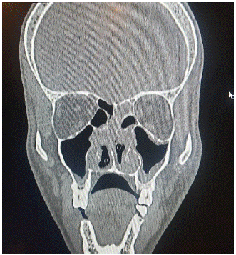
A 41 Year old male patient came with chief complaints of spontaneous bleeding from bilateral nostrils since 1 day. No history of nasal obstruction, No h/o URTI, No h/o nose pricking, trauma, No h/o difficulty in breathing. On examination External nasal framework – normal, Columella and Vestibule – normal, Anterior rhinoscopy – DNS to left present. No PNS/TMJ tenderness. INVESTIGATIONS: Hematological parameters all within normal limits. (Bleeding parameters are within normal limits) CT PNS –Mucosal thickening in bilateral ethmoidal, maxillary and sphenoid sinuses. Feature suggestive of Chronic sinusitis.
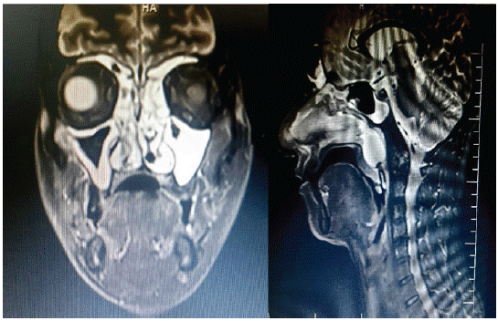
Screening MRI
Mucosal thickening in B/L ethmoid, maxillary, sphenoid and left frontal sinuses. Posterior nasal pack noted insitu.
Treatment
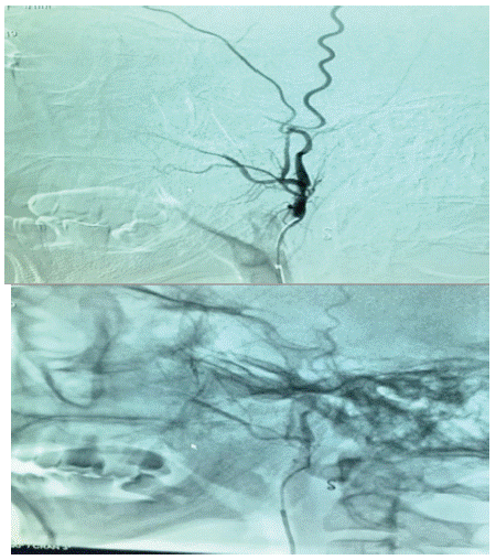
Anterior nasal packing was done under aseptic precaution, patient continued to bleed. Hence posterior nasal packing was done, in spite of 48 hrs of anterior and posterior nasal packing patient continued to bleed profusely. He was posted under GA for cauterization of the bleeding points. He was reposted under GA; the pack was removed and he had torrential bleeding from B/L post nasal cavities and posterior wall of nasopharynx. In spite of cauterizing all the areas and posterior wall of nasopharynx, patient continued to bleed. Hence Sphenopalatine artery ligation was not attempted. After repacking and after patient came out of anesthesia, reference was given to Interventional radiologist to determine the vessel causing bleeding and to embolize the same. Under strict aseptic precautions, arterial access was obtained through the right common femoral artery using Modified Seldinger's technique. Selective angiograms of ECA branches revealed mucosal staining predominantly in the posterior nasopharynx supplied by branches from both ascending pharyngeal arteries, ascending palatine branch of facial arteries and both IMA branches which were selectively cannulated and embolized using PVA particles and Gel foam. Right ascending pharyngeal artery was coil embolized after particulate embolization. Check angiogram revealed stasis within the targeted vessels. Patient tolerated the procedure well.
Discussion
Epistaxis is one of the emergencies [7] which may prove life threatening if not managed appropriately. There may be many etiological factors responsible for the nasal bleeding but in most of the cases these bleeds are idiopathic. Majority of the nasal bleeds are managed easily with Anterior and/or Posterior (AP) nasal packing. However, the failure rate of AP packing is about 0%-52% [8]. The optimal treatment of nasal bleeds refractory to the AP packing is still controversial [9]. Traditionally the standard treatment for uncontrolled nasal bleeds was the surgical ligation of the bleeding vessels [10]. In 1974, Sokoloff was the first one who introduced the embolization technique for the treatment of epistaxis. Then over the period of time, technology as well as the level of experience improved in the field of interventional radiology and embolization became one of the accepted treatment options for epistaxis. Similar study was done in Aga Khan University Hospital, Karachi where the primary method to deal with intractable epistaxis is angioembolization. They achieved overall success rate of 87.5%, comparable to the international literature, which ranges from 71 to 100%. This is also comparable with the average success rate of trans-antral ligation of Internal Maxillary Artery (IMA), which is 87%. Polyvinyl alcohol (PVA) particles and gel foam were used as an embolizing material. The PVA particles vary in their size, from 45 to 150 and even up to 700 microns. In our series PVA size was between 250 to 350 microns. These particles must be directed in antegrade fashion to the arteriolar bed and care must be taken to avoid the reflux to the proximal arterial branches [11].
Conclusion
Nasal bleed refractory to conservative management can be treated either surgically or by embolization. But angioembolization may be the treatment of choice for intractable nasal bleeding due to high success rate without any serious complications, short hospital stays, avoidance of general anaesthesia and less discomfort to the patients.
Informed Consent
Separate informed consent was taken prior to CT scan, MRI and surgery.
References
- Schaitkin B, Strauss M, Houck JR. Epistaxis: medical versus surgical therapy: a comparison of efficacy, complications, and economic considerations. Laryngoscope. 1987; 97: 1392-6.
- Small M, Murray JA, Maran AG. A study of patients with epistaxis requiring admission to hospital. Health Bull (Edinb). 1982; 40: 20-9.
- Leppanen M, Seppanen S, Laranne J, Kuoppala K. Microcatheter embolization of intractable idiopathic epistaxis. Cardiovasc Intervent Radiol. 1999; 22: 499-503.
- Pope LE, Hobbs CG. Epistaxis: an update on current management. Postgrad Med J. 2005; 81: 309-14.
- Willems PW, Farb RI, Agid R. Endovascular treatment of epistaxis. AJNR Am J Neuroradiol. 2009; 30: 1637- 45.
- Strach K, Schrock A, Wilhelm K. Endovascular treatment of epistaxis: Indications, management, and outcome. Cardiovasc Inter-vent Radiol. 2011; 34: 1190-8
- Hussain G, Iqbal M, Shah SA, Said M, Sanaullah, Khan SA, et al. Evaluation of aetiology and efficacy of management protocol of epistaxis. J Ayub Med Coll Abbottabad. 2006; 18: 63-6.
- Cannon CR. Effective treatment protocol for posterior epistaxis: a 10-year experience. Otolaryngol Head Neck Surg. 1993; 109: 722-5.
- Elahi MM, Parnes LS, Fox AJ, Pelz DM, Lee. Therapeutic embolization in the treatment of intractable epistaxis. Arch Otolaryngol Head Neck Surg. 1995; 121: 65-9.
- Small M, Maran AGD. Epistaxis and arterial ligation. J Laryngol Otol. 1984; 98: 281-48.
- Tseng EY, Narducci CA, Willing SJ, Sillers MJ. Angiographic embolization for epistaxis: a review of 114 cases. Laryngoscope. 1998; 108: 615-9.