Case Report
Austin J Otolaryngol. 2023; 9(1): 1130.
Undifferentiated Sinonasal Carcinoma with Lymph Node Metastases: A Case Report
Bencheikh; Benbouzid; Oujilal; Essakalli; Noureddine J*
Avicenne University Hospital Center, Specialized Hospital, Morocco
*Corresponding author: Jelloul Noureddine Department of Otolaryngology Head and Neck Surgery, Specialized Hospital, AVICENNE University Hospital Center, Rabat, Morroco. Email: noureddinejelloul00@gmail.com
Received: November 13, 2023 Accepted: December 21, 2023 Published: December 28, 2023
Introduction
Undifferentiated sinonasal carcinoma (SNUC) is a rare malignant tumor that originates from the Schneiderian membrane lining the nasal cavities and paranasal sinuses. It often presents with rhinological symptoms, while other ocular or neurological signs can be either initial or secondary manifestations [1,2]. CT and MRI provide precise locoregional assessment [2,14]. Diagnosis relies on anatomopathological examination [2,3], and the treatment primarily involves surgery and radiochemotherapy (RECFOR.4). This tumor poses management challenges due to its typically late diagnosis and significant extent, which can hinder complete tumor resection. This study aims, along with a literature review, to outline the histoclinical characteristics of SNUC, revisit prognostic factors, and establish an appropriate therapeutic approach for this tumor.
Materials and Methods
Clinical Case
We present the case of a 70-year-old female patient with no significant medical history, admitted due to rhinosinusitis symptoms dominated by left-sided nasal obstruction accompanied by recurrent ipsilateral epistaxis evolving for 2 months. Nasal endoscopy revealed a budding process within the left nasal cavity, straddling the inferior and middle turbinates and curving along the posterior edge of the nasal septum.
Cervical examination identified multiple bilateral laterocervical lymph nodes. Neurological and ophthalmological examinations were unremarkable. The rest of the ENT examination showed no particular findings.
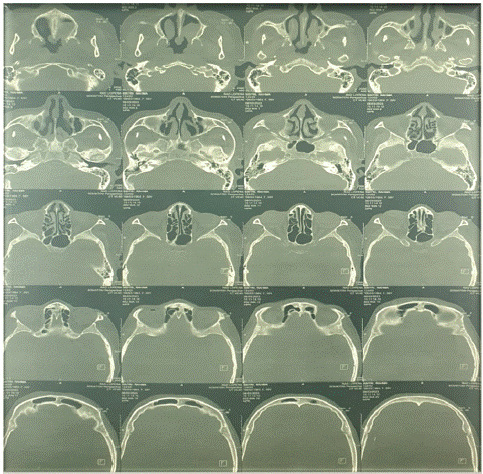
Facial CT scan exhibited a tissue-density lesion within the left nasal cavity, showing discrete enhancement after contrast agent injection. The lesion straddled the middle and inferior turbinates without expansion into the choanal or associated bone lysis (Figure 1).

Figure 1: CT scan cervical.
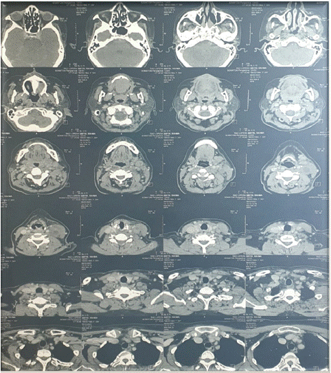
Both CT and cervical ultrasound revealed significant bilateral cervical lymphadenopathy, predominantly on the left side and of suspicious appearance (Figure 2).

Figure 2: CT scan cervical.
A tumor biopsy confirmed Undifferentiated Sinonasal Carcinoma.
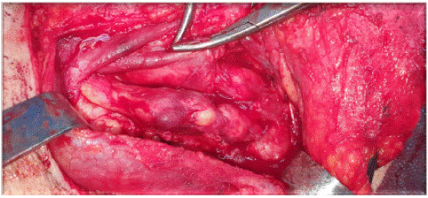
Initially, the patient underwent a principle-based cervical lymph node dissection (Figure 3), involving the right side with territories IIa, IIb, III, IV, and V, and the left side with territories IIa, III, IV, including ligature of the internal jugular vein adherent to the mass of lymph nodes.

Figure 3: Lymph node metastases.
Intraoperative frozen section analysis of the right lymph node dissection favored metastatic nodes of an undifferentiated carcinoma, with some nodes showing necrosis.
Subsequently, the patient underwent endoscopic endonasal surgery. Intraoperative examination revealed a budding formation straddling the inferior and middle turbinates. We achieved tumor resection via an endonasal approach.
The resection was macroscopically complete. Final histological examination confirmed a similar morphological aspect to that observed in the biopsy.
The diagnosis of undifferentiated sinonasal carcinoma was thus reaffirmed.
Discussion
Undifferentiated carcinomas, first identified in 1986, are rare and aggressive. Chambers et al, in a retrospective study of 318 American cases, manage to estimate the incidence at 0.2 new cases per million inhabitants per year. SNUC would represent less than 2% of malignant naso-sinus tumors (Chambers 2015). The increase in published cases in recent years corresponds more to improved diagnostic capabilities than to an actual increase in the disease's frequency. In most series, this tumor affects men more than women and can occur at any age with an average of around 53 years [5]. No risk factor has been clearly identified in the literature. Risk factors commonly found in other malignant tumors of naso-sinus origin have not yet demonstrated a causal link with SNUC (RECFOR.6).
Clinically, in 75% of cases, the tumor is revealed by rhino-sinus signs, primarily nasal obstruction (20%), followed by epistaxis (17%), headaches (12%), and facial pain (7.1%) at the time of diagnosis [6]. Clinical examination should investigate lymph node involvement, present in 8 to 16% of cases [6].
Ophthalmological examination is mandatory due to the frequency of ocular signs (20%) (5). the ophthalmological examination of our patient was normal. Examination of the cervical lymph nodes is also systematic, especially since SNUC is lymphophilic [7]. Our patient presented with multiple palpable bilateral lateral cervical lymph nodes.
Radiologically, standard sinus radiology is of no interest in the initial assessment of naso-sinus neoplasms (RECFOR.14). Imaging of this type of cancer mainly involves Computed Tomography (CT) and MRI, which are complementary [14]. CT, with coronal and axial sections before and after contrast injection, is the preferred examination, highlighting a homogenous opacity that moderately enhances after contrast injection [14]. It precisely defines the exact limits of intracranial extension and detects involvement of the anterior skull base. More recently, UICC published its 8th version of the TNM classification using CT and MRI:
• T1 = Nasal and/or sinus tumor leaving an air space between the tumor and the cribiform plate.
• T2 = Tumor coming into contact with the cribiform plate, possibly eroding it.
• T3 = Extracranial intradural tumor and/or orbital involvement.
• T4 = Intracranial intradural tumor.
In our study, radiological staging was done according to the TNM classification. Thus, our patient was classified as T1N2c.
For diagnosis, the importance of a comprehensive immunohistochemical panel and, if necessary, molecular analyses [8] should be emphasized. This helps rule out certain differential diagnoses (olfactory neuroblastoma, poorly differentiated neuroendocrine carcinoma, etc.).
Due to the rarity of the disease, the aggressive behavior of the tumor, and poor outcomes, defined treatment protocols and recommendations are lacking. According to recommendations (RECFOR.1.4.9.10), the treatment of SNUC depends on whether the tumor is operable or not. If the tumor is operable (T1, T2), complete macroscopic and microscopic excision surgery with clear margins followed by postoperative concurrent radiochemotherapy (professional consensus.13) is the standard treatment for SNUC.
In the case of inoperable tumors (T3, T4), it is recommended to consider induction chemotherapy and, if there is a good response, discuss potential concurrent radiochemotherapy (RECFOR.1.4.9.10.13).
Place and modalities of radiothérapie: Preoperative radiotherapy, although advocated by a few centers, is not a standard approach [3,8]. This type of radiotherapy targets the tumor bed (64.5–71.3 Gy at 2.0–2.25 Gy/fractions, five fractions per week) as well as the lymph node regions (64–70 Gy at 2.0–2.2 Gy/fraction). The radiation dose can range from 65 to 75 Gy in cases of significant tumor volume. Elective nodal irradiation of the neck was administered at a dose of 70-75 Gy [15].
Place and modalities of chemotherapy:
In the REFCOR cohort, significant reduction in recurrence rates was observed among patients who received neoadjuvant chemotherapy, with a 2-year recurrence-free survival rate of 92%, compared to 46% for patients who underwent surgery without induction chemotherapy and 25% for patients who did not receive any pre- or post-operative chemotherapy (de Bonnecaze 2018).
For patients with N0 status, it is not recommended to propose cervical lymph node dissection. Instead, prophylactic irradiation of the cervical lymph node regions is suggested to enhance regional disease control and potentially improve survival (RECFOR.9).
For patients diagnosed with N+ status, as in the case of our patient and which constitutes a poor prognostic factor, treatment of the cervical lymph node regions is warranted systematically, either through cervical radiotherapy or lymph node dissection if tumor surgery is being considered (RECFOR.9.11).
SNUC is associated with a very poor prognosis. Survival is linked to clinical factors, undifferentiated nature, TNM classification, and therapeutic strategy (REFCOR.12). Local and locoregional recurrences can be either early or late, necessitating lifelong monitoring of these patients. Reiersen et al's meta-analysis of 167 patients found intraorbital involvement in 32%, skull base invasion in 25%, with one-third of those invading brain tissue, accounting for 8% at the time of diagnosis (Reiersen 2012). The disease is predominantly staged as T4 at the time of diagnosis (Xu 2013 75%, Bonnecaze 2018 81%, Morand 2017).
It is important to note that our case was classified as T1N2c SNUC. Therefore, our data support elective neck treatment through bilateral lymph node dissection followed by adjuvant radiotherapy.
Conclusion
SNUC is a rare tumor with a relatively grim prognosis. Several areas of uncertainty remain in the management of this disease, including the sequencing of various therapeutic modalities, optimal chemotherapy agents and dosages, as well as the optimal radiation dose and target.
For medically inoperable patients, radiochemotherapy is recommended, while those who are resectable or potentially resectable should undergo curative resection. The extent of resection is a reliable predictor of good tumor control, unlike nerve invasion, which is an unfavorable factor for local control. Given the scarcity of data on this rare tumor, the limited reports in the literature may provide valuable assurance that surgery, in addition to radiochemotherapy, can offer better outcomes in the context of SNUC.
References
- Wang L, Wang J, Tang T, Yan L, Song X. Definitive-intent (chemo) radiotherapy for sinonasal undifferentiated carcinoma. Br J Radiol. 2023; 96: 20220244.
- Agaimy A, Hartmann A, Antonescu CR, Chiosea SI, El-Mofty SK, Geddert H, et al. SMARCB1 (INI-1)-deficient sinonasal carcinoma: A series of 39 cases expanding the morphologic and clinicopathologic spectrum of a recently described entity. Am J Surg Pathol. 2017; 41: 458-71.
- Amit M, Abdelmeguid AS, Watcherporn T, Takahashi H, Tam S, Bell D, et al. Induction chemotherapy response as a guide for treatment optimization in sinonasal undifferentiated carcinoma. J Clin Oncol. 2019; 37: 504-12.
- Mody MD, Saba NF. Multimodal therapy for sinonasal malignancies: updates and review of current treatment. Curr Treat Options Oncol. 2020; 21: 4.
- Al-Mamgani A, van Rooij P, Mehilal R, Tans L, Levendag PC. Combined-modality treatment improved outcome in sinonasal undifferentiated carcinoma: single-institutional experience of 21 patients and review of the literature. Eur Arch Otorhinolaryngol. Eur Arch Otorhinolaryngol. 2013; 270: 293-9.
- Reiersen. Morand. 2012. p. 2017.
- Thariat J, Moya Plana A, Vérillaud B, Vergez S, Régis-Ferrand F, Digue L, et al. Bull Cancer. Diagnosis, prognosis and treatment of sinonasal carcinomas (excluding melanomas, sarcomas and lymphomas). 2020; 107: 601-11.
- Ejaz A, Wenig BM. Carcinome indifférencié sinonasal: caractéristiques cliniques et pathologiques et discussion sur la classification, la différenciation cellulaire et le diagnostic différentiel. Adv Anat Pathol. Adv Anat Pathol. 2005; 12: 134-43.
- Faisal M, Seemann R, Lill C, Hamzavi S, Wutzl A, Erovic BM, et al. Elective neck treatment in sinonasal undifferentiated carcinoma: systematic review and meta-analysis. Head Neck. J Neurol Surg B. 2019; 80: 088-095.
- de Bonnecaze G, Verillaud B, Chaltiel L, Fierens S, Chapelier M, Rumeau C, et al. Clinical characteristics and prognostic factors of sinonasal undifferentiated carcinoma: a multicenter study. Int Forum Allergy Rhinol. 2018; 8: 1065-72.
- Baglin AC, Benlyazid A, Borel C, et al. Tumeurs malignes primitives des fosses nasales et des sinus. Recommandations pour la Pratique Clinique. juillet 2009.
- Eggesbø HB. Imaging of sinonasal tumours. Cancer Imaging. Cancer Imaging. 2012; 12: 136-52.
- Radiotherapy for sinonasal undifferentiated carcinoma. Am J Otolaryngol. 2014; 35: 141-6.