Case Report
Austin J Pediatr. 2015;2(1): 1017.
Moyamoya Syndrome Presenting as Stroke in a Toddler with Down Syndrome
Arora R¹*, Kannikeswaran N¹, Bhaya N¹ and Sivaswamy L²
¹Division of Pediatric Emergency Medicine, Wayne State University, USA
²Division of Pediatric Neurology, Wayne State University, USA
*Corresponding author: Rajan Arora, Division of Pediatric Emergency Medicine, Department of Pediatrics, Children’s Hospital of Michigan, Wayne State University, 3901 Beaubien Blvd, Detroit, Michigan, 48230, USA.
Received: April 20, 2015; Accepted: May 14, 2015; Published: May 15, 2015
Abstract
Pediatric stroke is a rare but important clinical entity. Lack of experience with childhood stroke, atypical presentations, and a wider differential diagnosis often leads to delayed diagnosis of stroke in children. This case illustrates the association of Moyamoya Syndrome (MMS) with Down Syndrome (DS) in a 2 year old who presented to our emergency department with acute onset hemiparesis.
Keywords: Down syndrome; Stroke; Moyamoya
Case Presentation
A 22 month old toddler with known diagnosis of DS presented with a one day history of acute onset left sided weakness. The weakness was not associated with seizures or alteration in sensorium. Review of symptoms was negative for fever, URI, vomiting, diarrhea, rash, trauma, ingestion or any other recent illness. The child had a similar episode 2 weeks previously which was transient and resolved spontaneously over a couple of hours. Apart from mild developmental delay, she was an otherwise healthy child with no history of cardiac disease.
On examination she was alert and playful. Her vital signs included temperature of 37°C, HR of 96/min, RR of 28/min and BP of 98/66 mmHg. Neurological examination was consistent with left sided hemiparesis with up going plantar response on the affected side. Cranial nerves were grossly normal with no facial asymmetry. Examination of other systems was unremarkable.
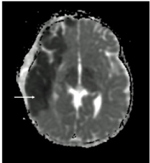
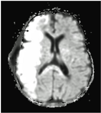
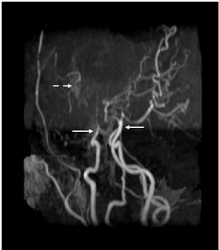
Initial work up including blood counts, comprehensive metabolic panel, coagulation profile, lipid profile and hemoglobin electrophoresis studies were within normal limits. Magnetic Resonance Imaging of the child’s brain revealed a fresh infarct in the right middle cerebral artery territory (Figures 1 and 2). Magnetic Resonance Angiography (MRA) (Figure 3) of the brain showed complete occlusion of bilateral middle cerebral arteries with significant collateral formation consistent with diagnosis of moyamoya. Echocardiogram was normal. Screening for other causes of stroke including autoimmune (Anti-nuclear antibody, double stranded DNA, cardiolipin antibody screen), prothrombotic (protein C, S and antithrombin III deficiency, lupus anticoagulant, factor V Leiden mutation) and metabolic disorders (homocystinuria) were normal.

    Figure 1:  MRI Apparent Diffusion Coefficient showing dark signal (arrow)
corresponding to area of new infarct.

    Figure 2:  MRI Diffusion Weighted Imaging showing bright signal (arrow) in
area of new infarct.

    Figure 3:  Magnetic Resonance Angiography revealing occlusion of bilateral
internal carotid arteries (solid arrows) and formation of collaterals (dotted
arrow).
Patient was started on low dose (3mg/kg/day) aspirin therapy. Single Photon Emission Computed Tomography (SPECT) imaging further revealed diffuse asymmetric baseline activity and abnormal poor perfusion in bilateral frontal parietal regions suggesting multiple areas at risk for further stroke. In view of poor perfusion reserve and risk for further strokes she underwent surgical reanastomosis and experienced moderate recovery of motor function. Post operatively she again suffered a left sided stroke and focal seizure for which she was started on Levetiracetam. Presently she has a marked left sided weakness for which she is currently receiving physical and occupational therapy to help with her rehabilitation.
Discussion
Pediatric stroke, though a relatively rare phenomenon, is increasingly being recognized as an important cause of childhood morbidity and mortality. International data suggest an incidence of 2 to 5/100,000 children per year [1]. Significant delays often exist in timely diagnosis of stroke in children. Such delays are often attributed to lack of experience with pediatric stroke, frequent non-focal presentations, broader differential diagnosis and poor sensitivity of computed tomography scanning for the diagnosis of pediatric acute ischemic stroke [2].
Stroke in children may present differently in comparison to adults and manifestations could be age specific. Infants are more likely to present with seizures and altered mental status whereas older children tend to have hemiparesis or other focal neurologic deficits. A total of 70 to 80% of the children present with hemiparesis, with or without facial palsy, or dysphasia [3]. The etiology of childhood stroke is extensive and not limited to atherosclerosis and cardiovascular disease as in their adult counterparts. Arteriopathies are thought to be most frequently associated with pediatric acute ischemic stroke and may be acute, transient, or progressive. Common arteriopathies include focal cerebral arteriopathy of childhood, moyamoya, cervicocephalic arterial dissection, and sickle cell disease [4]. Other reported risk factors of childhood stroke include congenital or acquired heart disease, infectious/parainfectious etiologies such as vasculitis, hematological disorders, coagulopathies, arteriosclerosis, connective tissue and metabolic disorders (Fabry disease, mitochondrial disorders and Menkes syndrome).
Children with DS are at increased risk for cerebral infarction due to multiple reasons. Thromboembolism secondary to an underlying cardiac malformation accounts for the vast majority of cases. An increased susceptibility to bacterial infection, including meningitis, septicemia, and sub-acute bacterial endocarditis also contribute towards cerebrovascular occlusion and stroke in this patient population [5]. Other less commonly reported mechanisms include leukemia and upper cervical subluxation associated with atlanto-axial instability. MMS is a rare cause of stroke in DS, with estimated incidence in this subgroup to be approximately three times when compared to the general population [5,6]. However a recent study based on data from the Nationwide Inpatient Sample reported the prevalence of DS in patients with coexisting MMS to be 26-fold greater than the prevalence of DS among live births (145 per 100,000) suggesting that in the white and Hispanic patient populations, DS is a risk factor of much greater magnitude for reasons that are poorly understood [7].
Children who are suspected of having a stroke warrant urgent neuroimaging. MRI is the ideal modality, though CT brain without contrast is also helpful in excluding hemorrhage and mass effect if MRI is not readily available. There are no well validated guidelines addressing further work up for the assessment of pediatric stroke. Laboratory work up should be undertaken to identify specific underlying causes of stroke such as cardiac disease, infections, vasculitis, thrombophilia, and metabolic disorders with guidance from multidisciplinary sub-specialty consultation.
Moyamoya Disease (MMD) is an idiopathic entity characterized by a progressive non-inflammatory, non-vasculitic and nonatherosclerotic occlusion of bilateral intracranial arteries in absence of any associated disease. MMS or phenomenon is the term used when an underlying disease is thought to be responsible for extensive collateralization and angiographic appearance similar to that seen in the primary disease. The conditions associated with MMS include atherosclerosis, sickle cell anemia, neurofibromatosis and DS. The slowly progressive occlusion of blood vessels allows for the development of small anastomotic collaterals which appear on an angiogram or a MRA scan as a characteristic “puff of smoke”, giving the disease it’s Japanese name.
MMD has a bimodal onset, peaking in the first and the fourth decades of life. The average age of presentation of patients with MMS with trisomy 21 has been reported to be 5–7 years [8,9]. Hemiparesis happens to be the most common presentation in both the groups. In children, the strokes tend to be ischemic, whereas they are more often hemorrhagic in adults [10]. Children often present with recurrent transient ischemic attacks which may alternate sides. In addition to motor deficits, these patients may present with aphasia, sensory impairment, involuntary movements (chorea), headache, seizures, or cognitive impairment. Many of these episodes can be triggered by hyperventilation, crying, coughing, straining, dehydration or fever.
The exact etiology of the moyamoya phenomenon is unknown but several hypotheses have been proposed. Genetic mode of inheritance is suggested based on a higher incidence of the disease in Asian populations and familial occurrence in both Asians and whites [11]. Mutations in the mediators of angiogenesis and connective tissue development such as fibroblast growth factor, transforming growth factor 1, elastin and infectious agents like Epstein-Barr virus have also been implicated as a contributing factor [11,12]. Lastly, patients with DS are inherently predisposed to vascular disease. This is reflected in abnormal nail-bed capillary morphology, high pulmonary vascular resistance, and primary intimal fibroplasia [13]. Similar vascular dysplasia in the brain may result in a structural defect which forms the basis for MMS.
Diagnosis of MMS requires visualization of the cerebral blood vessels. MRA is an excellent non-invasive alternative to conventional angiography. However if results are equivocal on MRA one should pursue angiography. Intracranial changes may include narrowing of vessels of the circle of Willis, areas of previous stroke, and intracerebral hemorrhage. MRA studies depicting absent or reduced flow in the major arteries of the brain coupled with abnormally prominent flow voids from basal ganglia and thalamic collateral vessels are virtually diagnostic of moyamoya [14]. The Research Committee on the Pathology and Treatment of Spontaneous Occlusion of the Circle of Willis have laid down criteria and guidelines for diagnosis and treatment of MMD [15]. Perfusion and cerebral flow studies like Positron Emission Tomography (PET) and SPECT scan are necessary to identify surgical candidates and in planning the surgical procedure itself.
Various treatments modalities for pediatric stroke associated with moyamoya have been explored, although none are ideal. Emergency department care involves supplemental oxygen for hypoxemic patients, antipyretics if febrile and adequate hydration with isotonic solutions. Medical management includes antithrombotic therapy, correction of underlying cause if present and general supportive measures. Daily low dose aspirin therapy is recommended for those patients with normal perfusion reserve on PET. Surgical treatment is indicated in patients with poor reserve on PET scan or those with cognitive decline or recurrent or progressive neurological deficits. Cerebral revascularization surgery with the pial synangiosis technique seems to confer long-lasting protection against additional strokes in this patient population [16].
MMD results in recurrent strokes with gradual neurologic and cognitive deterioration in 50% to 60% of patients if untreated. Mortality rates of up to 4.3% are seen in MMD [14]. The prognosis for patients with moyamoya is variable and difficult to predict. Overall prognosis depends upon a host of factors including the rate and severity of vascular blockade, effective collateral circulation, the age at onset of symptoms, and the severity of disability resulting from a stroke. However, if left untreated, the disease invariably progresses, producing clinical deterioration and potentially irreversible neurological deficits over time.
Conclusion
Pediatric stroke is a rare but important clinical entity. General practitioners and emergency physicians should be able to recognize common presenting features and risk factors associated with pediatric stroke. MMS should be considered as a differential diagnosis in patients with DS presenting with new-onset focal weakness or any other neurological abnormalities. Increased awareness will help in timely diagnosis and treatment of children with stroke syndromes.
References
- Amlie-Lefond C, Sébire G, Fullerton HJ. Recent developments in childhood arterial ischaemic stroke. Lancet Neurol. 2008; 7: 425–435.
- Rafay MF, Pontigon AM, Chiang J, Adams M, Jarvis DA, Silver F, et al. Delay to diagnosis in acute pediatric arterial ischemic stroke. Stroke. 2009; 40: 58–64
- Steinlin M, Pfister I, Pavlovic J, Everts R, Boltshauser E, Capone Mori A, et al. The first three years of the Swiss Neuropaediatric Stroke Registry (SNPSR): a population-based study of incidence, symptoms and risk factors. Neuropediatrics. 2005; 36: 90–97.
- Fox CK, Fullerton HJ. Recent advances in childhood arterial ischemic stroke. Curr Atheroscler Rep. 2010; 12: 217–224.
- Fung CW, Kwong KL, Tsui EYK, Wong SN. Moyamoya syndrome in a child with Down syndrome. Hong Kong Med J. 2003; 9: 63–66.
- Rison RA. Fluctuating hemiparesis secondary to moyamoya phenomenon in a child with Down syndrome: a case report. Cases J. 2008; 1: 240.
- Kainth DS, Chaudhry SA, Kainth HS, Suri FK, Qureshi AI. Prevalence and characteristics of concurrent down syndrome in patients with moyamoya disease. Neurosurgery. 2013; 72: 210-215.
- Boggs S, Hariharan SL. An uncommon presentation of stroke in a child with trisomy 21. Pediatr Emerg Care. 2008; 24: 230-232.
- Worley G, Shbarou R, Heffner AN, Belsito KM, Capone GT, Kishnani PS. New onset focal weakness inchildren with Down syndrome. Am J Med Genet A. 2004; 128A: 15-18.
- Cramer SC, Robertson RL, Dooling EC, Scott RM. Moyamoya and Down syndrome: clinical and radiological features. Stroke. 1996; 27: 2131-2135.
- Gosalakkal JA. Moyamoya disease: a review. Neurol India. 2002; 50: 6-10.
- Yamamoto M, Aoyagi M, Tajima S, Wachi H, Fukai N, MatsushimaY, et al. Increase in elastin gene expression and protein synthesis in arterial smooth muscle cells derived from patients with moyamoya disease. Stroke. 1997; 28: 1733-1738.
- Dai AI, Shaikh ZA, Cohen ME. Early-onset Moyamoya syndrome in a patient with Down syndrome: case report and review of the literature. J Child Neurol. 2000; 15: 696-699.
- Scott RM, Smith ER. Moyamoya disease and moyamoya syndrome. N Engl J Med. 2009; 360: 1226–1237.
- Research Committee on the Pathology and Treatment of Spontaneous Occlusion of the Circle of Willis Guidelines for diagnosis and treatment of moyamoya disease (spontaneous occlusion of the circle of Willis). Neurol Med Chir (Tokyo). 2012; 52: 245–266.
- Jea A, Smith ER, Robertson R, Scott RM. Moyamoya syndrome associated with Down syndrome: outcome after surgical revascularization. Pediatrics. 2005; 116: e694-e701.