Case Report
Austin Pediatr. 2019; 6(1): 1070.
Primary Obstructive Megaureter with Giant Ureteral Stone Presenting as Orchialgia and Undescended Testis
Telli O*, Hanci B, Dinçer E, Karaca Y, Bulut M, Sevinç A and ÖzcanT
Doctor, Kartal Lütfi Kirdar Training and Research Hospital, Kartal/Istanbul, Turkey
*Corresponding author: Onur Telli, Pediatric Urology, Kartal Lütfi Kirdar Training and Research Hospital, Kartal/Istanbul, Turkey
Received: September 02, 2019; Accepted: October 01, 2019; Published: October 08, 2019
Introduction
Primary Obstructive Megaureter (POM) is relatively uncommon in adults that only 15% of megaureters, Nevertheless, surgical intervention is indicated in POM in cases of deterioration of split renal function and significant obstruction. Most of the cases remains asymptomatic for years until some symptoms may occur in the event of any complication such as urolithiasis, renal insufficiency, recurrent urinary infections, and pain [1]. Here in, we present a case with giant ureter stone presented as orchialgia after diagnosis of undescended testis.
Case Presentation
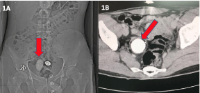
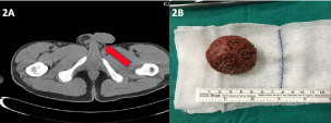
A 15 years-old male patient presented to our outpatient clinic with non-specified chronic orchialgia. He had taken only painkillers occasionally to relieve his pain and no relevant medical history. A physical examination revealed that he has left undescended testes. Scrotal ultrasonography showed left atrophic testis at the deep inguinal ring. Routine urinalysis showed normal blood urea nitrogen, creatinine, and positive leukocytes. Serum levels of a-fetoprotein and β-human chorionic gonadotrophin were within the normal ranges. Interestingly, X-Ray abdominal radiography evaluation showed a radiopaque stone, 5 cm in diameter, in right side of pelvic bone (Figure 1A). CT scan showed mild renal pelvis hydronephrosis with right megaureter, and approximately 5 cm stone at distal ureter with narrow ending (Figure 1B). Secondly, a left undescended testis was seen with an empty scrotum in CT (Figure 2A). Diuretic renography performed and right kidney had a non-obstructed megaureter with % 46 split function. The patient underwent ureterorenoscopy under general anesthesia. After dilatation the of ureterovesical junction, 5 cm of non-obstructed stone was seen in the distal part of ureter. Ureteroneocystostomy was performed resecting a dynamic distal end of the ureter and stone was removed (Figure 2B). Patient had left simple inguinal orchiectomy in same session. Histopathologic examinations of the specimens confirmed as atrophic–benign tissue and fibrotic uretero-vesical junction.

    Figure 1: 1 A) X-Ray abdominal radiography showed a radiopaque stone, 5
cm in diameter, in right side of pelvic bone. 1B) CT scan right megaureter,
including 5 cm stone at distal ureter.

    Figure 2: A) A left undescended testis was seen with an empty scrotum in CT.
2B) 5 cm in diameter stone.
Discussion
POM is a functional or anatomical abnormality involving the ureterovesical junction. Diagnosis of POM depends on the absence of secondary causes of lower ureteral obstruction with a ureter greater than 7 mm in diameter. [2] However, most of the cases without recurrent febrile urinary tract infections or an obstruction shown in diuretic renography could be follow-up without surgery. Hemal et al. reported 30% of 55 patients with POM had urinary tract calculi located in the ureter. [3] In current case, the patient had 46% split function and non-obstructed diuretic renogram. Initial Differential Renal Function (DRF) below 40%, or a drop in DRF of 5% on serial scans, and an increasing dilatation on serial Ultrasound (US) scans are considered suggestive of obstruction. We believe that stone was developed in the dilated portion of the ureter due to stasis of urine that was not severe enough to cause thinning of the renal parenchyma. Additional factors contributing to stone formation could be identified by metabolic evaluation.
On the other hand, the patient had referred orchialgia that may cause by ureteric stone. Sometimes, the pain associated with ureter stones can extend into the groin area and cause testicular pain. However, it is without scrotal swelling or redness and there is no pain when touching the testes. Referring pain to the testis could be explained by the ureter lying in contact with the genitofemoral nerve at the level of L4-5 vertebra. Testis afferents of the autonomic ganglia can cross over the ureteric autonomic afferents (L1,2) from the same somatic segment. [4]
Clinicians be aware of scrotal pain may arise not only from the testicles, but can involve adjacent Para testicular structures, such as the epididymis and vas deferens, or pain may be referred from ureteric stones.
References
- Shukla AR, Cooper J, Patel RP, Carr MC, Canning DA, Zderic SA, Snyder HM 3rd. Prenatally detected primary megaureter: a role for extended followup. J Urol. 2005; 173: 1353-1356.
- Rosenblatt GS, Takesita K, Fuchs GJ. Urolithiasis in adults with congenital megaureter. Can Urol Assoc J. 2009; 3: 77-80.
- Hemal AK, Ansari MS, Doddamani D, Gupta NP. Symptomatic and complicated adult and adolescent primary obstructive megaureter--indications for surgery: analysis, outcome, and follow-up. Urology. 2003; 61: 703-707.
- Jarvi KA, Wu C, Nickel JC, Domes T, Grantmyre J, Zini A. Canadian Urological Association best practice report on chronic scrotal pain. Can Urol Assoc J. 2018 2018; 12: 161–172.