Review Article
Austin Pediatr. 2020; 7(2): 1078.
The Hydatid Cyst of the Pulmonary Infundibulum: A Rare Pediatric Localization
Rherib C1*, Oudrhiri M1, Ghissasi J2, Benbrahim F1, Mahraoui C1 and Hafidi1 NEL3
1Department of Pediatrics, Pediatric Infectious Diseases Division, Rabat children's hospital, Morocco
2Cardiovascular surgery service, Ibn Sina Hospital Center, Morocco
3Laboratory of Medical biotechnology, Morocco
*Corresponding author: Chaima Rherib, Department of Pediatrics, Pediatric Infectious Diseases Division, Rabat children's hospital, Morocco
Received: August 04, 2020; Accepted: August 27, 2020; Published: September 03, 2020
Abstract
Hydatid Cysts (HC) of the pulmonary infundibulum are unusual even in endemic countries. Clinical and radiological signs are notspecific, the diagnosis is often difficult. It is one of the worst localizations for the disease because of the risk of rupture and hematogenous spread. Treatment is essentially surgical. We report a case of hydatid cyst of the pulmonary infundibulum, in an 8 year old child. Through this case we are going to highlight of diagnosis difficulties and treatment challenges of cardiovascular localization.
Introduction
Hydatid disease is a human parasitic infestation caused by the larval stage of Echinococcus granulosus. The liver and the lungs are the most common locations. Cardiac involvement is rare and accounts for 0.5–2% of all hydatid disease [1]. Hydatid Cysts (HC) of the pulmonary infundibulum are unusual even in endemic countries. It is one of the worst locations because of the risk of rupture and hemorragie [2,3].
We report the observation of an 8-year-old child with hydatic cyst of the pulmonary infundibulum.
Case Presentation
The patient is an 14 -year-old child, who had contact with dogs, and had presented one month before his admission to the hospital with right basithoracic pain and dyspnea evolving in a feverish feeling without weightloss.
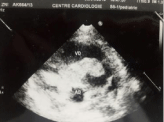
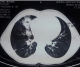
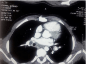
The physical examination on admission identified a febrile child with right pleural effusion syndrome and a systolic murmur on auscultation. Chest x-ray and chest CT showed multiple right basithoracic opacities with a normal heart shape, echocardiography objectified a pedunculated tumor appended to the pulmonary infundibulum without valvular involvement (Figure 1). The radiological assessment was completed by a thoracic CT angiography showing a hypodense lesional process at the level of the right ventricle and the emergence of the trunk of the pulmonary artery associated with multiple structures of liquid densities not enhanced by the contrast product at the pulmonary level (Figure 2). The abdominal ultrasound did not show any liver damage and the brain scan was normal. The biological assessment carried out had objectified a hypereosinophiliain the blood count and a positive hydatid serology.
An antiparasitic treatment based on albendazole was administrated, at a dose of 10 mg / kg / day. Cardiac surgical treatment under extra-corporeal circulation has been indicated, the exploration of the pulmonary artery, has shown a huge intra-arterial cyst limited to the infundibulum with unaffected heart valves (Figure 3). Postoperative follow up was simple. The evolution was marked by an asymptomatic right proximal pulmonary embolism (Figure 4). The antiparasitic treatment was continued.

Figure 1: Ultrasound appearance of an intracardiac hydatid cyst.

Figure 2: Chest scanner showing pulmonary hydatidosis.

Figure 3: Image of the cystic membrane after excision.

Figure 4: CT aspect in favor of a hydatid embolism.
Discussion
Hydatid disease is a parasitic infection caused by echinococcus granulosus. The hydatid cyst is a major health problem in undeveloped and developing countries. Human beings are only incidental intermediate hosts of this parasitic agent [4]; Cardiac hydatic cysts are rare and most frequently located in the left ventricle (55–60%), the right ventricle (15%), the interventricular septum (7–9%), the left atrium (8%), the right atrium (3–4%) and the pulmonary artery (7%). Echinococcosis is a tissue infestation and affects most frequently the liver with second most frequent site is the lungs.
The presence of hydatid cysts in the heart cavities potentially carries the risk of embolism. The left ventricle is involved in more than half of the cardiac cysts [5]. However, as in our patient, the right ventricle is rarely involved [6].
The clinical manifestations depend on the size, location, the vicinity of the valve orifice areas and the conduction tissue, during the early stages the disease is asymptomatic and may be discovered accidentally. A voluminous cyst causes symptoms, signs and complications by compressing and displacing the adjacent structures, or infiltrating tissues or by rupture in hollow organs. Rupture of the cyst is not rare and it isa serious complication that may cause death by anaphylactic shock [7].
Medical Imaging is a fundamental element for positive diagnosis, The chest X-ray, although it remains an important initial examination, The most frequent sign found is cardiomegaly often can be normal if hydatid cyst is small or intracavitary; Cardiac ultrasound remains the most useful exam with a positive diagnosis it allows to visualize the heart masses and to specify their aspects [2]. Eosinophilia is a suggestive but not specific element for the parasitic infection.
In our case, the transthoracic echocardiography was the main examination that helped to make the diagnosis for this atypical presentation. The evolution of the cardiac hydatid cyst is unpredictable; it can involute and calcify or rupture and be responsible for real complications; acute complications may include rupture of a cyst in a cavity of the heart. The complications in these cases are due to migration of hydatid fragments. Thus symptoms and signs of a hydatid pulmonary embolism reveal rupture in the right cavities similar to those of a pulmonary thromboembolism whose severity depends on the size and location of the embolus or emboli. With rupture in the left cavities, the symptoms and signs depend on the site of the intraarterial hydatid embolus, which often lies in the cerebral arteries, the abdominal aorta and its terminal branches, the mesenteric arteries, or the upper and lower limb arteries [8].
Treatment is mainly based on surgery with open heart under ECC, which allows systemic arterial clamping preventing any embolism migration and minimizing the risk of dissemination in the event of a breach of the cyst. After sterilization puncture of the hypertonic saline cyst, its removal is followed by resection of the salient dome or perikystectomy [9].
The prognosisis conditioned by the involvement of the pulmonaryartery, valvular and heart mass [9]; Medical treatment with albendazolepre and postoperative adjuvant helps to limit the accidental risk of hydatic release [10].
Given the risk of dissemination and recurrence, clinical, serological, and radiological surveillance is essential.
Conclusion
Cardiac localization of hydatid disease is rare. The clinical polymorphism, latency, and severity of complications are the essential characteristics. Echocardiography, CT and lately MRI represent the main examinations for early diagnosis. Treatment is primarily surgical. The risk of recurrence is present, hence the importance of biological and radiological monitoring. Good prevention in endemic countries is key to the global eradication of this parasitic infection.
References
- Bakkali A. Les kystes hydatiques cardiaques à propos de 17 cas opérés. Ann CardiolAngeiol (Paris). 2017.
- Jerbi S, Romdhani N, Tarmiz A. Kyste Hydatique emboligène du Cœur droit. Ann Cardiol Angeiol. 2008; 57: 62-65.
- Bouhaouala MH, Hendaoui L, Charfi MR. Hydatidosethoracique. EMC. Radiodiagnostic – Coeur –Poumon, 2007; 32-470-A-20.
- Abid A. Intracavitary cardiac hydatid cyst. Cardiovascular Surgery. 2003; 11: 521-525.
- Kaplan M, Demitras M, Cimen S. Cardiac hydatic cysts with intracavary expansion. Ann Thorac Surg. 2001; 71: 1587-1590.
- Orhan G, Bastopcu M, Aydemir B, Ersoz MS. Intracardiac and pulmonary artery hydatidosis causing thromboembolic pulmonary hypertension. Eur J Cardiothorac Surg. 2018; 53: 689-690.
- Niarchos C, Kounis GN, Frangides CR, Koutsojannis CM, Batsolaki M, Gouvelou-Deligianni GV. Large hydatic cyst of the left ventricle associated with syncopal attacks. International Journal of Cardiology. 2007; 118: e24-e26.
- Thameur H, Abdelmoula S, Chenik S, et al. Cardiopericardial hydatid cysts. World J Surg. 2001; 25: 58-67.
- El Khattabi, Afif, Berrada, Rhissassi, Ichane Z. Bouayad Hydatidose pulmonaire multiple avec localisation cardiovasculaire . Revue des maladies respiratoires. 2011; 28: 686-690.
- Mayoussi C, El Mesnaoui A, Lekehal B, Sefiani Y, Benosman A, Bensaid Y. Lescomplications artérielles de la maladie hydatique. J Mal Vasc. 2002; 27.