Case Report
Phys Med Rehabil Int. 2014;1(3): 4.
Furunculosis in a Transtibial Amputee
M Jason Highsmith1*, Scott Cummings2 and James T Highsmith3
1University of South Florida, USA
2Next Step Bionics and Prosthetics, USA
3Total Skin and Beauty Dermatology Center, University of Alabama at Birmingham, USA
*Corresponding author: M Jason Highsmith, School of Physical Therapy & Rehabilitation Sciences, Morsani College of Medicine, University of South Florida, USA
Received: August 27, 2014; Accepted: September 23, 2014; Published: September 25, 2014
Abstract
Dermatoses are present in 15-41% of persons with lower limb amputation. Cysts are reportedly the third most prevalent dermatologic condition in this population whereas folliculitis and furuncles are not hierarchically ranked. This case report describes the post-transtibial amputation course of a man with recurrent development of furuncles. Management included recurrent periods of prosthetic disuse and cutaneous surgery. It is likely that self-management of prosthetic factors and hygiene were contributors to his skin maladies as he was undergoing significant residual limb volume change, learning to use his prosthesis, care for himself, and cope with limb loss.
Keywords: Amputee; Cyst; Dermatoses; Folliculitis; Furunculosis; Prosthesis
Introduction
There are approximately 1.8M persons with amputation living in the U.S. Of these, approximately 378,000 have transtibial amputation[1]. Persons with amputation who utilize prostheses require ongoing care of prosthetists to fabricate, fit, align and maintain their artificial limbs to maximize mobility, function and quality of life. Even with regular prosthetic care to maintain device fit and function, dermatoses are prevalent in this population [2-4]. The prevalence of skin maladies among lower limb amputees reportedly ranges from 15 to 41%. There is no consensus regarding the type and frequency of specific dermatoses affecting persons with amputation. A recent study of 745 prosthesis users found cysts to be ranked as the third most prevalent dermatologic diagnosis affecting the residual limb while folliculitis and furunculosis were not specifically ranked[5]. Evidence to guide practitioners in the management of these diagnoses in persons with amputation is lacking. This report presents a case that was initially diagnosed as folliculitis, then with cystic lesions, and finally as furunculosis demonstrating specific treatment management options that can be applied to patients with similar findings.
Case Presentation
In 2009, a 30 year-old male (height: 1.85m; mass: 70.3kg) sustained a crush injury to his right foot-ankle while employed as a truck driver and manual laborer. He underwent partial-foot amputation and secondarily experienced rapid onset of complex regional pain syndrome (CRPS) affecting the right ankle and foot. Partial-foot prosthesis, including a lower-leg orthotic section was provided. However, due to the severity of his CRPS symptoms, an elective transtibial amputation (TTA) with traditional long posterior flap closure was performed 1year after the primary amputation. The TTA resulted in a residual limb that was 30% of the sound leg length with good soft tissue coverage distally. The patient's goals were to return to work and be able to enjoy outdoor activities.
Six days post-TTA, the patient was fit with an adjustable, customfit preparatory prosthesis. Physical therapy commenced including ambulation with prosthetic side partial weight-bearing. Eight weeks post-TTA, his residual limb had matured sufficiently and his functional level plateaued with the preparatory prosthesis so he was fit with a definitive, endoskeletal prosthesis. The prosthesis included a custom molded, total surface bearing socket with a cushion gel liner, energy storing foot and suction sleeve suspension. The prosthetist instructed the patient in daily stump and prosthetic cleansing to include soap and water. Rubbing alcohol was substituted two times per week to disinfect the liner and sleeve.
Six weeks following definitive prosthesis fitting, the patient was wearing the limb full-time daily and ambulating independently. Due to daily volume-loss and fluctuations, he used 13-18 sock plies to optimize socket fit. The patient began to remark that material bulk of the combined liner, socks, and suspension sleeve reduced comfort with knee flexion. The patient also reported odor build-up in the liner and sleeve. Therefore, he self-initiated use of a prosthetic cleanser (i.e. water-based detergent with ammonium) to mitigate the problem.
Over the next 8 months (3.5-11.5 months post-TTA), the patient's residual limb volume and shape changed sufficient to require fitting of two replacement sockets. Following successful fitting of the second socket replacement, the patient began to complain of posterior knee irritation. This included development of elevated, folliculocentric masses in the popliteal region. Suspecting folliculitis, the prosthetist recommended conservative, over-the-counter (OTC) antibiotic ointment applied in accordance with product instructions. Combined OTC ointment and short-duration, episodic prosthetic disuse, enabled activity continuance. A new prosthesis including powered ankle, cushion liner, and sleeve suspension was fit several months after the first outbreak of component odor, knee discomfort and elevated popliteal masses. After 10 additional months of wearing various prostheses (11.5-21.5 months post-TTA) including a water prosthesis, the powered prosthesis, and the aforementioned socket replacements, the patient began to experience skin issues over the entirety of his residual limb. Particularly problematic were recurrent elevated, erythemic popliteal masses. The OTC antibiotic ointment was continued as needed in selected locations. Barrier cream (i.e. A&D Ointment. Merck & Co., Inc. Whitehouse Station, NJ. USA.) was utilized for generalized areas of irritation.
For the next 10 months (21.5-31.5 months post-TTA), bouts of residual limb dermatoses occurred interspersed with periods of healing partially attributable to prosthetic discontinuance and possibly with topical antibiotics. He then developed a cystic lesion in the popliteal region so painful that prosthetic use was not optional. The patient was referred to a dermatologist for consultation. The dermatologist formally diagnosed the condition as furunculosis and performed incision, drainage and biopsy. Cultures revealed a bacterial infection and the patient was treated with oral antibiotics.
At this point, the prosthetist changed the socket design to include a pin system, thereby eliminating the suspension sleeve and decreasing the amount of skin surface area contacting gel materials. Despite the prosthetic change, the patient's dermatologic problems increased in frequency and severity. A second furuncle formed which was also incised, drained and cultured and a wick placed. Prosthetic use was discontinued to permit healing. Cultures were negative for infection and healing continued over the next two weeks at which time the prosthesis was progressively re-integrated. The patient returned to function until a third bout of furuncles (i.e. carbuncles) emerged.
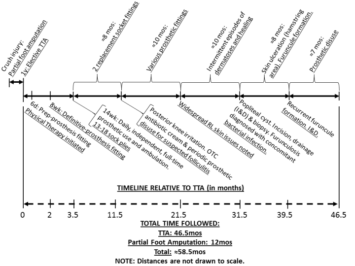
At 3 years following the fitting of his first definitive prosthesis, the patient's skin ulcerated in the distal hamstring area and furuncles formed again along the anterior and posterior aspects of the residual limb. Prosthetic use was discontinued again. Anterior and posterior furuncles were incised and drained and the residual limb was placed in a semi-rigid stump protector for 6 weeks post-operatively to permit healing. A total period of 7 months of prosthetic disuse was necessary, with the first portion involving use of the stump protector. Immediately prior to prosthetic re-integration, a second prosthetist and dermatologist team were consulted to objectively assess the case. At the extreme end of assessment, predisposition to furuncle development as a reaction to material irritants was considered. However the repeated furuncle development without introduction of new chemicals matched a diagnosis of furunculosis (or carbuncles) quite well. Further, review of the photographic history revealed linear excoriations and xerotic skin suggesting other factors contributing to dermatoses, inflammation, and skin breakdown. Therefore, hygiene, tissue hydration and prosthetic self-management (i.e. volume change, perspiration management) were determined to be worthy focal points of management. See Figure 1 for the post-amputation timeline of development of dermatoses, management and follow up.

Figure 1: ##
Treatment Plan
Daily bathing in the evening was recommended to avoid excess moisture accumulation in the morning which could compromise suspension. Chlorhexidine 4% antimicrobial skin cleanser (i.e. Hibiclens. Molnlyckle Healthcare. Gothenburg, Sweden.) was recommended to wash the residual limb 2 to 3 times per week. The goal of this therapy was to decrease cutaneous and follicular microbes in an effort to minimize infections. For daily washing of the residual limb, 2.5-4.0% benzyl peroxide (BPO) wash was recommended. BPO is an extremely powerful agent that improves acne and folliculitis through a multitude of pathways including reducing follicular hyperkeratosis while destroying surface and ductal bacteria, including some yeasts, by way of free oxygen radical formation[6-8]. The same protocol using chlorhexidine and BPO should be used with the prosthetic components that contact the residual limb skin, that is, the liner and sleeve. Bathing parameters that contribute to xerosis and pruritis were discussed and modified. Bathing recommendations included use of lukewarm water, as opposed to hot water, for no more than 15min and to ensure all the topical cleanser was fully rinsed away. After bathing, blot drying as opposed to frictional rubbing were all emphasized in their importance to minimizing xerosis and pruritis [9]. Further, the "soak and smear" technique was recommended to moisturize the limb whereby following blot drying and leaving a small bit of moisture on the skin, a non-comedogenic OTC moisturizer (i.e. Cataphil. Galderma Laboratories, L.P. USA.) applied prior to going to bed for the evening to prevent moisture loss overnight[10].Of note, non-comedogenic moisturizer was recommended in this case as they are manufactured to avoid clogging pores.
The intervention plan also called for prosthetic and behavioral considerations. Prosthetically, a flexible interface and rigid frame design was recommended so that the interface would be more adjustable in the event of rapid volume change or development of dermatoses (i.e. ulceration, furuncle). Additionally, during prosthetic wear, if it became apparent that the fit had changed due to volume loss throughout the day or that perspiration had accumulated, the prosthesis must be removed and the limb dried and sock ply adjusted to re-establish a proper fit as soon as possible.
Results
The patient was educated with the aforementioned instructions both orally and via instructional handouts. Seven months following the incision and drainage of the cystic lesions as described above, the prosthesis with new socket, was re-introduced. Specifically, the interface portion of the prosthesis included a flexible interface and rigid frame, cushioned liner and suction sleeve instead of pin suspension. At two months follow-up of full-time prosthetic use, no issues were reported relative to adherence to his hygiene plan. Additionally, the patient reports clear skin without dermatoses, independent prosthetic use, return to work and recreational pursuits, and continues with routine prosthetic follow-up as needed.
Discussion
In this case, once the patient's volume began to stabilize (˜11.5months post-TTA), dermatologic issues began. However,his volume changes fluctuated throughout the observation period and was a constant factor affecting his skin. This was evident in the various adjustments, multiple sock plies, replacement fittings and sockets, as well as complete system changes. Furthermore, the prosthesis to skin interface increased friction, elevated temperatures, increased sweating, and led to decreased skin integrity and inflamed hair follicles.
Initial folliculitis may have resulted from friction pushing the hair shaft retrograde into the dermis resulting in inflammation. Alternatively or concurrently, local resident flora microbes could have invaded the compromised skin from in or around the follicle or even from a distant site, such as the nares, introduced to the area after scratching. Less likely, a foreign body could have been traumatically introduced into the affected area.
Another unlikely but possible contributing factor to the initial residual limb folliculitis may have been telogen effluvium (TE). TE occurs after periods of stress (such as an amputation) when many hair follicles stop growing in the anagen phase causing the affected hair shafts to fall out as the follicle rapidly converts to the resting cycle, or telogen phase. Several months after the inciting event the hairs may return to normal and begin growing again. However, hair shafts may curl into the skin or fail to exit the skin normally as they grow resulting in folliculitis analogous to hair re growth in shaving, a wellknown risk factor for folliculitis. We point this out as unlikely due to the fact that TE typically affects the scalp. However it should be noted that TE has been documented to affect anywhere on the body though not specifically noted in this case[11].
Nonetheless, the patient experienced a deeper recurrent infection of the hair follicles, termed furunculosis and eventually led to the development of two cystic lesions that were most likely abscesses, necessitating incision and drainage. Ultimately, his regimen above was created after carefully reviewing his history, objective laboratory findings, including previous physician notes and cultures, as well as various clinical photographs throughout the observation period. During this re-evaluation with a multi-disciplinary team, several key points were identified that may have exacerbated his condition in addition to the artificial interface and volume changes. For example, the initial cleanser used was an ammonium based cleanser with a high pH value that could worsen bacterial infections. Secondly, the topical antibiotic initially recommended was an ointment known by dermatologists to be follicularly occlusive and could easily exacerbate folliculitis. The vehicle used is critical and carefully selected to optimize results. For instance, gels, lotions, foams, or solutions are typically used on hair bearing areas and would be a better choice compared with an ointment. However, recurrent bouts of furunculosis and possible abscesses led us to attempt to alter resident flora with potent topical antimicrobial cleansers. Xerosis was also overlooked which is a common cause of pruritis and often manifests with itching and scratching, further compromising skin integrity. Lastly and perhaps most important, overall suboptimal skin hygiene and prosthetic selfcare were identified and corrected.
Since prosthetic reintroduction in this case, a few key factors seem vital to the success identified upon follow-up. First, this case patient with amputation had to learn to care for himself in order to use his prosthesis successfully. This included an understanding of which prosthetic materials and components optimize success.Success in this case included maximizing comfort and minimizing adverse skin reactions. Additionally, learning to manage volume and perspiration throughout the day were of great importance as failing to do so quickly lead to increased focal stress and skin irritation. Finally, learning and adapting to a new hygiene plan for the body, the residual limb and the prosthesis was still another skill set that had to be learned to improve prosthetic success. Coping with limb loss is enormous in the psychologic and emotional realms. This is greatly confounded given all of the technical elements of self-care that accompany learning to use a prosthesis.
Case studies are always limited in the ability to generalize findings. Future work could include retrospective review of a sample of persons with lower limb amputation who experienced folliculitis, cysts or furuncles to determine which interventions optimally provided symptom resolution. Beyond this, prospective research could be considered.
Conclusion
This case report provided details on the course of management leading to proper diagnosis of furunculosis and eventually to resolution and return to prosthetic use and daily function. Additionally, the case underscores the importance of a multi-disciplinary approach and consultation in difficult cases. Consultation in this case enabled provision of valuable education to the patient and to healthcare providers regarding their unique insights into care of complex, multidisciplinary cases such as persons with amputation. The recurrent episodes of disabling, inflammatory furuncles may ultimately be unavoidable given today's prosthetic materials. However, it seems that education translating to sound self-management and hygiene practices may provide considerable relief, possibly resolution and sustainment of prosthetic use, function and quality of life.
References
- Ziegler-Graham K, MacKenzie EJ, Ephraim PL, Travison TG, Brookmeyer R. Estimating the prevalence of limb loss in the United States: 2005 to 2050. Archives of physical medicine and rehabilitation. Mar 2008; 89: 422-429.
- Highsmith JT, Highsmith MJ. Common skin pathology in LE prosthesis users. JAAPA : official journal of the American Academy of Physician Assistants. Nov 2007; 20: 33-36.
- Highsmith MJ, Highsmith JT, Kahle JT. Identifying and managing skin issues with lower-limb prosthetic use. In Motion. 2011; 21: 41-43.
- Bui KM, Raugi GJ, Nguyen VQ, Reiber GE. Skin problems in individuals with lower-limb loss: literature review and proposed classification system. Journal of rehabilitation research and development. 2009; 46: 1085-1090.
- Dudek NL, Marks MB, Marshall SC, Chardon JP. Dermatologic conditions associated with use of a lower-extremity prosthesis. Archives of physical medicine and rehabilitation. Apr 2005; 86: 659-663.
- Bojar RA, Cunliffe WJ, Holland KT. The short-term treatment of acne vulgaris with benzoyl peroxide: effects on the surface and follicular cutaneous microflora. The British journal of dermatology. Feb 1995; 132: 204-208.
- Haider A, Shaw JC. Treatment of acne vulgaris. JAMA : the journal of the American Medical Association. Aug 11 2004; 292: 726-735.
- Fulton JE, Jr., Farzad-Bakshandeh A, Bradley S. Studies on the mechanism of action to topical benzoyl peroxide and vitamin A acid in acne vulgaris. Journal of cutaneous pathology. 1974; 1: 191-200.
- Norman RA. Xerosis and pruritus in the elderly: recognition and management. Dermatologic therapy. 2003; 16: 254-259.
- Gutman AB, Kligman AM, Sciacca J, James WD. Soak and smear: a standard technique revisited. Archives of dermatology. Dec 2005; 141: 1556-1559.
- Tosti A, Pazzaglia M. Drug reactions affecting hair: diagnosis. Dermatologic clinics. Apr 2007; 25: 223-231.