
Clinical Image
Austin J Pulm Respir Med 2014;1(3): 1015.
A Middle Aged Lady with Pyrexia and Infiltrates on Chest Radiograph
Talha Mahmud*
Department of Pulmonology, Shaikh Zayed Hospital, Pakistan
*Corresponding author: Talha Mahmud, Department of Pulmonology, Shaikh Zayed Hospital, PGMI, Lahore, Pakistan
Received: June 02, 2014; Accepted: July 31, 2014; Published: Aug 05, 2014
Case Scenario
A 55 year old non smoker house wife, residing in the premises of a cotton factory, came to accident and emergency department with one week history of dyspnea and chest pain associated with cough productive of purulent sputum occasionally stained with blood. She was normotensive (blood pressure 124/85 mmHg), tachycardic (pulse 110/m), tachypneoic (respiratory rate 28/min) & had fever of 39oC. Her chest examination revealed coarse crackles on the left upper side anteriorly while remaining systemic examination was unremarkable.
Besides other relevant investigations, her chest radiograph-PA view was taken:
- Look at the chest radiograph given below and name 04 important radiological findings?
- What is your clinico-radiological diagnosis?
Answers with Explanations
- Four radiological findings include
- Homogenous opacity in left middle & lower zones suggestive of community acquired pneumonia.
- Presence of air bronchograms within homogenous opacity again suggestive of pneumonia.
- Cavity in left upper radiological zone (cavity/abscess complicating pneumonia).
- Silhouette sign (used to help localizing the anatomical location of the abnormality within the lung) is positive [1,3]. Though the opacification is involving the lower lung field (but it is not in the lower lobe) with obliteration of left heart border and sparing of left hemidiaphragm shadow suggestive of consolidation in lingular lobe and not the lower lobe [1,2]. These findings were confirmed after the lateral radiograph (given below) was taken that showed clear left lower lobe shadow and opacity involving the lingular lobe, extending into the posterior segment of left upper lobe along with a cavitary lesion in left upper lobe [3].
- Clinico-radiological diagnosis
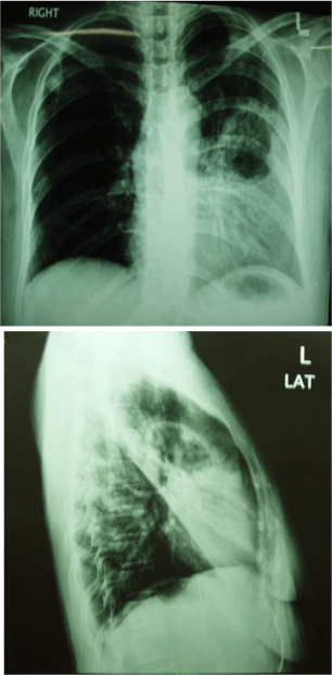
Figure 1:
Community acquired pneumonia involving left lingular lobe complicated by abscess/cavity in left upper lobe.
*This patient grew no organism on sputum culture (probably because she took antibiotics before coming to hospital) and the sputum smears were also negative for TB (ZN staining). She was managed empirically in hospital with IV azithromycin and ceftriaxone for 5 days followed by 9 days of oral cefpodoxime with complete clinical and radiological resolution of radiological infiltrates.
References