Special Article - Pulmonary Critical Care & Sleep Medicine
Austin J Pulm Respir Med 2016; 3(1): 1040.
Use of Home Ventilators for Ventilatory Support during Magnetic Resonance Imaging
Ogna A1,3*, Ambrosi X¹, Prigent H², Falaize L2,3, Leroux K4, Annane D¹, Carlier R5, Orlikowski D1,3 and Lofaso F²
¹Service de Réanimation Médicale et Unité de Ventilation à Domicile, AP-HP, Hôpital Raymond Poincaré, France
²Service de Physiologie-Explorations Fonctionnelles, APHP, Hôpital Raymond Poincaré, France
³INSERM CIC 14.29, AP-HP, Hôpital Raymond Poincaré, France
4ASV Sante, France
5Service d’Imagerie médicale, AP-HP, Hôpital Raymond Poincaré, France
*Corresponding author: Ogna A, Service de Réanimation Médicale et Unité de Ventilation à Domicile, AP-HP, Hôpital Raymond Poincaré 92380 Garches, France
Received: April 03, 2016; Accepted: May 18, 2016; Published: May 23, 2016
Abstract
Purpose: Magnetic Resonance Imaging (MRI) is a valuable diagnostic tool for neuroimaging in the Emergency and Critical Care setting, but its use may be limited in acutely and chronically ventilated patients, who cannot maintain the supine position in spontaneous breathing for the duration required for the procedure, as it may be the case in acute and chronic neurological and neuromuscular diseases with diaphragm involvement.
We aimed to evaluate the performance of home life support ventilators used with a longer circuit, allowing the application of ventilatory support during MRI. The study hypothesis was that home ventilators are accurate in delivery the set ventilatory parameters despite a modified circuit.
Materials and Methods: Four non-MRI-compatible life-support home ventilators were tested on a bench using 3 circuits of 4.8 m length and 3 ventilation settings.
Results: We found measurable differences in the efficacy of the ventilation delivered to the test lung, which was influenced from the used ventilator, the type of circuit and the ventilation parameters. In the volumetric setting with unvented circuit, the difference between set VT and delivered VT ranged between -10% and +3%. In the barometric setting, only the ventilators providing automatic compensation for circuit compliance and resistance were reliable in the delivery of the set inspiratory and end-expiratory pressures.
Conclusion: The use of home ventilators during MRI may represent a valuable alternative when a MRI-compatible ventilator is not available, but may require an adjustment of the ventilatory setting, and a systematic verification of the parameters effectively delivered to the patient.
Keywords: Respiratory failure; Home ventilators; Magnetic resonance imaging; Critical care; Bench evaluation
Abbreviations
ICU: Intensive Care Unit; MRI: Magnetic Resonance Imaging; PC-CMV: Pressure-Controlled Mechanical Ventilation Mode; VC-CMV: Volume-Controlled Mechanical Ventilation Mode; VT: Respiratory Tidal Volume
Introduction
In recent years, Magnetic Resonance Imaging (MRI) has been increasingly performed as a diagnostic exam, and currently represents an essential diagnostic tool for acute and chronic diseases affecting the neurological system [1-5]. Among the situations where neuroimaging is indicated, the execution of a MRI may be problematic in patients presenting acute or chronic respiratory failure and who are unable to sustain the supine position in spontaneous breathing for the exam’s duration. These situations may be present in the Emergency and Critical Care setting, right where neuroimaging may be required. For example, patients with neuromuscular disorders or cervical spinal cord injuries may present a restrictive respiratory failure, requiring ventilatory support in the acute phase or in the long term, especially in case of diaphragmatic involvement [6-8]. The use of mechanical ventilation during MRI may allow to overcome this problem, which is shared with other intensive or critical care patients requiring ventilatory support. The application of mechanical ventilation during MRI imaging raises an issue, since the unique electromagnetic environment of MRI requires dedicated medical devices, but only very few ICU- and transport-ventilators are MRI-compatible [9] and the acquisition of such an expensive ventilator may not be warranted if the projected use is infrequent. A possible alternative may consist in leaving the ventilator in the MRI control room, where non-MRIspecific devices are allowed, and to ventilate the patient using a longer circuit [10]. This would allow to use portable life-support ventilators during MRI, whose performance in this setting has however not yet been tested.
The aim of our bench study was to evaluate the performance of life-support home ventilators for the use with a longer circuit, allowing the application of ventilatory support during MRI. The study hypothesis was that home ventilators are accurate in delivery the set ventilatory parameters despite a modified circuit in the volumetric ventilation mode, but an adaptation of the setting may be necessary in the barometric ventilatory mode to compensate for the increased circuit resistance.
Methods
Four life-support home ventilators were studied: VIVO 60 (BREAS, Sweden), Astral 150 (ResMed, France), PB 560 (Covidien, USA) and Trilogy 100 (Philips Respironics, USA). A volumecontrolled setting (VC-CMV with Tidal Volume (VT) 500 ml) was tested in the different configurations available for each ventilator: double limb circuit (VIVO 60, Astral 150, PB 560), single limb circuit with expiratory valve (VIVO 60, Astral 150, PB 560), single limb vented circuit (VIVO 60, Trilogy 100). Three standard circuits of 22 mm diameter were assembled in series for a total circuit length of 4.8 m; in the MRI room of our hospital, this length allowed to place the ventilator in the control room and to reach the patient lying in the MRI, passing the circuit in a waveguide feed through in the Faraday cage. A calibrated expiratory leak (Whisper Swivel II, Philips Respironics) was used in the vented setting. Two pressure-controlled setting (PC-CMV at 20 cm H2O and 15 cm H2O respectively, PEEP 5 cmH2O) were also tested with the single-limb vented circuit (VIVO 60, Astral 150, Trilogy 100).
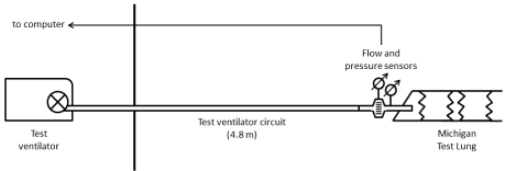
The test ventilator was connected via the test circuit to a lung model (Michigan Dual Adult Test Lung TTL 2600i, Michigan Instruments, USA). The compliance of the lung model was set at 30 mL/cmH2O, and the airway resistance at 5 cm H2O/L.s (Pneuflo Rp5, Michigan Instruments, Grand Rapids, Michigan), corresponding to a restrictive adult pattern. Flow and pressure signals were captured near the test lung, using a Fleischman pneumotachograph (Fleisch, Switzerland) and an analog/digital system (MP150, Biopac Systems, USA) (Figure 1). For each ventilator and each setting, the effectively delivered VT and pressures were recorded over 15 respiratory cycles, after a stabilization time of 2 minutes.

Figure 1: Experimental setup.
Statistical analysis was conducted using R3.1.2 statistical software (R Core Team). We used t-test to compare expected and measured values for the same ventilator, and ANOVA to assess differences between the four test ventilators.
Results
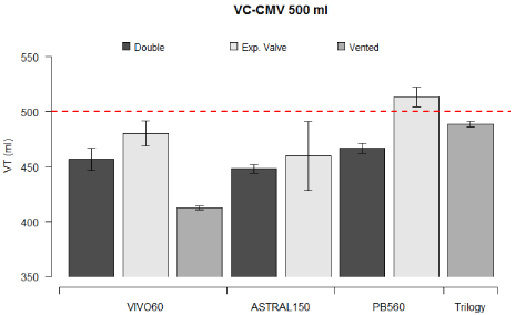
Figure 2 shows the mean VT provided by the 4 ventilators in the Volume-Controlled settings (VC-CMV) with VT 500 ml. The difference between set VT and delivered VT ranged between -52 ml and +14 ml (-10% to +3% of the set VT, all p<0.001) for the two unvented configurations (p<0.001 for the difference between the test ventilators, in both double-limb and expiratory valve configuration). For the 3 ventilators tested in both configurations, the single-limb setting with expiratory valve showed slightly better results than the double limb configuration.

    Figure 2: Ventilation parameters effectively delivered to the test lung in the
volumetric setting.
Volume-controlled setting (VC-CMV) with tidal volume (VT) 500 ml and 3
types of circuits: double-limb (Double), single-limb with expiratory valve
(Exp. Valve) and single-limb with calibrated expiratory leak (Vented). Bars
represent the mean delivered VT, whiskers the 95% confidence interval. The
dashed horizontal line represents the set VT.
In the single-limb, vented VC-CMV test, the Trilogy 100 ventilator provided accurate volume delivery, with a mean delivered VT of 489±1 ml (2% lower than the set VT, p<0.001), whilst the VIVO 60 showed the lowest values of the whole test in this configuration (mean VT 413±1 ml, -17% of the set VT, p<0.001).
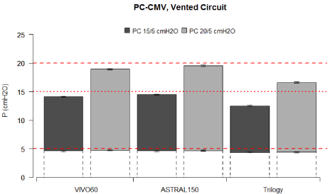
In the barometric (PC-CMV) setting using a single-limb vented circuit (Figure 3), both VIVO 60 and Astral 150, which provide automatic compensation for circuit compliance and resistance after a calibration maneuver, were reliable in delivering the set inspiratory and end-expiratory pressures, with measured values 2 to 6% lower than the set values (all p<0.001). In contrast, the inspiratory pressure values delivered by the Trilogy 100 were 17% lower than the set pressures (12.5±0.1 and 16.6±0.1 cm H2O respectively in the 15 and 20 cm H2O settings, both p<0.001). As a consequence, the VT effectively delivered by the Trilogy 100 was significantly lower than the VT delivered by both VIVO 60 and Astral 150 (273±1 ml vs. 307±1 ml and 308±2 ml respectively for the 15 cm H2O setting, p<0.001, and 356±1 ml vs. 403±2 ml and 405±1 ml respectively for the 20 cm H2O setting, p<0.001).

    Figure 3: Ventilation parameters effectively delivered to the test lung in the
barometric, vented setting.
Pressure-controlled setting (PC-CMV) with inspiratory pressure 15 cm H2O
(PC 15/5) and 20 cm H2O (PC 20/5), expiratory pressure 5 cm H2O; singlelimb
circuit with calibrated expiratory leak. Bars represent the mean delivered
pressure, whiskers the 95% confidence interval. The dashed horizontal lines
represent the set pressures (P).
Discussion
Our bench study shows that the use of home ventilators during MRI is feasible after adaptation of the circuit, with effectively delivered parameters differing less than 10% from the set parameters in most settings, which is considered as an acceptable accuracy. However, the choices of ventilator, type of circuit and ventilation parameters all have a measurable impact on the efficacy of ventilation in this configuration.
MRI represents a valuable diagnostic tool, both in the acute setting, such as patients with an acute neurological injury [1-3] as well as in the chronic setting, such as neuromuscular disease patients [4,5], but the necessity to lie supine during several minutes may preclude its use in patients with limited respiratory autonomy [6,7]. Our study provides a solution for these patients, indicating that they could be ventilated during MRI with life-support home ventilators; by the means of a longer circuit that allows placing the device outside the MRI room. The same strategy could be applied for the transport of critically ill patients to the MRI, as it has been proposed several years ago using ICU-ventilators [10]. This may however require an adjustment of the ventilator settings and presuppose to verify the parameters effectively delivered to the patients, since the hydraulic characteristics of the circuit are substantially modified. According to our observations, the use of an expiratory valve provided lightly better result than the double limb circuit in the volumetric setting. This probably results from the lower total volume of the circuit (and thus lower circuit compliance), reducing the inaccuracy due to volume loss in the circuit during pressurization. This advantage is however counterbalanced by reduced monitoring possibilities in the absence of expiratory circuit. The Trilogy 100 ventilator, which only offers the single-limb, vented configuration, was accurate in delivering VCCMV in this setting. In the barometric single-limb vented setting, the ventilators proposing an automatic compensation for circuit compliance and resistance (VIVO 60 and Astral 150) proved to be more accurate in delivering the set pressure.
A limitation of our study lies in the necessity to have waveguide feed through in the Faraday cage to pass the circuit, and in the arbitrary choice of the tested circuit length which, although adapted for the MRI room of our hospital, may be insufficient in radiology departments with a different architecture.
Conclusion
In conclusion, the use of life-support home ventilators during MRI may represent a valuable alternative when a MRI-compatible ventilator is not available, but may require an adjustment of the ventilatory parameters, depending on the choice of the ventilator, the type of circuit and the ventilation mode which is used.
Acknowledgement
A. Ogna, L. Falaize, K. Leroux, Pr. R. Carlier and Pr. D. Annane declare that they have no conflict of interest. X. Ambrosi received financial support for a congress inscription fee from ResMed France. Pr. F. Lofaso and H. Prigent have no conflict of interest related to the present work to disclose; the Service de Physiologie-Explorations Fonctionnelles of Garches received research funds from ResMed France, not related to the present work. Pr. D. Orlikowski has no conflict of interest related to the present work. The CIC 14.29 of Garches received research funds from BREAS Medical, not related to the present work.
References
- Stocchetti N, Le Roux P, Vespa P, Oddo M, Citerio G, Andrews PJ, et al. Clinical review: neuromonitoring - an update. Crit Care. 2013; 17: 201.
- Stevens RD, Hannawi Y, Puybasset L. MRI for coma emergence and recovery. Curr Opin Crit Care. 2014; 20: 168-173.
- Morandi A, Rogers BP, Gunther ML, Merkle K, Pandharipande P, Girard TD, et al. The relationship between delirium duration, white matter integrity, and cognitive impairment in intensive care unit survivors as determined by diffusion tensor imaging: the VISIONS prospective cohort magnetic resonance imaging study. Crit Care Med. 2012; 40: 2182-2189.
- Mercuri E, Jungbluth H, Muntoni F. Muscle imaging in clinical practice: diagnostic value of muscle magnetic resonance imaging in inherited neuromuscular disorders. Curr Opin Neurol. 2005; 18: 526-537.
- Costa AF, Di Primio GA, Schweitzer ME. Magnetic resonance imaging of muscle disease: a pattern-based approach. Muscle Nerve. 2012; 46: 465-481.
- Simonds AK. Chronic hypoventilation and its management. European respiratory review: an official journal of the European Respiratory Society. 2013; 22: 325-332.
- Polkey MI, Lyall RA, Moxham J, Leigh PN. Respiratory aspects of neurological disease. J Neurol Neurosurg Psychiatry. 1999; 66: 5-15.
- Annane D, Orlikowski D, Chevret S. Nocturnal mechanical ventilation for chronic hypoventilation in patients with neuromuscular and chest wall disorders. The Cochrane database of systematic reviews. 2014; 12: CD001941.
- Chikata Y, Okuda N, Izawa M, Onodera M, Nishimura M. Performance of ventilators compatible with magnetic resonance imaging: a bench study. Respir Care. 2015; 60: 341-346.
- Rotello LC, Radin EJ, Jastremski MS, Craner D, Milewski A. MRI protocol for critically ill patients. Am J Crit Care. 1994; 3: 187-190.