
Special Article - Pulmonary Tuberculosis
Austin J Pulm Respir Med 2017; 4(2): 1056.
Musculoskeletal Tuberculosis
Sanel S*
Assistant Professor, Department of Orthopedic Surgery and Traumatology, Medical School of University of Maltepe, Istanbul, Turkey
*Corresponding author: Selim Sanel, Assistant Professor, Department of Orthopedic Surgery and Traumatology, Medical School of University of Maltepe, Istanbul, Turkey
Received: August 04, 2017; Accepted: September 07, 2017; Published: September 14, 2017
Introduction
Tuberculosis is still one of the most common causes of death worldwide and most commonly seen in crowded communities with malnutrition and poor sanitanition. Tuberculosis may be found on all continents and highest rate of new infection is in South and Southeast Asia. But highest rates of infection and mortality are in sub-Saharan Africa [1].
Skeletal tuberculosis is an ancient disease and signs of spinal tuberculosis have identified in Egyptian mummies. Molecular studies have shown Mycobacterium tuberculosis complex DNA in ancient body specimens [2,3].
The disease is transmitted by inhalation or ingestion of Mycobacterium tuberculosis or Mycobacterium bovis. It can be cleared by the host, lead to primary infection or can later be reactivated from a latent infection. Spread of the disease may be lymphogenous, hematogenous, or contagious extension to other tissues organ systems. The clinical manifestations vary if the disease is isolated musculoskeletal tuberculosis or miliary tuberculosis.
Miliary disease has a rapid course and constitutional symptoms include fever, chills and cough with accompanying pleuratic pain, weight loss and fatigue. The patient may have acute or chronic symptoms [4].
Populations at risk include individuals with Acquired Immune Deficiency Syndrome (AIDS) or other immune deficiencies, patients with chronic renal failure, substance abusers, homeless or incarcerated individuals and immigration from developing countries.
Pathophysiology
The primary focus of disease is visceral (lungs, kidneys, lymph nodes) and musculoskeletal involvement occurs via hematogenous spread. Once deposited at a site, the organisms are ingested by mononuclear cells. Mononuclear cells then coalesce into epitheloid cells, and a tubercle is formed when lymphocytes formaring around a group of epitheloid cells. Caseation then develops within the center of the tubercle. The host inflammatory response intensifies, resulting in exudation and liquefaction and a cold abscess is formed. A cold abscess is composed of serum, leukocytes, caseation, bone debris, and bacilli. The outcome depends on the characteristic and sensitivity of the organism, the status of the host immune system, the stage of the disease at presentation, and the treatment. The range of end results may include resolution with minimal or no morbidity, healed disease with residual deformity, walled off lesions with calcification of caseous tissue, a low grade chronic granular lesion, and local or miliary spread of the disease that may result in death [5].
Extra pulmonary involvement is noted in approximately 14% of patients, with 1% to 8% having osseous disease. Approximately 50% of patient with osseous tuberculosis have pulmonary involvement. The most common site of osseous involvement is the spine (30-50%) especially in elderly individuals. But in developing countries young adults and children can also be affected.
The most common involvement site of the spine is thoracal and thoracolumbar segment, but it may be seen in any region of the spine. Usually, active spinal lesions involve a particular segment; two vertebral bodies and the corresponding disc.
According to some authors tuberculosis bacilli requires high oxygen pressure and affect these areas because of the generous arterials and venous supply.
A peridiscal presentation occurs in approximately 80% of patients, with the anterior vertebral body affected and contiguous progression through anterior longitudinal ligament and eventual extension to adjacent vertebrae. Less frequently lesions occur centrally in the vertebral body. These lesions are more difficult to diagnose and may mimic tumor or contribute to significant spinal deformity.
Patients may have intramedullary granulomas, arachnoiditis, segmental collapse with anterior wedging and gibbous formation (Pott disease). The posterior elements of the spine are rarely the only sites affected. Perispinal abscess with sinus extension to the skin also may arise and extend through tissue planes to reach intraperitoneal structures. They have been reported to occur as far distally as the popliteal fossa (Figure1). Patients present with pain, weakness and in the late stages paralysis.
Appendicular joint involvement typically affects the major weight hearing joints of the lower extremities. Lesions involve the articular cartilage; the trabecular zones of the bone are affected, with subchondral involvement affecting the weight bearing capability of the joint, which may progress to significant accelerated joint surface degeneration [1,5].
Ankle, foot and upper extremity joints are less frequently involved.
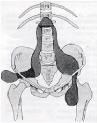
Figure 1: Perispinal abscess reaching distally.
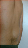
Figure 2: Kyphotic deformity.
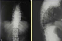
Figure 3: Plain radiograms of involved spine.
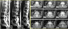
Figure 4: MRI (left) and CT (right) images of spinal tuberculosis.
Clinical Findings
Low-grade fevers, night sweats, weight loss, anorexia, malaise are constitutional symptoms [6]. Musculoskeletal complaints include swelling, stiffness and pain. Osseous involvement is associated with tenderness, soft tissue swelling and restriction of movement. Findings in spinal involvement include back pain/tenderness, a neurologic deficit and a kyphotic deformity (Figure 2). Swelling and tenderness over a synovial bursa or tendon sheath is seen less frequently. Lymphadenopathy is common and sinuses are frequently observed [7].
Laboratory Findings
- Patients may have anemia, pancytopenia, or thrombocytopenia.
- White blood cell count is frequently normal, ESR is elevated.
- Tuberculosis skin testing maybe helpful in diagnosis, but 20-30% false negative rates can be seen. Immune compromised patients frequently have an uncertain skin test result.
- The exact diagnosis is demonstration of tuberculosis acid-fast bacilli from a tissue or fluid source.
- Bone culture; taken from infected material can be helpful in 60 to 80% of cases.
- Transbronchial biopsy specimens in patients with pulmonary involvement are positive in 70 to 80% of patients.
- Molecular subtyping also has been used to assess infection patterns and sensitivities to medications.
Imaging
A plain x-ray of chest should be ordered when the diagnosis is suspected or established. Plain radiographs of involvement joints and spine may demonstrate pathological findings according to the stage of the disease (Figure 3).
MRI and CT also can provide more detail and delineate the disease in earlier phases (Figure 4)
CT or Ultrasound guided fluoroscopy can assist in obtaining appropriate tissues or fluid samples for additional studies.
Bone scan or gallium scan can detect 88 to 96% of osseous tuberculosis lesions.
Arthrography is less frequently used for diagnosis.
Clinical Manifestations
Skeletal tuberculosis forms are spondylitis (Pott disease), arthritis and osteomyelitis.
Tuberculosis Osteomyelitis: Tuberculosis osteomyelitis cases are less than 5% of osteoarticular tuberculosis cases. The duration of symptoms range from days to months, and coexisting visceral disease is uncommon [8,9]. Sinuses are common and abscess formation may occur. Bluish discoloration, undermined edges, sero-sanguinous discharge, matted draining lymph nodes and fixation to bone are features of the tuberculosis sinus. Tuberculosis osteomyelitis can be seen in any bone like ribs, skull, phalanx, pelvis, and long bones. The most common presentation is a solitary lytic lesion with a sclerotic rim. The lesion may be diaphyseal or metaphyseal, and may penetrate the physis or adjacent joint. Brodie’s abscess, chronic osteomyelitis, granulomatous lesions, and neoplasms must be considered in differential diagnosis [10,11]. Multiple cystic tuberculosis (one or more large oval areas of rarefaction, children), disseminated skeletal tuberculosis (multiple osseous and/or articular sites, compromised host), closed multiple diaphysitis (swelling in forearms and legs in compromised children) and tuberculous dactylitis (metacarpal or phalanx) are unusual forms of skeletal tuberculosis [12-15]. Biopsy from inside the granulamatous focus or synovium adjacent to the cyst is recommended if applicable to diagnose the disease [16]. Curettage of the lesion without bone grafting is recommended [5,10,17].
![]()
Tuli Classification. The Natural History of Tuberculous Arthritis Progresses Through 5 Stages
Clinical Findings
Radiographic Findings
Treatment
Anticipated Outcome
Stage I (Synovitis)
1.) Soft tissue swelling
2.) 75% motion preserved1.) Soft tissue swelling
2.) Osteopenia1.) Chemotherapy
2.) Rest
3.) ROM
4.) SplintingNormal or minimal residua
Stage II (early arthritis)
1.) Soft tissue swelling
2.) 25-50% loss of mation1.) Soft tissue swelling
2.) Marginal joint erosions
3.) Diminution in joint space1.) Chemotherapy
2.) Rest
3.) ROM
4.) Splinting
5.) Synovectomy50-70% mobility
Stage III (advanced arthritis)
1.) 75% loss of motion
1.) Marginal erosions
2.) Cysts
3.) Significant loss of joint space1.) Chemotherapy
2.) Osteotomy
3.) Arthrodesis
4.) ArthroplastyStable, painless joint after salvage, with or without motion
Stage IV (advanced arthritis)
1.) 75% loss of motion
2.) Subluxation or dislocation1.) Joint destruction
1.) Chemotherapy
2.) Osteotomy
3.) Arthrodesis
4.) ArthroplastyStable, painless joint after salvage
Stage V (Ankylosis)
1.) Ankylosis
1.) Ankylosis
1.) Chemotherapy
2.) Osteotomy
3.) Arthrodesis
4.) ArthroplastyStable, painless
Table 1: Tuli classification, the natural history of Tuberculous Arthritis progresses through 5 stages.
Tuberculosis Arthritis: The invasion of joint by the tuberculous bacillus may occur by direct hematogenous infection of the synovial membrane or by indirect spread from a focus in an adjacent bone in the metaphysis or epiphysis. The synovial membrane reacts first by secreting excessive fluid and later by proliferation, thickening, studding of its inner surface with tubercles, and fibrosis of its outer surface. The tuberculous granulation tissue soon covers the hyaline cartilage as a pannus that eventually destroys the underlying articular cartilage and subchondral bone. Joint destruction may be associated with subluxation or dislocation. Progression of disease leads to increasing amount of caseous necrotic material and tuberculous exudates and soon increased intraarticular pressure results with perforation of the joint capsule. Acute inflammation absent tuberculous abscess (cold abscess) forms. They spread by dissecting along tissue planes between muscles or muscle sheaths, being limited by the deep fascia. With increased tension inside the compartment deep fascia is perforated and the abscess becomes subcutaneous. Tuberculous abscess is lined by a thick fibrous tissue. If untreated abscess ruptured outside the skin with sinuses. Tuberculous arthritis is often monoarticular (90%). Tuli classification (Table 1) shows the stages, clinical and radiographic manifestations of the disease and recommended treatment with expected outcome in patients with articular disease [18]. Chemotherapy is recommended for all patients with active disease. During the early stages of disease, the goal is to obtain as well as possible range of motion. Range of motion exercises begins with relief of symptoms and splinting, serial casting, traction, bracing helps to prevent deformities. Weight bearing is allowed after adequate alignment and motion are achieved and the disease has been effectively controlled by chemotherapy. For patients in the later stages of disease, the aim is to hold the joint in functional position with cast or splint, as ankylosis is expected. Corrective periarticular osteotomies, excisional arthroplasty, prosthetic reconstruction may be required for the patients with not functional joints [19,20]. Current evidence suggests that there should be minimum 10 years disease free interval in between completion of treatment and prosthetic implantation. Prophylactic chemotherapy may allow earlier implantation and can salvage a prosthetic joint when reactivation of infection has been observed.
Tuberculosis of Spine: Tuberculosis of the vertebral column was first described by Percivall Pott as a painful kyphosis of the spine associated with paraplegia. The condition is often referred to as Pott’s disease [21].The spine is the most common site of skeletal tuberculosis which accounts for 50% of cases. From the most common segment to the less are the lumbar, upper dorsal, cervical, and sacral regions. But the disease may be seen in any segment of the spine [7]. Skipped lesions may occur rarely. Males and females are equally infected but incidence of infection seems to increase with age. A good outcome can be expected if the disease is diagnosed before the appearance of spinal deformity and neurologic symptoms.
Pathologically the infection is characterized by acid-fast-positive, caseating granulomas with or without pus. Tubercles composed of monocytes and epitheloid cells, forming minute masses with central caseation in the presence of Langerhans-type giant cells, are typical on microscopic examination. Abscesses expand, following the path of least resistance, and contain necrotic debris. Skin sinuses form, drain and heal spontaneously. Bone reaction to the infection varies from intense to no reaction. In the spine infection spare the intervertebral discs and spreads beneath th anterior and posterior longitudinal ligamens. Epidural infection is more likely to result in permanent neurological damage.
In the beginning of the disease, weakness, malaise, night sweats, fever and weight loss are predominant symptoms. Pain is a late symptom associated with bone collapse and paralysis. Anterior abscess formation in the neck may cause Millar asthma with symptoms of dysphagia, stridor, hoarseness because of recurrent laryngeal nerve palsy. Sudden death with cervical infection has been reported after invasion to the great vessels. Neurological signs usually occur late. Rectal tone and motor function are good prognostic.factors. Laboratory findings are, anemia, hypoproteinemia, and mild elevation of ESR and CRP. Skin testing may be helpful but is not diagnostic. Early radiographic findings are one or more disc space narrowing and localized osteopenia. Later findings include vertebral collapse called ‘‘concertina collapse’’ by Seddon because of its accordion resembling appearance. Atypical radiographic findings include involvement of the posterior elements, circumferential involvement, lateral vertebral translation, involvement of a single vertebra or multiple vertebrae, and the spinal tumor syndrome. Abnormal findings that support tuberculosis on Magnetic Resonance Imaging (MRI) are multiple levels of involvement, relative sparing of the vertebral disc, a large paravertebral abscess, subligamentous spread, multicentric involvement, and heterogenous signal with rim enhancement. Myelogram is also helpful like MRI in diagnosing the spinal tumor syndrome (Intradural, extradural or intramedullary tubercular granulomas).
Radiographic or CT guided biopsy and culture of the organism is the definitive diagnosis. Thoracoscopic, laparoscopic or open biopsies are other biopsy options. Tissue diagnosis is difficult to achieve. Chen et al. found that on biopsy, the smear was positive in only 15%, and that histology was ‘‘typical’’ in 60% and ‘‘compatible’’ im 36%. Delayed or missed diagnosis is common [22].
Pyogenic and fungal infections, secondary metastatic disease, primary tumors of bone, sarcoidosis, giant cell tumors of bone, and bone deformities such as Scheuermann disease must be considered in differential diagnosis.
In general if the neurologic dysfunction develops gradually with short duration the prognosis of recovery is good. Complete paraplegia, rapid development, flaccid paralysis, longer duration of symptoms, and late onset of disease are indicators of poor prognosis.
Management Principles
The primary treatment objectives for tuberculosis of bone include halting the infection, limiting deformity, maintaining mobility and reducing discomfort.
Multidisciplinary approach by an affiliated team which consists of infectious disease and pain management specialists, nurses, physical therapists, occupational therapists and orthotics.
Chemotherapy should be approximately 90% effective in eradicating the infection, provided that patient compliance is ensured. Inadequate treatment and for lack of compliance will result in the emergence of resistant organisms.
The management of the recurrence is challenging, expensive and may fail to eradicate the disease.
The current trend treatment protocol has been to use intermittent dosing (2-3 times for week) and to decrease the overall duration of therapy from 18 months to 9 months.
In spinal disease our recommended duration of therapy is 18 months and 12 months in osteoarticuler tuberculosis.
Treatment is divided in two phases. During first phase 4 agents are administered for 2-3 months and in the second phase 2-3 agents are administered over 4-6 months.
Multidrug resistance is very low (1-2%) but resistance to a single agent is 13%.
The treatment of relapse typically involves 5 agents in the first phase and 3 agents at the second phase.
Antitubercular drugs have been grouped into first and second line agents [23,24]. First line agents include Isoniazid, Rifampicin, Streptomycin, Pyrazinamide, Ethambutol and Thiacetazone. Second line agents Capreomycin, Kanamycin, Ethiaramide, Cycloserine and Paraamino-salicylic acid. These agents can penetrate a tuberculosis abscess [25]. Sinuses should heal within 6 to 12 weeks unless there has been a secondary bacterial infection.
Multidrug resistance should be considered if there is no clinical improvement after 4 weeks of treatment. Streptomycin and ciprofloxacin can be used if the infection is caused by a multidrug-resistant organism.
More than 90% of patients respond to nonsurgical treatment.
Operations applicable to bone and joint tuberculosis include [1]:
- Arthrotomy including biopsy, synovectomy, and curettage and bone grafting of articular erosions.
- Curettage and bone grafting of extraarticular skeletal lesions.
- Resection of joints.
- Resection of bones.
- Evacuation or excision of soft tissue abscesses.
- Arthrodesis.
- Amputation.
- Arthroscopic debridement of knee joint.
Immobilization (casting, bracing) and drug therapy are mainstays of treatment of spinal tuberculosis. Immobilization itself may prevent further bony destruction and progressive kyphosis. Methods for surgical treatment of spinal tuberculosis can be summarized assurgical debridement, posterior or anterior stabilization with allograft fusion.
Most authorities agree that effective antibiotic therapy should be started before surgery for tuberculosis. When surgery was done without adequate chemotherapeutic coverage miliary dissemination of the disease has been reported.
Paraplegia is the most serious complication of the spinal tuberculosis with an incidence rate of 10-29%. Younger children are more likely to become paraplegic. Level and type of paraplegia can be determined by radiography, MRI and myelography. Early anterior decompression and spinal stabilization is strongly recommended. Delay in treatment may result in permanent paraplegia.
Summary
Musculoskeletal tuberculosis may be seen with some frequency in various regions of the world, especially in South and Southeast Asia. Although satisfactory results can still be achieved with salvage procedures in patients presenting with late stages of disease, early diagnosis and adequate treatment maximizes the treatment outcomes. Chemotherapy is extremely effective as long as the appropriate regimen is prescribed, and the patient compliance is ensured. Treatment should not be limited to the patient but chemoprophylaxis should be considered in family members and other close contacts who have a positive tuberculosis skin test. Surgical intervention is most commonly required to establish the diagnosis, and to treat the musculoskeletal complications of the disease, especially in cases with delayed presentations.
References
- Mihalko MJ. Tuberculosis and other unusual infections. Campbell’s Operative Orthopaedics. Elsevier Mosby. 2013; 23: 773-782.
- Hershkovitz I, Donoghue HD, Minnikin DE, Besra GS, Lee OYC, Galili E, et al. Detection and molecular characterization of 9000 year-old Mycobacterium tuberculosis from a Neolithic settlement in the Eastern Mediterranean. PLoS One. 2008; 3: e3426.
- Donoghue HD, Lee OY, Minnikin DE, Besra GS, Taylor JH, Spigelman M. Tuberculosis in Dr Granville’s mummy: a molecular re-examination of the earliest known Egyptian mummy to be scientifically examined and given a medical diagnosis. Proc Biol Sci. 2010; 277: 51-56.
- Jacob JT, Mehta AK, Leonard MK. Acute forms of tuberculosis in adults. Am J Med. 2009; 122: 12-17.
- Tuli SM. General principles of osteoarticular tuberculosis. Clin Orthop Relat Res. 2002; 398: 11-19.
- Hodgson SP, Ormerod LP. Ten-year experience of bone and joint tuberculosis in Blackburn 1978-1987. J R Coll Surg Edinb. 1990; 35: 259.
- Spiegel DA, Singh GK, Banskota AK. Tuberculosis of the musculoskeletal system. Techniques in Orthopaedics. 2005; 20: 167-178.
- Kumar K, Saxena MBL. Multifocal osteoarticular tuberculosis. Int Orthop (SICOT). 1988; 12: 135-138.
- Martini M, Adjrad A, Bouddjemaa A. Tuberculous osteomyelitis. A review of 125 cases. Int Orthop (SICOT). 1986; 10: 201-207.
- Huang CH. Extra-articular tuberculous osteomyelitis. A report of 11 cases. Int Orthop (SICOT). 1996; 20: 169-171.
- Wang MNH, Chen WMC, Lee KS, Chin LS, Lo WH. Tuberculous osteomyelitis in young children. J Ped Ortho. 1999; 19: 151-155.
- Hsieh CK, Miltner LJ, Chang CP. Tuberculosis of the shaftof the large long bones of the extremities. J Bone Joint Surg. 1934; 16a: 545-563.
- Aggarwal AN, Dhammi IK, Jain AK. Multifocal skeletal tuberculosis. Tropical Doctor. 2001; 31: 219-220.
- Babhurkal SS, Pande SK. Unusual manifestations of osteoarticular tuberculosis. Clin Orthop Rel Res. 2002; 398: 114-120.
- Shannon FB, Moore M, Houkom JA, Waecker NJ Jr. Multifocal cystic tuberculosis of bone. J Bone Joint Surg. 1990; 72a: 1089-1092.
- Versfeld GA, Solomon A. A diagnostic approach to tuberculosis of bones and joints. J Bone Joint Surg. 1982; 64b: 446-449.
- Vohra R, Kang HS, Dogra S, Saggar RR, Sharma R. Tuberculous osteomyelitis. J Bone Joint Surg. 1997; 79b: 562-566.
- Tuli SM. Tuberculosis of the Skeletal System: Bones, Joints, Spine and Bursal Sheaths. 2004.
- Babhulkar SS, Pande S. Tuberculosis of the hip. Clin Orthop Rel Res. 2002; 398: 93-99.
- Eskola A, Santavirta S, Konttinen YT, Tallroth K, Lindholm ST. Arthroplasty for old tuberculosis of the knee. J Bone Joint Surg. 1988; 70b: 767-769.
- Boachie-Adjei O, Squillante RG. Tuberculosis of the spine. Orthop Clin North Am. 1996; 27: : 95-104.
- Chen Wj, Chen CH, Shih CH. Surgical treatment of tuberculous spondylitis, 50 patients followed for 2-8 years. Acta Orthop Scand. 1995; 66: 137-142.
- Bastian I, Colebnders R. Treatment and prevention of multidrug-resistant tuberculosis. Drugs. 1999; 58 :633-661.
- Shenbekar A, Babhulkar S. Chemotherapy for osteoarticular tuberculosis. Clin Orthop Rel Res. 2002; 398: 20-26.
- Tuli SM, Mischra S. Penetration of antitubercular drugs in cold abscesses of skeletal tuberculosis and in tuberculous joint aspirates. Ind J Orthop. 1983; 17: 14-18.