Case Series
Austin J Radiol. 2015;2(1): 1010.
MR Findings of Primary Female Genital Tract Lymphoma: Cases Report and Literature Review
He Zhang*
Department of Radiology, Fudan University, China
*Corresponding author: He Zhang, Department of Radiology, Obstetrics and Gynecology Hospital, Fudan University, No.419, Fangxie, Shanghai, P.R. China
Received: January 07, 2015; Accepted: February 17,, 2015; Published: February 18,, 2015
Abstract
Primary female genital tract lymphoma is a rare etiology with extremely rare occurrence. Imaging findings of primary female genital tract lymphoma are mostly reported in case report. Here, we reported three cases in our single institution with focus on the conventional MRI and Diffusion-Weighted Imaging (DWI) findings. In this cohort case series, there were no additional abnormal findings in other organs except in female genital tract. Lymphadenopathy was observed in one case and tumor extended beyond the uterus was observed in another case. The tumor enhancement and DWI findings were not different from other female malignant tumors. The final histological diagnosis will be needed to determine the etiology of disease.
Introduction
Primary lymphoma involving female genital tract is extremely rare. It is reported that the incidence is about 0.2 - 1.1% of all primary extranodal lymphoma [1]. Primary non-Hodgkin lymphoma of ovary accounts for ~1.5% of all ovarian tumors. Most of primary lymphoma in gynecological tract has been published as “cases report” type when searched the database on Medline [2-4]. Owing to the capability of multi-planar imaging and better soft tissues resolution, Magnetic Resonance Imaging (MRI) has its great advantages in characterizing gynecological tumors [5]. However, only few studies described the imaging findings of lymphoma of ovary and uterus at MRI [6-8]. In this short report, we reported three patients in our single institution with pathologically proved primary non-Hodgkin lymphomas of ovary and uterus in our institution. All of them underwent full MRI examinations before surgery.
Case 1
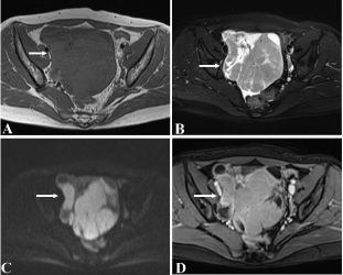
A 31-year-old woman presented with 01 month history of pelvic mass and abdominal distension. There were no vaginal discharge and bleeding. No other disease history has been recorded. The patient had a MRI scan before the surgery. On MRI, there was tubal mass along the surface of loop of small intestine (Figure 1). The mass appeared solid without clear margin with the surrounding tissues. The tumor displayed the intermediate signals on both T1WI (Figure 1A) and T2WI (Figure 1B) images and slightly high signals on DWI images (Figure 1C). On contrast-enhanced images, the mass displayed mild enhancement (Figure 1D). Pelvic images did not show free fluid or lymphadenopathy. The patient underwent laparotomy one week later. The gross findings revealed that the right ovary obviously enlarged in the size of 12×9×9 cm, which displayed as a gray, fragile mass with some necrotic sis areas in it. The final histological diagnosis was the B - cell lymphoma. On immunohistological analysis, the mass tested strong positive for CD10 antibody, mild positive for Ki67 and P53.

    Figure 1:  A 31-year-old woman was pathologically proven with the B - cell
lymphoma (Case 1).
The homogeneous, solid mass displayed as iso-intensity signals on both
T1WI(A) and fat-suppressed T2WI (B)(arrow); On DWI images(C), it showed
the restricted diffusion in the main body of tumor and on contrast enhanced
images(D), the mass manifested as mild enhancement.
Case 2
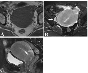
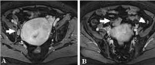
A 65-year-old woman had a menopause 16 years ago and complained of abdominal distension for about half year. There were no virginal discharge and bleeding. No other surgery has been recorded. On MRI (Figure 2), the uterine signals appeared as homogeneously intermediate signals on both T1WI (A) and T2WI images (B). The uterus diffusely enlarged with ambiguous functional zone with normal endometrium (C). On contrast-enhanced images (Figure 3), the uterine displayed obviously homogenous enhancement and enlarged lymphadenopathy was observed on the right part of pelvis (Figure 3A). Surgical findings showed apart from the enlarged uterus, there was a mass observed among the intestine loop with normal bilateral ovary structure which could be retrospectively detected on enhanced MR images (Figure 3B). The final pathological diagnosis was uterine lymphoma.

    Figure 2:  A 31-year-old woman was pathologically proven with the B -
cell lymphoma (Case 2). (A) The mass displayed as intermediate signals
as normal uterus on T1WI; There was detected an enlarged lymph node
(arrow) besides the right of uterus on fat-suppressed T2WI images; The
mass infiltrated muscular layers with ambiguous margin with junctional zone
(arrow) on sagittal fat-suppressed T2WI images(C).

    Figure 3:  A 31-year-old woman was pathologically proven with the B - cell
lymphoma (Case 2).
On post-enhanced T1WI images, the mass showed mild enhancement
with observing enlarged lymphadenopathy(A); At the upper slice, the mass
extended beyond from uterus was retrospectively noticed (arrow) and the
enlarged adenopathy in the bilateral paracolical gutter(arrowhead) was
detected.
Case 3
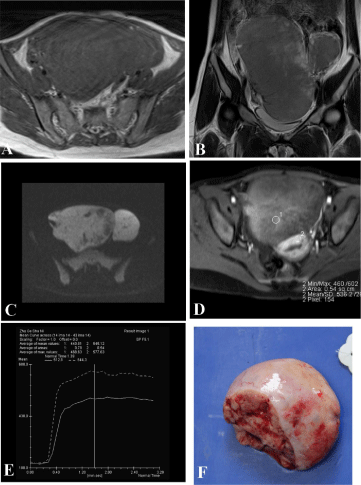
A 21-year-old woman complained of pelvic mass for a couple of weeks. There were no virginal discharge and bleeding. No surgical history has been recorded. Her Cancer Antigen (CA) 125 level was about 56.1U/mL and CA199 level was about 19.8U/mL. On MRI (Figure 4), the giant solid mass occupied the right adnexal region compressing the uterus and left ovary to the other side. The mass manifested as intermediate signals on both T1WI (A) and T2WI (B) images and slightly high signals on DWI images(C). The mass showed mild enhancement less than normal myometrium (D/E). The gross specimen revealed a round, solid mass with intact capsule (F) and the final histological diagnosis was B-cell lymphoma involving the bilateral ovaries and the right fallopian tube. The immunohistological analysis indicated strong positive for Ki67 stain.

    Figure 4:  A 21-year-old woman was pathologically proven with the B - cell
lymphoma (Case 3).
The mass displayed as totally solid mass and displayed as intermediate signals
on T1WI(A); On T2WI, there have some patchy high signals at the periphery of
tumor body, most of which showed intermediate signals(B); On DWI images,
the tumor showed relatively high signals(C); On contrast-enhanced images,
the tumor displayed as the mild enhancement when compared with normal
uterus(D/E); the resected sample showed a homogeneous, solid mass with
small foci of necrosis components with intact capsule.
Discussion
Primary genital tract lymphoma is extremely rare, in which the B-cell phenotype is the predominant type with better prognosis than T-cell lymphoma [9]. There are four subtypes of lymphoma involve the ovary: diffuse large B-cell lymphoma, Burkitt’s Lymphoma (BL), lymphoblastic lymphoma or an plastic large cell lymphoma [10]. The distinction between primary and secondary lymphoma is relatively difficult when solely based on imaging findings, while treatment and prognosis varies [8,9]. In this study, all three participants have no other systems involved except for pelvic adenopathy in one case. MRI has the most advantage of staging or assessing advanced lymphoma extension to adjacent structure [6].
In this series, all tumors displayed as the homogeneously solid mass without any necrosis or fat/lipid component in it. On post-enhanced MRI images, they showed mild and/or moderate enhancement compared with normal myometrium. It was noticed that no obvious pelvic fluid was detected on all cases. However, in one case with uterine lymphoma, the extra-uterus extension was observed, appearing as mesenteric mass abutting the uterine dome which was overlooked at the initial reading. Our findings were consistent with Ferrozzi’s report that primary lymphoma of genital tract appeared as solid mass with isointensity signals on both T1WI and T2WI [7]. On post-contrast images, the tumor always display mild and moderate enhancement, indicating its difference from other malignant ovarian tumors.
Considering the differential diagnosis, some ovarian solid masses, like sex cord-stromal ovarian tumors and germ cell tumors, must be included for the possible diagnosis. The coma or fibro-the coma is the most benign ovarian solid tumors encountered in the clinical unit. The characteristic signals of the coma are always low signals on any MRI protocol because there is little of water molecule among fibrous components, making it easily differentiated from other ovarian tumors [8]. For the young patients, germ cell tumors are another common ovarian disease with elevated AFP level, which may provide a useful clue for correct diagnosis. Upon bilateral solid ovarian masses, metastatic tumors should be also excluded and carefully look for clinical medical history is necessary to make a proper diagnosis [8]. As for uterine lymphoma, the differential diagnosis must include endometrial cancer, endometrial stroma sarcoma. Endometrial cancer always originates from endometrium, while primary lymphoma of the cervix or uterus arises in the stroma. Endometrial stroma sarcoma, an indolent neoplasm with a favorable prognosis involving long-term survival, in patients in whom the normal endometrium was wellpreserved, invariably appeared as solid masses with homogeneous signals and avid enhancement on MRI [11]. However, in both uterine lymphoma and endometrial stroma sarcoma, functional zone cannot be clearly demonstrated. Therefore, the histological diagnosis is needed.
Conclusion
In a word, owing to the rare occurrence and atypical imaging findings, primary female genital tract lymphoma make a great challenge for diagnosticians to establish a correct evaluation before invasive surgery. MRI can provide excellent soft tissue resolution; clearly delineate tumor margin and extension, giving the promising information for both clinicians. Fibroma, the coma, germ cell tumors, endometrial cancer, endometrial stroma sarcoma and metastatic tumors all should be considered as the differential diagnoses. The final diagnosis should be established on the histological diagnosis in relation with clinical medical history.
Acknowledgment
I appreciated Wang Hui Ph.D from biomedical research imaging center in department of radiology from the University of North Carolina at Chapel Hill for language editing. I thank tissue-bank department in Obstetrics & Gynecology hospital, Fudan University to provide the gross picture (Figure 4) of the lesion in case 3.
References
- Yadav BS, George P, Sharma SC, Gorsi U, McClennan E, Martino MA, et al. Primary non-Hodgkin lymphoma of the ovary. Semin Oncol. 2014; 41: 19-30.
- Mandato VD, Palermo R, Falbo A, Capodanno I, Capodanno F, Gelli MC, et al. Primary diffuse large B-cell lymphoma of the uterus: case report and review. Anticancer Res. 2014; 34: 4377-4390.
- Samama M, van Poelgeest M. Primary malignant lymphoma of the uterus: a case report and review of the literature. Case Rep Oncol. 2011; 4: 560-563.
- Sellami-Dhouib R, Nasfi A, Mejri NT, Doghri R, Charfi L, Sassi S, et al. Ovarian ALK+ diffuse large B-cell lymphoma: a case report and a review of the literature. Int J Gynecol Pathol. 2013; 32: 471-475.
- Crawshaw J, Sohaib SA, Wotherspoon A, Shepherd JH. Primary non-Hodgkin's lymphoma of the ovaries: imaging findings. Br J Radiol. 2007; 80: 155-158.
- Alves Viera MA, Cunha TM. Primary lymphomas of the female genital tract: imaging findings. Diagn Interv Radiol. 2014; 20: 110-115.
- Ferrozzi F, Tognini G, Bova D, Zuccoli G. Non-Hodgkin lymphomas of the ovaries: MR findings. J Comput Assist Tomogr. 2000; 24: 416-420.
- Jung SE, Lee JM, Rha SE, Byun JY, Jung JI, Hahn ST. CT and MR imaging of ovarian tumors with emphasis on differential diagnosis. Radiographics. 2002; 22: 1305-1325.
- Ahmad AK, Hui P, Litkouhi B, Azodi M, Rutherford T, McCarthy S, et al. Institutional review of primary non-hodgkin lymphoma of the female genital tract: a 33-year experience. Int J Gynecol Cancer. 2014; 24: 1250-1255.
- Bianchi P, Torcia F, Vitali M, Cozza G, Matteoli M, Giovanale V. An atypical presentation of sporadic ovarian Burkitt's lymphoma: case report and review of the literature. J Ovarian Res. 2013; 6: 46.
- Zhang GF, Zhang H, Tian XM, Zhang H. Magnetic resonance and diffusion-weighted imaging in categorization of uterine sarcomas: correlation with pathological findings. Clin Imaging. 2014; 38: 836-844.