Case Report
Austin J Radiol. 2015; 2(6): 1036.
A Giant Meningioma of the Posterior Fossa Extending to the Para Pharyngeal Space, MRI Findings
Ozdil Baskan¹* and Sema Demirci²
¹Department of Radiology, Istanbul Medipol University, Turkey
²Department of Neurology, Istanbul Medipol University, Turkey
*Corresponding author: Ozdil Baskan, Department of Radiology, School of Medicine, Istanbul Medipol University, Goztepe Cikisi Bagcilar, Turkey
Received: September 05, 2015; Accepted: September 09, 2015; Published: September 11, 2015
Abstract
Meningiomas involving the jugular foramen and parapharyngeal space are extremely rare. They most commonly occur intracranially and then extend to the extra cranial region through the foramen of the skull base, such as jugular foramen. Preoperative diagnosis and extensions of the lesions by imaging may predict the post-operative morbidity.
We present a case of extra cranial spread of a giant posterior fossa meningioma extending into the internal jugular vein and perineural spaces, presenting as a cervical mass with encasement of the internal carotid artery and cranial nerves.
Meningiomas involving the skull base, the neural foramina, the jugular foramen can present surgical challenges. Extra cranial extension of meningioma is uncommon, and direct intraluminal spread within the internal jugular vein to the neck is a rare finding. Imaging has an important role in identifying the lesions, detecting tumour-related complications and in presurgical differential diagnosis, which is essential for optimizing treatment strategies. MRI is the main tool of meningioma imaging because of its superior soft-tissue resolution and multiplanar capabilities. Appropriate preoperative imaging differential diagnosis and treatment planning are essential in these patients.
Keywords: Atypical meningioma; Magnetic resonance imaging; Para pharyngeal space; Jugular foramen
Introduction
Meningiomas are the most common benign intracranial neoplasms, arising from the arachnoid “cap” cells of the arachnoid villi in the meninges. Extra cranial extension of meningioma is seen rarely. Intracranial meningiomas can spread outside the cranium by direct invasion of the skull, by extension through perineural spaces or vascular channels. Rare cases of meningiomas extending into the Internal Jugular Vein (IJV) and presenting as cervical and parapharyngeal masses.
Herein, we present a case of extra cranial spread of a giant posterior fossa meningioma extending into the internal jugular vein and perineural spaces, presenting as a cervical mass with encasement of the internal carotid artery and cranial nerves.
Case Report
A 54-year old woman with a history of sphenoid wing meningioma over a 5 year period, who had no treatment, presented with a progressive dysphasia for a year. Physical examination revealed a left neck mass. Her neurological examination revealed that there was bilateral ptosis prominent on the left, bilateral papilledema, diminished gag reflex and truncal ataxia.
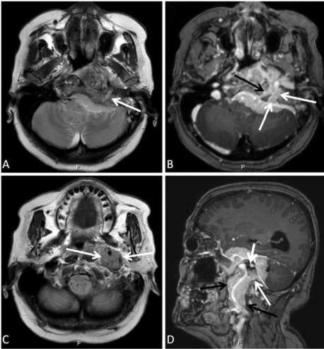
Cranial Magnetic Resonance Imaging (MRI) examination was performed on a 3T scanner (Achieva, Philips Best, Netherlands). MRI revealed a large posterior fossa mass involving sellar and parasellar area, extending directly to sphenoid sinus and extradural extension through the jugular foramen and hypoglossal canal with an extra cranial component in the left infratemporal fossa and parapharyngeal space (Figure 1). The tumour extended into the internal auditory meatus, too. It is iso-to hyperintens on T2- weighted images and isointens on the T1-weighted images, with intense contrast enhancement (Figure 1). T1-weighted, gadoliniumenhanced MRI demonstrated a uniformly enhanced tumor in the left jugular foramen invading the juguler bulb and extending through the jugular foramen into the internal jugular vein, and downward to the parapharyngeal space of the neck with encasement of the internal carotid artery (Figure 1).

    Figure 1:  A, B. Axial T2 weighted and contrast enhanced axial T1 weighted images at the same level show the lack of the normal flow void in the left jugular
vein secondary to extension of the meningioma (white arrows) and invasion of the hypoglossal canal (black arrow). C. Axial T2 weighted image at the level of C2
vertebra showing the lesion encasing the carotid vessels (arrows). D. Contrast enhanced sagittal T1 weighted image shows the parapharyngeal mass encasing the
carotid vessels (black arrows), and a component in the jugular foramen (white arrows), internal acoustic canal (short white arrow).
Discussion / Conclusion
Meningiomas are the common neoplasms of the central nervous system, account for 15% to 18% of all primary intracranial tumors [1,2]. Meningiomas, arise from the lining cells of the arachnoid villi, are often located in the dural venous sinuses and their large tributary veins and most of them affect the parasagittal, falx, convexity, and sphenoid wing, where cerebrospinal fluid is returned to the venous system [3]. Although meningiomas are the common neoplasms of the central nervous system, extra cranial meningiomas are extremely rare. They constitute about 2% of all meningiomas [4].
Hoye et al. [5] suggest a classification of extra cranial meningiomas into four groups.
Type 1; Direct extra cranial extension of the primary intracranial meningiomas from the skull, 1. The neural foramina (foramen ovale, the jugular foramen etc.), 2. along the calvarial sutures and pneumatic cells 3. through the haversian canals of the skull.
Type 2; involves extra cranial growth from the arachnoid within the cranial nerve sheaths.
Type 3; Extra cranial growth from ectopic or embryonic arachnoid cell rest, meningiomas having no demonstrable connection with foramina or cranial nerves. The predilective locations of this type include orbita and optic nerve, the nasal cavity, the paranasal sinuses and the nasopharynx.
Type 4; Distant metastases from intracranial meningioma. The liver and the lung are the most common sites of metastases, but metastasizing meningiomas are not always pathologically malignant.
Nager et al. had reported that [6] the most common sites are the orbit (8%) and external table of the calvaria (7%). Extension and presentation into parapharyngeal space is exceedingly rare, accounting for only 1.4% of all intracranial meningiomas. The most frequent pathway to the parapharyngeal space is through the jugular foramen.
Our case, the posterior fossa meningioma was in continuity through the jugular foramen with a component that filled the parapharyngeal space and also had extensions to the foramen ovale, hypoglossal canal, was thought to be classified as type 1 according to Hoye’s classification, the most common of the four presentations [7]. Meningiomas can infiltrate the dura and adjacent venous sinuses, but it is very uncommon for them to extend through the sinuses into the extra-cranial draining veins. Our patient had direct extension of the tumour into the IJV by invasion of the sigmoid sinus.
Meningiomas range from isointense to hypo intense on T1- weighted images, and from isointense to hyper intense on T2- weighted images. They enhance strongly and homogeneously after gadolinium injection. The extensions of the lesion to the foramina, invasions to the dural sinuses and relations to the vascular structures can be well delineated with the MRI. A study by Shimono et al. [8] demonstrated differences in MR signal intensity and contrast enhancement between the intra- and extra cranial components of JF meningiomas. The signal intensities of the intracranial component of JFM were significantly higher than those of the extra cranial component on T1-, T2-, and post contrast T1-weighted images. We noted these different signals that are the best visualized on the noncontrast T2-weighted images (Figure 1).
Meningiomas involving the skull base, the neural foramina, and the jugular foramen can present surgical challenges. Although the treatment choice is surgery, resection can be associated with compromise of multiple cranial nerves and is also associated with an increased local recurrence rate [9]. Imaging has an important role in identifying the lesions, detecting tumour-related complications and in presurgical differential diagnosis, which is essential for optimizing treatment strategies. Evaluation of the cranial nerve involvement by imaging may predict the post-operative morbidity. MRI is the main tool of meningioma imaging because of its superior soft-tissue resolution and multiplanar capabilities. Evaluation by MRI and Magnetic Resonance Angiography (MRA), Magnetic Resonance Venography (MRV) should be considered in such conditions, thus offer important aids to correct diagnosis and to verify the tumor location and extensions.
Extra cranial extension of meningioma is uncommon, and direct intraluminal spread within the internal jugular vein to the neck is a rare finding. Appropriate preoperative imaging, differential diagnosis and treatment planning are essential in these patients.
References
- Dhingra S, Mohindra S, Gupta SK, Shankar A. Intracranial meningioma presenting as a parapharyngeal tumour – A unique extracranial presentation. Int J Pediatr Otorhinolaryngol. 2011; 6: 325–328.
- Wara WM, Sheline GE, Newman H, Townsend JJ, Boldrey EB. Radiation therapy of meningiomas. Am J Roentgenol Radium Ther Nucl Med. 1975; 123: 453-458.
- Farr HW, Gray GF, Vrana M, Panio M. Extracranial meningioma. J Surg Oncol. 1973; 5: 411-420.
- Friedman CD, Costantino PD, Teitelbaum B, Berktold RE, Sisson GA Sr. Primary extracranial meningiomas of the head and neck. Laryngoscope. 1990; 100: 41-48.
- Hoye SJ, Hoar CS Jr, Murray JE. Extracranial meningioma presenting as a tumor of the neck. Am J Surg. 1960; 100: 486-489.
- Nager GT, Heroy J, Hoeplinger M. Meningiomas invading the temporal bone with extension to the neck. Am J Otolaryngol. 1983; 4: 297-324.
- Rogers L, Gilbert M, Vogelbaum MA. Intracranial meningiomas of atypical (WHO grade II) histology. J Neurooncol. 2010; 99: 393-405.
- Shimono T, Akai F, Yamamoto A, Kanagaki M, Fushimi Y, Maeda M, et al. Different signal intensities between intra- and extracranial components in jugular foramen meningioma: an enigma. AJNR Am J Neuroradiol. 2005; 26: 1122-1127.
- Gilbert ME, Shelton C, McDonald A, Salzman KL, Harnsberger HR, Sharma PK, et al. Meningioma of the jugular foramen: glomus jugulare mimic and surgical challenge. Laryngoscope. 2004; 114: 25-32.