Case Presentation
Austin J Radiol. 2017; 4(3): 1076.
Osteochondroma of Lumbar Spine – Multi Modality Imaging of a Case
Kulbhushan Vishnoi, Indiran Venkatraman* and Beno Jefferson P
Department of Radiodiagnosis, Sree balaji Medical College and Hospital, India
*Corresponding author: Indiran Venkatraman, Department of Radiodiagnosis, Sree balaji Medical College and Hospital, India
Received: October 30, 2017; Accepted: December 12, 2017; Published: December 22, 2017
Abstract
Osteochondroma is a benign outgrowth of bone and cartilage and is one of the most common bone tumors that usually occurs in the long bones but rarely involves the spine [1]. In spine, it occurs most commonly in the cervical and upper dorsal segments [2]. It shows male preponderance and an average age presentation is approximately 32+4.6 years [3]. Lumbar osteochondroma can be asymptomatic or cause symptoms like pain, radioculopathy/myelopathy, or cosmetic deformity. Osteochondromas are also known as exostosis, the most common primary bone tumors comprising one-third of all benign bone tumors. They can be solitary (90% of cases) or multiple in the form of hereditary multiple exostosis [4]. Here we report a case of 53 year old male, came with left abdominal pain, in which lumbar osteochondroma was an incidental finding on Computed Tomography (CT).
Keywords: Osteochondroma; Spinal; Exostosis
Case Presentation
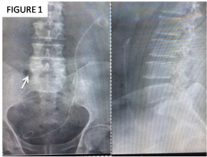
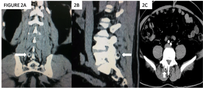
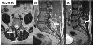
A 53 year old male came to the casualty with complaints of pricking left abdomen pain since one week, which had aggravated one day prior. The patient was evaluated for the same. His physical examination, neurological examination and blood investigations were unremarkable. Ultrasound abdomen revealed left ureteric calculus and cholelitheasis. CT Abdomen done to confirm the diagnosis before undergoing lithotripsy, showed an incidental finding of osteochondroma arising from right pars interarticularis of L5 vertebra was observed. CT showed a well defined bony lesion protruding posteriorly from the right pars interarticularis of L5 vertebra (measuring ~ 1.4 x 1.1 cm) with continuity of cortex and medulla with the involved vertebra. Radiograph also showed a bony mass protruding posteriorly, probably arising from the L5 vertebra. Magnetic resonance imaging showed a bony outgrowth with fatty marrow seen arising from right pars interarticularis of L5 vertebra and pointing posteriorly. There was no large cartilage cap or soft tissue component. He underwent ureteric stenting for the calculi and was relieved of his symptoms. He was just reassured with respect to the spinal osteochondroma (Figure 1).

    Figure 1:  Radiograph of Lumbar spine AP and Lateral view shows increase
in bone density in the right lateral aspect of the L5 vertebra (Arrow).
Discussion
Osteochondromas are developmental lesions rather than true neoplasms. Lumbar osteochondromas are very rare, accounting for only 3-4% of spinal osteochondromas [5]. Marrow and cortical continuity with the underlying parent bone defines the lesion [6]. It results mainly from the separation of a fragment of epiphyseal growth plate cartilage, which then herniates through the periosteal bone cuff that normally surrounds the growth plate [7].
Osteochondromas are better visualized on computed tomography scan [8]. MRI is useful to determine the extent of neurologic structures compromise and it identifies lesions that look suspicious of malignant transformation.
Osteochondromas are often present in young patients and their growth usually arrests after puberty with closure of the epiphysis [9].
Spinal involvement is more common in hereditary multiple extostoses. In this condition, thoracic and lumbar vertebrae are commonly affected, while the solitary type mostly affects the cervical spine particularly the atlantoaxial region. Sacral involvement is rare (Figure 2A, B and C).

    Figure 2:  A, B and C: Computed tomography image of lumbar spine shows
bony outgrowth with cortical and trabeculations and fatty marrow seen arising
from right pars intra-articularis of L5 vertebra.

    Figure 3:  A, B and C: Magnetic resonance imaging shows bony outgrowth
with fatty marrow seen arising from right laminar of L5 vertebra and pointing
posteriorly.
Spinal osteochondromas occur mainly in the posterior elements of vertebra due to presence of secondary ossification centers in the neural arch (Spinous processes, transverse processes, articular processes and endplate of vertebral bodies) [10]. The aberrant cartilage tissue in these second ossification centers can give rise to osteochondromas.
Diagnosis of spinal exostosis may be difficult on plain radiography owing to the complex anatomy. In ~ 15% of patients radiographs are normal [11].
CT findings are characterized by a paraspinal, dumbbell or eccentrically located, round, sharply outlined mass in the spinal canal with bonelike density with scattered calcifications and osteosclerotic changes in neighbouring bone and lacking contrast enhancement.
MRI is more accurate than CT but mainly to access the malignant transformation of the tumor. In 90% of cases malignant transformation leads to a chondrosarcoma, this usually develops in the cartilage cap of the osteochondroma. Malignancy is generally suspected if cap thickness >2 cm, but the diagnosis is only confirmed with a biopsy of the lesion (Figure 3A, B and C) [12].
Conclusion
Osteochondromas are benign tumors formed from bone with a cartilage cap. Osteochondromas rarely involve the spine, but when they occur they can be asymptomatic or cause symptoms, like pain, radiculopathy or myelopathy, or, even, cosmetic deformation. The radiologic exam of election for diagnosis is CT scan. When symptomatic the treatment of choice is surgical resection. The most concerning complication of osteochondromas is malignant transformation, fortunately a rare event. Surgical treatment involves resection of the tumor, and it relieves the symptoms almost immediately. There are low rates of complication and relapse following surgery.
References
- Qasem SA, Deyoung BR. Cartilage-forming tumors. Seminars in Diagnostic Pathology. 2014; 31: 10–20.
- Gille O, Pointillart V, Vital JM. Course of spinal solitary osteochondromas.Spine. 2005; 30: 13–19.
- Gaetani P, Tancioni F, Merlo P, Villani L, Spanu G, Rodriguez y Baena R. Spinal chondroma of the lumbar tract: case report. Surgical Neurology. 1996; 46: 534–539.
- Murphey MD, Choi JJ, Kransdorf MJ, Flemming DJ, Gannon FH. Imaging of osteochondroma: variants and complications with radiologic-pathologic correlation. Radiographics. 2000; 20: 1407–1434.
- Bess RS, Robbin MR, Bohlman HH, Thompson GH. Spinal exostoses: analysis of twelve cases and review of the literature. Spine (Phila Pa 1976). 2005; 30: 774–780.
- Thiart M, Herbrst H. Lumbar osteochondroma causing spinal compression.SA Orthopaedic Journal Winter. 2010; 9: 44–46.
- Resnick D, Kyriakos M, Greenway GD. Philadelphia: PA: Saunders; Osteochondroma. In: Resnick D. 3rd edition. Diagnosis of bone and joint disorders. 1995; 5: 3725–3746.
- Edelman RR, Hesselink JR, Zlatkin MB. Philadelphia: Saunders Elsevier. Clinical magnetic resonance imaging. 2006; 3: 2320–2327.
- Shim JH, Park CK, Shin SH, Jeong HS, Hwang JH. Solitary osteochondroma of the twelfth rib with intraspinal extension and cord compression in a middleaged patient. BMC Musculoskelet Disord. 2012; 13: 57.
- Fiumara E, Scarabino T, Guglielmi G, Bisceglia M, D’Angelo V. Osteochondroma of the L-5 vertebra: a rare cause of sciatic pain. Case report. J Neurosurg. 1999; 91: 219–222.
- Murphey MD, Andrews CL, Flemming DJ, Temple HT, Smith WS, Smirniotopoulos JG. From the archives of the AFIP. Primary tumors of the spine: radiologic pathologic correlation. Radiographics. 1996; 16: 1131–1158.
- Strovski E, Ali R, Graeb DA, Munk PL, Chang SD. Malignant degeneration of a lumbar osteochondroma into a chondrosarcoma which mimicked a large retropertioneal mass. Skeletal Radiology. 2012; 41: 1319–1322.