
Case Report
Sarcoma Res Int. 2014;1(2): 2.
A Clinical Case of Synovial Sarcoma of the Larynx as a Giant Vocal Fold Polyp
Magdalena Chirila1,2*, Vera Dinescu1, Maria Petri1 and Cristina Tiple1
1Department of Otorhinolarynhology, Iuliu Hatieganu University of Medicine and Pharmacy, Romania
2Department of Otorhinolarynhology, Emergency County Hospital, Romania
*Corresponding author: Magdalena Chirila, Department of Otorhinolarynhology, Iuliu Hatieganu University of Medicine and Pharmacy, Emergency County Hospital, Romania
Received: September 01, 2014; Accepted: October 13, 2014; Published: October 15, 2014
Abstract
Synovial cell sarcoma of the larynx is a very rare malignant tumor. We present the case of a 62-years-old man with a monophasic synovial sarcoma of the larynx, clinically presented as a vocal fold giant polyp. CO2 laser assisted surgery could be use as less invasive but effective approach than laryngectomies for a limited lesion, particularly for a vocal fold tumor with free surgical borders.
Keywords: Monophasic synovial sarcoma; Microlaryngoscopy; Laryngectomy
Introduction
Synovial cell sarcoma represents a rare group of malignant tumors, particularly in the head and neck region (3-9%) and the larynx is the least frequent site of occurrence [1]. Only 20 cases of laryngeal synovial sarcoma have been described in the English literature to date [1-3]. Synovial sarcoma typically affects young individuals of the second to fourth decade in the extremities and has a male preponderance [4].
In this paper we present the case of a 62-years-old man with a monophasic synovial sarcoma of the larynx, clinically presented as a vocal fold giant polyp.
Case Report
A 62-years-old man presented in March 2012 at the Otorhinolaryngological Department of our hospital with severe dysphonia for a couple of months. A video laryngoscopy revealed a giant polypoid mass arising from the anterior 2/3 of left vocal fold. The mobility of the vocal fold was preserved. Neck lymphadenopathy was absent clinically. The CT scan showed a polypoid mass on the left vocal fold with low to moderate heterogenous enhancement after the injection of contrast substance. No enlarged lymph node was present. The tumor was staged as T1NoMo.
We performed a biopsy under general anaesthesia in suspension microlaryngoscopy (Figure 1). The frozen section suggested we had a spindle cell tumor. Because the tumor respected the vocal fold ligament (Figure 2) a CO2 laser assisted type I cordectomy was performed (Figure 3). The final histological exam showed a sarcomatous area characterized by a cellular spindle cell infiltration without any special pattern. The immunohistochemical examination for vimentin antibody and CD99 was positive (Figure 4a and 4b). The sample confirmed the integrity of the surgical borders.
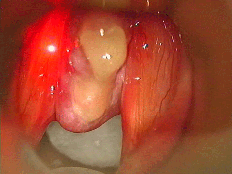
Figure 1: Giant left vocal fold “polyp” (suspension microlaryngoscopy).
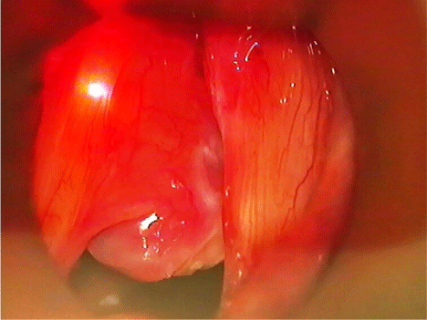
Figure 2: The vocal ligament seems not be involved.
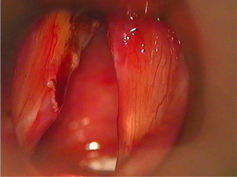
Figure 3: Type I CO2 laser assisted cordectomy.
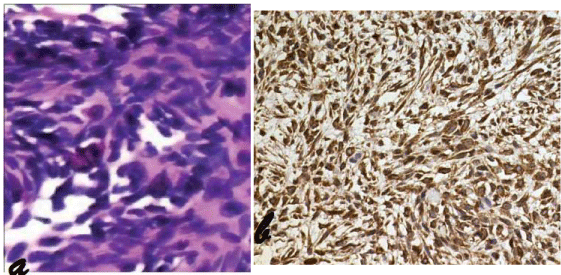
Figure 4: a) a highly cellular tumor with spindle-shaped cells (frozen section); b) Strongly positive IHC for vimentin (20 �).
The patient is free of disease 28 months after the initial diagnosis with a minimum glottic failure and slighty dysphonia (Figure 5).
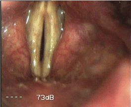
Figure 5: Larynx aspect at 28 months after initial diagnosis (video laryngoscopy).
Discussion
We add a new case of laryngeal synovial sarcoma with some specific clinical features. The clinical presentation as a huge polyp on the left vocal fold is unique in the English literature. The surgical treatment was comprehensive but functional and we had two reasons for that: first, the tumor respected the vocal fold ligament; second, the patient is a primary school teacher and he needs his voice. The integrity of the surgical borders was the fact which decided the follow-up schedule.
Laryngeal synovial sarcoma is an extremely rare form of laryngeal tumors, squamous cell carcinoma accounts for over 90% of all laryngeal malignancies [2]. In a comparative analysis with synovial sarcoma of the extremities Salcedo-Hernàndez et all [5] showed that head and neck sarcomas had similar survival rates compared to sarcomas of limbs. In 2013 Luna-Ortiz et al [3] described a 21 years old woman with an aryepiglottic folds sarcoma. They performed a spupraglottic laryngectomy with 2 years free disease and concluded that the organ preservation surgery seems to be indicated.
Prognosis for patients with synovial sarcoma is related to primary tumor extent, tumor grade, and size [6]. The optimal treatment for localized synovial sarcomas is complete surgical resection [6- 9]. Because of the lack of nodal metastasis, neck dissection is not necessary in the absence of nodal involvement [10]. When the surgical borders are involved, adjuvant chemotherapy or radiotherapy can be given [9,11,12].
Conclusion
In conclusion, laryngeal synovial sarcoma should be considered an aggressive tumor, like its counterpart in the extremities. CO2 laser assisted surgery could be use as less invasive but effective approach than laryngectomies for a limited lesion, particularly for a vocal fold tumor with free surgical borders.
References
- Bao YY, Wang QY, Zhou SH, Zhao K, Ruan LX, Yao HT. Poor outcome of comprehensive therapy in a case of laryngeal synovial sarcoma. Radiol Oncol. 2013; 47: 111-118.
- Saxby C, Bova R, Edwards M. Laryngeal synovial sarcoma: a rare clinical entity. Case Rep Otolaryngol. 2013; 2013: 578606.
- Luna-Ortiz K, Cano-Valdez AM, da Cunha IW, Mosqueda-Taylor A. Synovial sarcoma of the larynx treated by supraglottic laryngectomy: case report and literature review. Ear Nose Throat J. 2013; 92: E20-26.
- Jayachandra S, Chin RY, Walshe P. Synovial cell sarcoma of the larynx. Hematol Oncol Clin North Am. 2012; 26: 1209-1219.
- Salcedo-Hernández RA, Lino-Silva LS, Luna-Ortiz K. Synovial sarcomas of the head and neck: comparative analysis with synovial sarcoma of the extremities. Auris Nasus Larynx. 2013; 40: 476-480.
- Sato T, Hasegawa H, Sugasawa M, Yasuda M, Morita K, Nakahira M, et al. Free jejunal transfer for a 15-year-old girl with synovial sarcoma of the hypopharynx. J Plast Reconstr Aesthet Surg. 2011; 64: 1100-1103.
- Mhawech-Fauceglia P, Ramzy P, Bshara W, Sait S, Rigual N. Synovial sarcoma of the larynx in a 79-year-old woman, confirmed by karyotyping and fluorescence in situ hybridization analysis. Ann Diagn Pathol. 2007; 11: 223-227.
- Sridhar Reddy D, Shobhan Babu A, Lenin A. Synovial sarcomna of larynx-a rare site. Indian J Otolaryngol Head Neck Surg. 2007; 59: 51-52.
- Capelli M, Bertino G, Morbini P, Proh M, Falco CE, Benazzo M. CO2 laser in the treatment of laryngeal synovial sarcoma: a clinical case. Tumori. 2007; 93: 296-299.
- Papaspyrou S, Kyriakides G, Tapis M. Endoscopic CO2 laser surgery for large synovial sarcoma of the larynx. Otolaryngol Head Neck Surg. 2003; 129: 630-631.
- Bilgic B, Mete O, Oztürk SA, Demiryont M, Keles N, Basaran M. Synovial sarcoma: a rare tumor of larynx. Pathol Oncol Res. 2003; 9: 242-245.
- Alberty J, Dockhorn-Dworniczak B. Monophasic synovial sarcoma of the neck in an 8-year-old girl resembling a thyroglossal duct cyst. Int J Pediatr Otorhinolaryngol. 2002; 63: 61-65.