
Review Article
Austin J Sleep Disord. 2015;2(2): 1011.
Ocular Manifestations of Obstructive Sleep Apnea
Hatice Arda*, Duygu Gülmez Sevim, Ertugrul Mirza and Sarper Karakucuk
Department of Ophthalmology, Erciyes University Faculty of Medicine, Turkey
*Corresponding author: Hatice Arda, Department of Ophthalmology, Erciyes University Faculty of Medicine, Kayseri, 38039, Turkey
Received: March 11, 2015; Accepted: April 22, 2015; Published: April 24, 2015
Abstract
Obstructive sleep apnea (OSA) is a serious disorder characterized with repeated episodes of upper airway obstruction during sleep, resulting in nocturnal hypoxemia and hypercapnia which leads to many systemic and also ocular complications.
Various eye disorders reported to be associated with OSA. Floppy eyelid syndrome (FES), cornea disorders, glaucoma, non-arteritic anterior ischemic optic neuropathy, papilledema, central serous chorioretinopathy, and retinal vein occlusion are some of these disorders.
Keywords: Obstructive sleep apnea; Floppy eyelid syndrome; Optic neuropathy; Normal tension glaucoma
Abbreviations
OSA: Obstructive Sleep Apnea; PSG: Polysomnography; CPAP: Continuous Positive Airway Pressure; BiPAP: Bilevel Positive Airway Pressure; FES: Floppy Eyelid Syndrome; AHI: Apnea- Hypopnea Index; POAG: Primary Open Angle Glaucoma; NTG: Normal Tension Glaucoma; RNFL: Retinal Nerve Fiber Layer; IOP: Intraocular Pressure; NAION: Non-Arteritic Anterior Ischemic Optic Neuropathy; IIH: Idiopathic Intracranial Hypertension; CSCR: Central Serous Chorioretinopathy; RVO: Retinal Vein Occlusion
Introduction
Obstructive sleep apnea (OSA) is a serious disorder that leads to many systemic and ocular complications. It is characterized with repeated episodes of upper airway obstruction during sleep, resulting in nocturnal hypoxemia with the symptoms of excessive daytime sleepiness, disruptive snoring, and apnea episodes [1]. The prevalence of OSA is estimated to be approximately 9% in women and 24% in men [2]. Cardiovascular and neurological morbidity is a serious outcome of OSA with the fact that it is seen in approximately 60% to 70% of patients with stroke or ischemic heart disease [3]. The gold standard procedure in the diagnosis is the overnight polysomnography (PSG) [1]. The main medical treatment options for the condition are continuous positive airway pressure (CPAP) and bilevel positive airway pressure (BiPAP), if CPAP is not tolerated by the patient [4]. Modafinil is a novel wake-promoting agent, reported to be an effective adjunct therapy for residual excessive sleepiness in patients treated with CPAP [5]. Surgical management options are limited to those in whom the medical treatment is not well tolerated or failed to be successful.
Its ocular associations have been an issue of great interest due to the irreversible complications it may cause and may have been preventable, if the condition of OSA is diagnosed and treated properly beforehand or even after the diagnosis of the ocular findings. Various eye disorders have been reported to be associated with OSA including; floppy eyelid syndrome (FES), cornea disorders, glaucoma, nonarteritic anterior ischemic optic neuropathy (NAION), papilledema, central serous chorioretinopathy (CSCR), and retinal vein occlusion (RVO). This review aims to take the attention of the ophthalmologists on the possibility of ocular disorders that can be accompanied by sleeping disorders.
Floppy eyelid syndrome
Mostly seen in overweight, middle aged males with the complaint of foreign body sensation, burning, tearing, and redness; FES is characterized with the clinical findings of flaccid and easily everted upper lids, occurring spontaneously or with minimal traction, and chronic papillary conjunctivitis of the upper palpebral conjunctiva. Tarsal plaque biopsies of the patients with FES revealed the histopathological features as an increase in the elastolytic metalloproteinase enzymes and a subsequent decrease in the elastin fibers of the tissue [6,7]. In the literature the prevalence of FES in the OSA population has been reported to vary from 2% to 32% [8,9]. OSA is known to be seen mostly in overweight patients, so there is not a clear distinction regarding the etiology concerning whether FES and OSA is related directly or FES is mainly related to obesity. The prevalence of obesity in OSA has ranged from 60% to 70% [10,11], while the prevalence of obesity in FES patients has ranged from 43% to 92% [12,13]. In their review of patients with lax eyelid syndrome, Fowler and Dutton [14] stated that there was not a significant difference regarding the prevalence of OSA between patients who had obesity and FES and who had obesity but did not have FES. They also found that patients with OSA tended to have the prevalence of obesity significantly higher than those without OSA (76% vs. 20%). So they concluded that increased OSA prevalence among patients with FES was possibly associated with the concomitant obesity. On the contrary of the findings of this study, Ezra et al. [10] found a strong relationship between OSA and FES independent of weight. They explained that the possible mechanism might have been the changes in central nervous system arousability in OSA. Another possible explanation for the underlying mechanism of the association between OSA and FES is believed to be the increased venous pressure caused by right heart failure and apnea in patients with OSA [15].
Regarding the ocular surface findings along with FES, Acar et al. revealed low levels of Schirmer I values and tear break up times, and an increase in corneal staining and ocular surface disease index scores in patients with OSA, with the changes being correspondingly related to the severity of the disease based on apnea-hypopnea index (AHI) [16]. The authors suggested that the floppiness of the eyelid increases the inflammation in the ocular surface, causing a reduction in the amount and the quality of the tear and the symptoms of FES such as burning, itching and redness. Although there have been reports indicating a negative effect of CPAP treatment on ocular surface parameters because of the irritative properties of the treatment, in the second stage of their previous study, Acar et al. [17] reported that all of the latter impaired findings, including the stage of FES showed a significant improvement in patients with moderate and severe OSA, after 18 months of CPAP treatment. They concluded that the treatment should be implemented in an appropriate manner for at least one year in order to encounter the ocular surface irritation that is seen in the early stages of the therapy.
In their recent study to determine whether the presence of FES is associated with a higher prevalence of glaucoma in OSA patients, Muniesa et al. [18] found a significantly higher prevalence of glaucoma among the patients with FES (23.07%) compared to patients without FES (5.3%), and concluded that FES could be used as an indicator of glaucoma in patients with OSA.
Glaucoma
Several reports in the literature indicated possible associations between OSA and primary open angle glaucoma (POAG), normal tension glaucoma (NTG), visual field defects and reduced retinal nerve fiber layer thickness (RNFL). However, there have been contradictory reports regarding the relationship between OSA and glaucoma, with one study showing no difference [19], while another indicating the prevalence of glaucoma in OSA being approximately 4 times higher than the expected population rate [20].
The pathogenesis of ocular complications in OSA is most likely to have multifactorial origin. Vascular and mechanical factors have been thought to be involved in the pathological mechanism of the optic nerve involvement. Vascular factors are mainly the outcomes of repetitive or prolonged episodes of hypoxia; that include direct damage to optic nerve, oxidative stress and inflammation, increased vascular resistance, autonomic dysfunction, increased intracranial pressure and decreased cerebral perfusion (Figure 1) [21]. Repetitive prolonged upper airway obstruction and following arousal lead to an increase in the sympathetic tone, thus causing an activation in the rennin-angiotensin system. These factors and the accompanying effect of hypoxia trigger an increase in blood pressure and vascular resistance, causing damaging in the vascular endothelium. All of these consequences lead to the impairment of the autonomic function, an imbalance of vasodilatation and vasoconstriction. Furthermore the effect of the arousal periods and reperfusion lead to inflammation and oxidative stress, shown by the increased levels of reactive oxygen species and inflammatory markers [21]. In the etiologic scope, mechanical factors include supine position and obesity related increased intraocular pressure (IOP), and intracranial pressure at night, and the depletion of fiber in the trabeculum and lamina cribrosa [21]. To reveal the clinical impact of the pathogenesis of vascular pathology, Karakucuk et al. [22] noted that among the OSA patients with glaucoma, all of the patients had severe OSA and they revealed a strong correlation between AHI and IOP, and between mean defect in the visual field testing and resistivity index of the ophthalmic artery and central retinal artery determined with orbital Doppler ultrasonography.
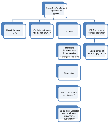
ON: Optic Nerve; ROS: Reactive Oxygen Species; RAA: Renin-angiotensinealdosterone; BP: Blood Pressure; ICP: Intracranial Pressure.
    Figure 1:  Vascular factors in the pathogenesis of ocular complications in obstructive sleep apnea.

    ON: Optic Nerve; ROS: Reactive Oxygen Species; RAA: Renin-angiotensinealdosterone; BP: Blood Pressure; ICP: Intracranial Pressure.
In their prospective photographic study of the ocular fundus, performed in the night of PSG, Fraser et al. [23] found no difference in rates of glaucomatous appearance or pallor of the optic disc between the OSA and the control groups.
In a recent study from Turkey, the authors revealed that POAG patients with OSA had significantly lower IOP levels than the patients without OSA [24]. This supports the fact that especially in NTG, and in patients showing damage progression despite the control in IOP, the presence of OSA should be kept in mind by the clinicians. A recent hypothesized mechanism for NTG is the imbalanced pressure between the two sides of lamina cribrosa, causing optic nerve fiber layer damage [25]. Accordingly with this theory, Xin et al. [26] found in their study that, cerebrospinal fluid pressure was significantly lower in the thinned RNFL group versus normal RNFL group among patients with OSA, regardless of body mass index and IOP. Among OSA patients in their study, Lin et al. [27] found that the glaucoma prevalence was significantly higher from the control group, with all of them being NTG, and the prevalence was in a strong relationship with the severity of the OSA.
In literature, thinning of the RNFL, which is associated with early stages of glaucomatous optic neuropathy had been shown to be related to OSA [28,29]. In a recent study, Shiba et al. [30] showed a significant negative correlation between AHI and nasal RNFL thickness, specifically. Visual field change, which is the sign of functional loss that appears after anatomical changes, had also been shown to be in relation with the severity of OSA in various studies [22,28,31].
Treatment of OSA has been shown to be effective in controlling the risk of glaucoma. In their cohort study, Chen et al. [32] pointed out that surgical treatment of OSA decreased the risk of glaucoma to the same levels with the control group, however patients treated with CPAP did not show a significant decrease in the hazard ratio of glaucoma.
Non-arteritic anterior ischemic optic neuropathy
Because of the similar known risk factors in their etiology, and the symptoms of NAION usually occurring at the awakening period, a possible relationship between NAION and OSA had been suspected. In their study to determine such relationship, Arda and her colleagues compared NAION patients for presence of OSA with a control group that have similar risk factors for NAION [33]. They determined that both groups had a high prevalence of OSA, suggesting that OSA being a contributing factor along with other risk factors, other than being an independent one. On the contrary of the findings of this study, Bilgin et al. [34] revealed a significant relationship between OSA and NAION in their study. The authors explained the different outcomes of the studies with the difference of the AHI criteria between the studies, with their study defining OSA with an AHI of = 20, with the previous study defining OSA with an AHI of = 5, causing false positive results. The importance of the possible relationship with OSA and NAION lies in the fact that, up to date there has not been a proven treatment method for NAION, so the main concern has been to minimize the risk of the involvement of the second eye. We suggest that an evaluation for OSA, at least in suspected patients, should be carried out in newly diagnosed NAION patients.
Papilledema
The intracranial pressure of patients with OSA is known to be elevated episodically, during the apneic episodes. Cerebral venous dilatation resulting from hypercapnia and raised venous pressure due to forced expiration have been suggested as the causative factors [35]. Various cases have been reported in the literature linking OSA with papilledema and idiopathic intracranial hypertension (IIH). In 2001, Marcus et al. [36] revealed that among the patients with IIH sleep-related breathing problems were common, but all of these patients were also obese. Lee et al. [37] also determined that OSA was common among men with IIH, and the symptoms of IIH tended to improve with the treatment of OSA. Treatment of OSA has been shown to be effective in resolving the symptoms of papilledema in various case reports [38,39].
On the other hand, there have been contradictory reports in the literature showing the association between IIH and OSA. In their study, Peter et al. [40] found no signs of papilledema in fundoscopic examination of 35 patients with OSA who had visual symptoms suggestive of papilledema. In a recent study conducted by Thurtell and his colleagues to determine whether OSA is an independent risk factor or a consequence of the other risk factors for OSA, especially obesity, for IHH, they revealed that the prevalence and severity of OSA showed no difference in IHH patients compared to control group with same demographic features including their BMIs [41].
Typically, IHH is seen in overweight females of childbearing ages. Although there has not yet been a consensus on the association between OSA and papilledema in the literature, we suggest that especially in male patients with older ages, OSA should be suspected in cases of IIH.
Keratoconus
Keratoconus is a non-inflammatory ectatic disease characterized by progressive changes in corneal collagen structure and organization, resulting in the thinning and steepening of the cornea [42]. Keratoconus has been linked to various conditions including FES. Since both OSA and keratoconus have been shown to be associated with FES, a possible direct association between OSA and keratoconus has been recently an issue of interest. Gupta et al. [43] and Saidel et al. [44] reported the prevalence of OSA among patients with keratoconus to be 18% and 19.6% respectively. Obesity is also a risk factor for both OSA and FES, and various studies showed an association between obesity and keratoconus [43-45]. The common underlying etiology covering all of the associations seems to be inflammation and the main mediator being matrix metalloproteinase which had been shown to increase in all of the mentioned conditions [46].
On the contrary of the findings of these previously published studies, recently Gencer et al. [47] published a study in a Turkish population showing no relationship between keratoconus and obesity, and indicated an insignificant risk for developing OSA in patients with keratoconus. The authors explained these contradictory results with the possible explanation of the all other studies being conducted in the United States, and obesity already being present approximately in the one third of the adult population in their general population which is a significant risk factor for OSA.
Central serous chorioretinopathy
Serous detachment of neurosensorial retina associated with increased sympathetic activity, elevated levels of serum steroids and type A personality is the case of CSCR. The underlying cause has been hypothesized to be the increased permeability of the choroidal vasculature and/or break-down of the outer retinal barrier [48,49]. The possible association between OSA and CSCR has been linked to the shared increased serum cortisol levels and sympathetic activity, resulting in vascular endothelial dysfunction in both conditions [50].
Leveque et al. [51] showed an association between OSA and CSCR, however in a very recent study, Brodie et al. [50] showed no significant association between these two conditions with their control group being gender/age/BMI matched with the patients. Brodie and his colleagues explained these contradictory results with their matching controls for BMI, a known risk factor for OSA, while the previous study had age and gender matched controls only. There is a limited number of studies in the literature to conclude a certain relationship between OSA and CSCR, thus further studies are needed.
Retinal vein occlusion
Leroux les Jardins et al. [52] reported the first possible association in the literature between RVO and OSA. The authors reported three cases of RVO with OSA, and hypothesized the possible mechanism as the slowdown of blood circulation caused by the hypoxemia and elevated nocturnal IOP in patients with OSA. In a cohort study by Chou et al. [53] OSA was shown to be an independent risk factor for RVO. Two other studies have also shown that sleep disorders might at least have a triggering affect in the pathogenesis of RVO [54,55]. There are two case reports in the literature, reporting two patients with bilateral RVO which is extremely infrequent, both having OSA [56,57]. Although the lack of scarce scientific evidence, we suggest due to possible common risk factors, patients with RVO should also be suspected and evaluated for OSA.
Conclusion
With the increasing prevalence of obesity worldwide, OSA seems to be getting more recognized in the general public. Due to its systemic associations and complications with high morbidity, it should be carefully and adequately recognized by the clinicians of any specialty as well as the ophthalmologists. A growing body of literature shows a relationship between OSA and various ocular problems. The limitation of this review is that the possible link between OSA and ocular manifestations are mostly depending on the observational and case series. There is a need for prospective randomized clinical trials and experimental studies to explain the underlying mechanisms of the associations between OSA and the related ocular pathologies. With the proper diagnosis and treatment of OSA, more importantly advising patients against the risk factors for OSA before the development of the disease, its complications may be preventable or the deteriorations may be reversed. In this literature we aimed to take both the ophthalmologists' and other clinicians' attention to the possible associations, thus making them refers the patients at risk for proper workup to each other.
References
- Guilleminault C, Abad VC. Obstructive sleep apnea syndromes. Med Clin North Am. 2004; 88: 611-630, viii.
- Young T, Palta M, Dempsey J, Skatrud J, Weber S, Badr S. The occurrence of sleep-disordered breathing among middle-aged adults. N Engl J Med. 1993; 328: 1230-1235.
- Xie W, Zheng F, Song X. Obstructive Sleep Apnea and Serious Adverse Outcomes in Patients with Cardiovascular or Cerebrovascular Disease: A PRISMA-Compliant Systematic Review and Meta-Analysis. Medicine (Baltimore). 2014; 93: e336.
- Grover DP. Obstructive sleep apnea and ocular disorders. Curr Opin Ophthalmol. 2010; 21: 454-458.
- Sheng P, Hou L, Wang X, Wang X, Huang C, Yu M, et al. Efficacy of modafinil on fatigue and excessive daytime sleepiness associated with neurological disorders: a systematic review and meta-analysis. PLoS One. 2013; 8: e81802.
- Netland PA, Sugrue SP, Albert DM, Shore JW. Histopathologic features of the floppy eyelid syndrome. Involvement of tarsal elastin. Ophthalmology. 1994; 101: 174-181.
- Schlötzer-Schrehardt U, Stojkovic M, Hofmann-Rummelt C, Cursiefen C, Kruse FE, Holbach LM. The Pathogenesis of floppy eyelid syndrome: involvement of matrix metalloproteinases in elastic fiber degradation. Ophthalmology. 2005; 112: 694-704.
- Karger RA, White WA, Park WC, Rosales AG, McLaren JW, Olson EJ, et al. Prevalence of floppy eyelid syndrome in obstructive sleep apnea-hypopnea syndrome. Ophthalmology. 2006; 113: 1669-1674.
- Mojon DS, Goldblum D, Fleischhauer J, Chiou AG, Frueh BE, Hess CW, et al. Eyelid, conjunctival, and corneal findings in sleep apnea syndrome. Ophthalmology. 1999; 106: 1182-1185.
- Ezra DG, Beaconsfield M, Sira M, Bunce C, Wormald R, Collin R. The associations of floppy eyelid syndrome: a case control study. Ophthalmology. 2010; 117: 831-838.
- Levinson PD, McGarvey ST, Carlisle CC, Eveloff SE, Herbert PN, Millman RP. Adiposity and cardiovascular risk factors in men with obstructive sleep apnea. Chest. 1993; 103: 1336-1342.
- Langford JD, Linberg JV. A new physical finding in floppy eyelid syndrome. Ophthalmology. 1998; 105: 165-169.
- McNab AA. Floppy eyelid syndrome and obstructive sleep apnea. Ophthal Plast Reconstr Surg. 1997; 13: 98-114.
- Fowler AM, Dutton JJ. Floppy eyelid syndrome as a subset of lax eyelid conditions: relationships and clinical relevance (an ASOPRS thesis). Ophthal Plast Reconstr Surg. 2010; 26: 195-204.
- Huerva V, Muniesa MJ, Ascaso FJ. Floppy eyelid syndrome in obstructive sleep apnea syndrome. Sleep Med. 2014; 15: 724-725.
- Acar M, Firat H, Acar U, Ardic S. Ocular surface assessment in patients with obstructive sleep apnea-hypopnea syndrome. Sleep Breath. 2013; 17: 583-588.
- Acar M, Firat H, Yuceege M, Ardic S. Long-term effects of PAP on ocular surface in obstructive sleep apnea syndrome. Can J Ophthalmol. 2014; 49: 217-221.
- Muniesa M, Sánchez-de-la-Torre M, Huerva V, Lumbierres M, Barbé F. Floppy eyelid syndrome as an indicator of the presence of glaucoma in patients with obstructive sleep apnea. J Glaucoma. 2014; 23: e81-85.
- Geyer O, Cohen N, Segev E, Rath EZ, Melamud L, Peled R, et al. The prevalence of glaucoma in patients with sleep apnea syndrome: same as in the general population. Am J Ophthalmol. 2003; 136: 1093-1096.
- Mojon DS, Hess CW, Goldblum D, Fleischhauer J, Koerner F, Bassetti C, et al. High prevalence of glaucoma in patients with sleep apnea syndrome. Ophthalmology. 1999; 106: 1009-1012.
- Pérez-Rico C, Gutiérrez-Díaz E, Mencía-Gutiérrez E, Díaz-de-Atauri MJ, Blanco R. Obstructive sleep apnea-hypopnea syndrome (OSAHS) and glaucomatous optic neuropathy. Graefes Arch Clin Exp Ophthalmol. 2014; 252: 1345-1357.
- Karakucuk S, Goktas S, Aksu M, Erdogan N, Demirci S, Oner A, et al. Ocular blood flow in patients with obstructive sleep apnea syndrome (OSAS). Graefes Arch Clin Exp Ophthalmol. 2008; 246: 129-134.
- Fraser CL, Bliwise DL, Newman NJ, Lamirel C, Collop NA, Rye DB, et al. A prospective photographic study of the ocular fundus in obstructive sleep apnea. J Neuroophthalmol. 2013; 33: 241-246.
- Balbay EG, Balbay O, Annakkaya AN, Suner KO, Yuksel H, Tunç M, et al. Obstructive sleep apnoea syndrome in patients with primary open-angle glaucoma. Hong Kong Med J. 2014; 20: 379-385.
- Ren R, Jonas JB, Tian G, Zhen Y, Ma K, Li S, et al. Cerebrospinal fluid pressure in glaucoma: a prospective study. Ophthalmology. 2010; 117: 259-266.
- Xin C, Zhang W, Wang L, Yang D, Wang J. Changes of visual field and optic nerve fiber layer in patients with OSAS. Sleep Breath. 2015; 19: 129-134.
- Lin PW, Friedman M, Lin HC, Chang HW, Wilson M, Lin MC. Normal tension glaucoma in patients with obstructive sleep apnea/hypopnea syndrome. J Glaucoma. 2011; 20: 553-558.
- Sergi M, Salerno DE, Rizzi M, Blini M, Andreoli A, Messenio D, et al. Prevalence of normal tension glaucoma in obstructive sleep apnea syndrome patients. J Glaucoma. 2007; 16: 42-46.
- Kargi SH, Altin R, Koksal M, Kart L, Cinar F, Ugurbas SH, et al. Retinal nerve fibre layer measurements are reduced in patients with obstructive sleep apnoea syndrome. Eye (Lond). 2005; 19: 575-579.
- Shiba T, Takahashi M, Sato Y, Onoda Y, Hori Y, Sugiyama T, et al. Relationship between severity of obstructive sleep apnea syndrome and retinal nerve fiber layer thickness. Am J Ophthalmol. 2014; 157: 1202-1208.
- Tsang CS, Chong SL, Ho CK, Li MF. Moderate to severe obstructive sleep apnoea patients is associated with a higher incidence of visual field defect. Eye (Lond). 2006; 20: 38-42.
- Chen HY, Chang YC, Lin CC, Sung FC, Chen WC. Obstructive sleep apnea patients having surgery are less associated with glaucoma. J Ophthalmol. 2014; 2014: 838912.
- Arda H, Birer S, Aksu M, Ismailogullari S, Karakucuk S, Mirza E, et al. Obstructive sleep apnoea prevalence in non-arteritic anterior ischaemic optic neuropathy. Br J Ophthalmol. 2013; 97: 206-209.
- Bilgin G, Koban Y, Arnold AC. Nonarteritic anterior ischemic optic neuropathy and obstructive sleep apnea. J Neuroophthalmol. 2013; 33: 232-234.
- McNab AA. The eye and sleep apnea. Sleep Med Rev. 2007; 11: 269-276.
- Marcus DM, Lynn J, Miller JJ, Chaudhary O, Thomas D, Chaudhary B. Sleep disorders: a risk factor for pseudotumor cerebri? J Neuroophthalmol. 2001; 21: 121-123.
- Lee AG, Golnik K, Kardon R, Wall M, Eggenberger E, Yedavally S. Sleep apnea and intracranial hypertension in men. Ophthalmology. 2002; 109: 482-485.
- Javaheri S, Qureshi Z, Golnik K. Resolution of papilledema associated with OSA treatment. J Clin Sleep Med. 2011; 7: 399-400.
- Kalyoussef E, Brooks NO, Quraishi H, Turbin R, Frohman L. Idiopathic intracranial hypertension in a child with obstructive sleep apnea cured by tonsillectomy/adenoidectomy. J Neuroophthalmol. 2013; 33: 413-414.
- Peter L, Jacob M, Krolak-Salmon P, Petitjean T, Bastuji H, Grange JD, et al. Prevalence of papilloedema in patients with sleep apnoea syndrome: a prospective study. J Sleep Res. 2007; 16: 313-318.
- Thurtell MJ, Trotti LM, Bixler EO, Rye DB, Bliwise DL, Newman NJ, et al. Obstructive sleep apnea in idiopathic intracranial hypertension: comparison with matched population data. J Neurol. 2013; 260: 1748-1751.
- Shetty R, Kaweri L, Pahuja N, Nagaraja H, Wadia K, Jayadev C, et al. Current review and a simplified "five-point management algorithm" for keratoconus. Indian J Ophthalmol. 2015; 63: 46-53.
- Gupta PK, Stinnett SS, Carlson AN. Prevalence of sleep apnea in patients with keratoconus. Cornea. 2012; 31: 595-599.
- Saidel MA, Paik JY, Garcia C, Russo P, Cao D, Bouchard C. Prevalence of sleep apnea syndrome and high-risk characteristics among keratoconus patients. Cornea. 2012; 31: 600-603.
- Carlson AN. Where are the older patients with keratoconus? Cornea. 2010; 29: 479-480.
- Pihlblad MS, Schaefer DP. Eyelid laxity, obesity, and obstructive sleep apnea in keratoconus. Cornea. 2013; 32: 1232-1236.
- Gencer B, Ozgurhan EB, Kara S, Tufan HA, Arikan S, Bozkurt E, et al. Obesity and obstructive sleep apnea in patients with keratoconus in a Turkish population. Cornea. 2014; 33: 137-140.
- Gass JD. Pathogenesis of disciform detachment of the neuroepithelium. Am J Ophthalmol. 1967; 63: 1-139.
- Levine R, Brucker AJ, Robinson F. Long-term follow-up of idiopathic central serous chorioretinopathy by fluorescein angiography. Ophthalmology. 1989; 96: 854-859.
- Brodie FL, Charlson ES, Aleman TS, Salvo RT, Gewaily DY, Lau MK, et al. Obstructive sleep apnea and central serous chorioretinopathy. Retina. 2015; 35: 238-243.
- Leveque TK, Yu L, Musch DC, Chervin RD, Zacks DN. Central serous chorioretinopathy and risk for obstructive sleep apnea. Sleep Breath. 2007; 11: 253-257.
- Leroux les Jardins G, Glacet-Bernard A, Lasry S, Housset B, Coscas G, Soubrane G. [Retinal vein occlusion and obstructive sleep apnea syndrome]. J Fr Ophtalmol. 2009; 32: 420-424.
- Chou KT, Huang CC, Tsai DC, Chen YM, Perng DW, Shiao GM, et al. Sleep apnea and risk of retinal vein occlusion: a nationwide population-based study of Taiwanese. Am J Ophthalmol. 2012; 154: 200-205.
- Glacet-Bernard A, Leroux les Jardins G, Lasry S, Coscas G, Soubrane G, Souied E, et al. Obstructive sleep apnea among patients with retinal vein occlusion. Arch Ophthalmol. 2010; 128: 1533-1538.
- Kanai H, Shiba T, Hori Y, Saishin Y, Maeno T, Takahashi M. [Prevalence of sleep-disordered breathing in patients with retinal vein occlusion]. Nihon Ganka Gakkai Zasshi. 2012; 116: 81-85.
- Turati M, Velez-Montoya R, Gonzalez-Mijares CC, Perez-Montesinos A, Quiroz-Mercado H, Garcia-Aguirre G. Bilateral central retina vein occlusion associated with obesity-hypoventilation syndrome (pickwickian syndrome). Retin Cases Brief Rep. 2009; 3:140-143.
- Govetto A, Domínguez R, Rojas L, Pereiro M, Lorente R. Bilateral and simultaneous central retinal vein occlusion in a patient with obstructive sleep apnea syndrome. Case Rep Ophthalmol. 2014; 5: 150-156.