Abstract
A 46 year-old female presented to a sports medicine clinic with an isolated complaint of gradually worsening calf pain and cramping. This eventually progressed to a state of near constant calf tetany. After several reasonable but ultimately incorrect diagnoses, the patient was successfully treated with surgery for a herniated lumbar disc. This case highlights the importance of including lumbar radiculopathy in the differential for lower-extremity complaints, especially in cases with atypical presentations. We also review the relevant literature on this topic, including other abnormal presentations and the current recommendations for diagnosis and treatment of lumbar disc herniation.
Keywords: Herniated lumbar disc; Calf tetany; Achilles tendinitis; Abnormal presentation
Abbreviations
MRI: Magnetic Resonance Imaging
Introduction
Acute herniated lumbar disc typically presents with a history of acute onset of pain in the lower back. Classic lumbar radiculopathy symptoms of pain, numbness, and tingling radiating throughout the distribution of the involved nerve root may also be present. A thorough physical exam will usually reveal diminished or altered sensation in the involved dermatome, hyperreflexia, and/or abnormal reflexes, such as a Babinski sign. There may also be weakness and atrophy of the involved muscles, especially in more chronic cases. Diagnosis is most commonly confirmed by a magnetic resonance imaging study of the lumbar spine.
However, atypical presentations can occur. Outside of the case presented in this study, recent literature contains several additional examples to illustrate the importance of maintaining a high index of suspicion for disc pathology when dealing with patients with lowerextremity complaints. We present the case of a herniated lumbar disc in which the patient had an isolated complaint of gradually worsening calf pain that progressed to outright tetany before the diagnosis was made. We also review the current recommendations for the treatment and evaluation of suspected lumbar disc herniations.
Case Presentation
A 46 year-old female who worked as a medical office assistant presented to a primary care sports medicine clinic complaining of several weeks of pain and cramping in her left calf. Her past medical history included obesity, diabetes, and dyslipidemia. She had no prior history of trauma, neurologic disease, or spinal pathology. She was diagnosed with Achilles bursitis or tendinitis with an associated calcaneal spur and treated for several weeks with oral anti-inflammatory medications, activity modification, and physical therapy. During this time, she was seen and treated by multiple physical therapists who continued to approach her care with the presumptive diagnosis of calf strain or Achilles tendinitis. The patient then visited a chiropractor that treated the patient with electronic stimulation, ultrasound, and laser therapy on her calf, as well as deep tissue massage and stretching. After this treatment regimen was unsuccessful, the chiropractor then arranged for a referral to the orthopedic surgery clinic out of concern for possible exerciseinduced compartment syndrome.
As the patient’s condition worsened, her primary employer, an urologist, became increasingly concerned about the status of her leg. After a brief physical exam that revealed extreme calf tightness, the urologist was worried that the patient might have a Deep Venous Thrombosis (DVT), which he ruled out via ultrasound. Sharing the chiropractor’s concern for possible compartment syndrome, he expedited the orthopedic referral.
At the time the patient was seen by the orthopedic surgeon, her symptoms had been present for over three months. Her physical examination revealed normal light touch sensation and pulses in the left leg, but tetanic contraction of the left gastrocnemius muscle. She had passive dorsiflexion only to 10 degrees beyond than neutral. The patient was then admitted to the hospital for further workup of her complaints, with a presumptive diagnosis of an upper-motorneuron lesion or possible multiple sclerosis. MRI scans of the brain, the cervical spine, and the thoracic spine were all normal, as well as a lower-extremity nerve conduction study.
A neurologist was consulted to help with making a diagnosis and a lumbar spine MRI was ordered. Ultimately, this lumbar spine MRI revealed the diagnosis - an L5-S1 neural foramina protrusion of a sub acutely herniated disc with abutment and deviation of the left S1 nerve root (Figure 1). A neurosurgeon was consulted for surgical management, and the patient was taken expeditiously to the operating room for a minimally invasive left L5-S1 discectomy and decompression. The patient reported immediate relief of her symptoms after surgery. While she still had some soreness in her calf (likely from the sustained contractions), she reported in the surgical recovery room that her “tightness” was completely relieved. She was able to walk with minimal problems on postoperative day one. On postoperative day two, she denied any calf pain and was discharged. She returned to work two weeks after her surgery. At a two-year follow-up, the patient had maintained complete relief of her left-sided symptoms and had no complaints at all regarding the left calf.
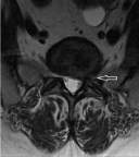
    Figure 1A: Pre-Op Axial MRI Image of the L5-S1 Level showing a left-sided
herniated disc with S1 nerve root impingement.
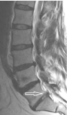
    Figure 1B: Pre-Op Sagittal MRI Image of the Lumbar Spine showing L5-S1
Level herniated disc.
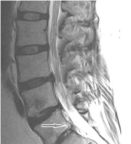
    Figure 1C: Post-Op Sagittal MRI Image of the Lumbar Spine showing
interval removal of L5-S1 herniated disc - which led to a complete resolution
of the patient’s symptoms.
Discussion
Many different healthcare providers, such as primary care physicians, sports medicine physicians, chiropractors, and orthopedic surgeons, etc, may be involved in the care of patients with lumbar disc herniations. Even with a thorough history and a proper physical exam, an atypical presentation of a lumbar disc herniation can be easily missed unless a high index of suspicion is maintained. This case provides a striking illustration of the difficulty of making an appropriate diagnosis when a common problem presents in a unique fashion.
There is a common misconception that lumbar disc herniations often have an inciting event in the clinical history. Without a so called “smoking gun,” it might seem reasonable to assume that the patient is suffering from more of a chronic, degenerative condition such as tendinitis or bursitis. In a recent study of 154 patients with MRI confirmed lumbar disc herniation done by Suri and colleagues, 62% of patients could not identify an inciting event [1]. Clinical history, therefore, may not always reliably lead the provider to the appropriate diagnosis.
Recent studies have suggested that even a proper physical exam can fail to elicit the signs of a disc herniation. A meta-analysis of over 7,000 patients by Al Nezari and colleagues suggested there is “limited diagnostic accuracy of all components of the neurological examination to detect a disc herniation in patients with suspected radiculopathy” [2]. All patients included were found to have disc herniation which was confirmed by surgery and/or imaging with CT or MRI. Testing for sensory deficits alone was 32-40% sensitive and 59-72% specific. Motor deficits were 22-40% sensitive and 62-79% sensitive. Motor atrophy was 31% sensitive and 76% specific. Reflex deficits were 25-27% sensitive and 75-78% specific. Another recent study by van der Windt, et al. found that in patients with suspected lumbar disc herniation, the straight leg raise test was the most sensitive at 92% with 28% specificity, while the crossed straight leg raise test was the most specific at 90% with 28% sensitivity [3]. The same study also determined that many of the following tests - especially in isolation - had poor to moderate diagnostic performance in differentiating low back pain from disc herniation: muscle weakness, muscle wasting, impaired reflexes, and sensory deficits.
Adding further to the difficultly of proper diagnosis is the wide range of symptoms with which a lumbar disc can present. There have been several unique presentations for herniated discs found in the literature (Table 1). In one case report by Rapport and colleagues, a 45-year old male presented for a positional headache and bilateral 6th cranial nerve palsies [4]. This was preceded by a “pop” in his back after performing a task which required heavy lifting. A cranial MRI confirmed intracranial hypotension with bilateral subdural collections, and an MRI of the lumbar spine revealed a T12-L1 disc herniation with fragments in the intradural space. The disc had penetrated the dural membrane, resulting in a cerebrospinal fluid leak that caused the cerebral hypotension. A complete resolution of symptoms followed discectomy and dural defect repair.
Another case reported by Khalatbari and colleagues involved a patient who presented for acute right lower quadrant pain radiating to the right groin which developed over the preceding 24 hours - suspicious for a possible urinary tract infection, hernia, or other intra-abdominal pathology [5]. The patient also experienced urinary frequency, urgency, and urethral discharge. This patient’s history included only mild chronic back pain. Neurological and gynecological exam were non-revealing with only right paraspinal upper lumbar tenderness as a positive finding. Lumbar MRI, however, identified a L1-L2 disc herniation. Discectomy resulted in complete relief of symptoms.
Occasionally, even cervical or thoracic disc pathology can mimic lumbar disc conditions [6-8]. One such report detailed a 42 year old female patient who presented with acute flaccid left lower limb monoplegia with increased deep tendon reflexes on the left sided ankle clonus, and a positive Babinski sign [9]. Additionally, she had pain and hyperesthesia involving the L1 dermatome and below, as well as increased urinary frequency. The remainder of the physical exam was unremarkable. MRI revealed a C6-C7 disc herniation. Surgery revealed several disc fragments and a penetrated dural sac. Following removal of all disc fragments, discectomy, an anterior cervical fusion, and closure of the dural tear using an epidural fat graft and fibrin glue, the patient began to improve immediately. Left lower limb motor function continued to improve over several weeks [9].
Once a lumbar disc is appropriately diagnosed, most authors recommend that conservative care should be attempted first, if appropriate. While determining exactly what percentage of patients will improve with non-operative treatment is difficult, most studies estimate that as high as 90% of patients will achieve satisfactory improvement on conservative therapy alone [10]. Current recommendations are at least six weeks of treatment, which include, but are not limited to: reduced physical activity, Non-Steroidal Anti-Inflammatory Drugs (NSAIDS), tramadol, or opioid pain medications, if other medications do not result in sufficient pain reduction. Physical therapy is an option and can be considered for mild stretching and massage. Pain relief therapies include ultrasound therapy, whirlpool therapy, and ice/heat pack therapy. For patients with no significant clinical progress after 3-4 weeks of the above treatment, epidural steroid injections can be considered. However, if a failure to respond to treatment within 6 weeks occurs, surgical treatment should be recommended. Urgent surgical evaluation should be obtained if signs of progressive neurological deficit, saddleanesthesia, or bowel/bladder incontinence are present [11]. In cases requiring surgical treatment, excellent clinical outcomes can be expected in a majority of patients.
In our reported patient, lumbar imaging should have been explored in a more timely fashion. The lack of inclusion of lumbar pathology in the differential diagnosis of her calf tetany resulted in a sense of “tunnel vision” for her providers, wherein everyone focused on the presumptive diagnosis of Achilles tendinitis/bursitis. Consideration of possible neuropathology and earlier lumbar imaging when the patient failed to show any real improvement may have resulted in a more prompt diagnosis and treatment for the patient. Hopefully, this report will add calf tetany to the list of conditions where spinal pathology should be considered, especially when patients fail to respond to their initial treatments.
Conclusion
To our knowledge, this is the first reported incidence of isolated sustained calf tetany as the presenting sign of a lumbar disc herniation. We would encourage all healthcare team members to maintain an appropriate index of suspicion for lumbar pathology in cases of calf pain and cramping and to pursue appropriate diagnostic testing after a patient has failed conservative management.
References
- Suri P, Hunter DJ, Jouve C, Hartigan C, Limke J, Pena E, et al. Inciting events associated with lumbar disc herniation. Spine J. 2010; 10: 388-395.
- Al Nezari NH, Schneiders AG, Hendrick PA. Neurological examination of the peripheral nervous system to diagnose lumbar spinal disc herniation with suspected radiculopathy: a systematic review and meta-analysis. The Spine Journal. 2013; 13: 657-674.
- van der Windt DA, Simons E, Riphagen II, Ammendolia C, Verhagen AP, Laslett M, et al. Physical examination for lumbar radiculopathy due to disc herniation in patients with low-back pain. Cochrane Database Syst Rev. 2010; 2: CD007431.
- Rapoport BI, Hartl R, Schwartz TH. Cranial neuropathy due to intradural disc herniation. Neurosurgery. 2014; 74: E561-565.
- Khalatbari M, Yahyavi S, Borghei-Razavi H, Ghalaenov H. Upper Lumbar Disc Herniation Presenting as Acute Abdomen. Acta Medica Iranica. 2009; 47: 427-429.
- Chan CK, Lee HY, Choi WC, Cho JY, Lee SH. Cervical cord compression presenting with sciatica-like leg pain. Eur Spine J. 2011; 20: S217-221.
- Giblin EM, Hochheiser GM. Thoracic disk herniation resulting in acutely progressing paraplegia in a pediatric patient. Pediatr Emerg Care. 2008; 24: 550-553.
- Linscott MS, Heyborne R. Thoracic intervertebral disk herniation: a commonly missed diagnosis. J Emerg Med. 2007; 32: 235-238.
- Menku A, Kamasak K, Gocmez C, Basarslan S, Dogu Y. Acute monoplegia associated with non-traumatic intradural cervical disk herniation. J Clin Exper Investigation. 2014; 5:120-123.
- Saal JA, Saal JS. Nonoperative treatment of herniated lumbar intervertebral disc with radiculopathy. An outcome study. Spine (Phila Pa 1976). 1989; 14: 431-437.
- Schoenfeld AJ, Weiner BK. Treatment of lumbar disc herniation: Evidencebased practice. Int J Gen Med. 2010; 3: 209-214.
- Jamieson DR, Ballantyne JP. Unique presentation of a prolapsed thoracic disk: Lhermitte’s symptom in a golf player. Neurology. 1995; 45: 1219-1221.
- Xiong Y, Lachmann E, Marini S, Nagler W. Thoracic disk herniation presenting as abdominal and pelvic pain: a case report. Arch Phys Med Rehabil. 2001; 82: 1142-1144.
- Rohde RS, Kang JD. Thoracic disc herniation presenting with chronic nausea and abdominal pain. A case report. J Bone Joint Surg Am. 2004; 86-86A: 379-81.
- Chaudhary KS, Bapat MR. Conus medullaris syndrome due to an intradural disc herniation: a case report. Indian J Orthop. 2008; 42: 94-96.
- Celik EC, Kabatas S, Karatas M. Atypical presentation of cauda equina syndrome secondary to lumbar disc herniation. J Back Musculoskelet Rehabil. 2012; 25: 1-3.
- Hakan T. Lumbar disk herniation presented with cauda equina syndrome in a pregnant woman. J Neurosci Rural Pract. 2012; 3: 197-199.
- Mahapatra AK, Gupta PK, Pawar SJ, Sharma RR. Sudden bilateral foot drop: an unusual presentation of lumbar disc prolapse. Neurol India. 2003; 51: 71- 72.