
Case Series
Austin Surg Case Rep. 2016; 1(1): 1004.
Management of Extrahepatic Biliary Disease of Pregnancy: A Continuous Dilemma
Li C¹, Kamine TH², Howell K³ and Odom SR4*
¹Department of Surgery, Harvard Medical School, USA
²Department of Surgery, Harvard Medical School, USA
³Harvard Medical School, USA
4Department of Surgery, Harvard Medical School, USA
*Corresponding author: Stephen R Odom, Department of Surgery, Beth Israel Deaconess Medical Center, 110 Francis Street, Suite 2-G, Boston, USA
Received: December 04, 2015; Accepted: February 03, 2016; Published: February 04, 2016
Abstract
Caustic ingestions are uncommon clinical encounters. We present three cases of intentional severe alkali ingestion in adults and discuss management and treatment of these complex and unusual lesions. Clinical presentation depends on the agent used, the quantity ingested, affected tissue and time to presentation. Airway is an initial concern and should be addressed promptly. Initial workup should include chest x-ray to rule out pneumomediastinum or pneumothorax. Staging requires upper endoscopy and sometimes, computed tomography. Viscous perforation or peritonitis requires urgent surgery. Evidence of full thickness necrosis generally warrants exploration. Operative intervention can include esophageal, gastric or, less commonly, small bowel resection. Feeding access should be obtained at the first operation. Steroids and antibiotics are not recommended. Definitive reconstruction is more complex and can be accomplished once the acute inflammatory reaction has resolved. Often, psychiatric treatment will be needed in intentional injuries of this nature.
Abbreviations
CT: Computed Tomogram; OR: Operating Room; EGD: Esophagogastroduodenoscopy; ICU: Intensive Care Unit; POD: Post Operative Day; HIV: Human Immunodeficiency Virus; HBV: Hepatitis B Virus; NGT: Nasogastric Tube
Introduction
There are between 5000-15000 cases of acid or alkaline caustic ingestions per year in the United States [1]. Most cases occur in the pediatric population as accidents and occasionally as a result of child abuse. In the adult population, ingestions most often occur in patients with a psychiatric background, such as those with depression, schizophrenia or more commonly in those who attempt suicide [2]. The severity of injury depends on several factors including corrosive properties of the agent, concentration, amount and duration of contact, as well as the intent of the patient [1,3]. Short-term outcomes include perforation and death, whereas long-term complications include dysphagia, strictures or even esophageal carcinoma [1].
Caustic agents causing the most significant damage often have a pH less than 2 or greater than 12, with a range of injuries from mucosal burns to esophageal or gastric perforations to death [4]. The most frequently documented alkaline agents involved are common household cleaners such as drain or dishwater cleaners and batteries [1]. In this review, we highlight management of the adult ingestion patient and three alkaline agents: household bleach, sodium hydroxide, and potassium hydroxide.
Case Series 1 (Potassium Hydroxide)
A 46year-old male with history of depression and 3 previous suicide attempts presented after being found down and agitated at home. A 62 fluid-ounce bottle of potassium hydroxide 45% was found next to him with 30 ounces left in the bottle. He was intubated in the field and brought to the emergency room. At admission, he was afebrile with normal vital signs. His physical exam was notable for partial thickness chemical burns of the bilateral posterior lower legs and bilateral buttocks. His labs were significant for a leukocytosis of 19.1 K/uL. A Computed Tomogram (CT) scan of his torso showed no obvious extravasation of oral contrast.
The patient was taken to the Operating Room (OR) for laryngoscopy, bronchoscopy, and upper endoscopy. The laryngoscopy revealed significant erythema and edema of the posterior oropharynx, but the mucosa appeared intact. The remainder of the bronchoscopy was normal. The Esophagogastroduodenoscopy (EGD) revealed severe caustic injury to the entire esophagus beginning with level 3a injury at 18cm and worsening to level 3b injury by the Gastroesophageal (GE) junction. The stomach similarly showed grade 3b injury concentrated near the antrum, characterized by disrupted mucosa without frank perforation. The duodenum appeared uninjured. A laparotomy was performed, and the stomach was noted to be thickened with some areas of necrosis. A total gastrectomy was performed, and the patient’s abdomen was temporarily left open. The following day, the patient returned to the OR and underwent repeat esophagoscopy which noted worsening burns and full-thickness necrosis of the esophagus. He then underwent right thoracotomy with esophagectomy and cervical esophagostomy as well as a feeding jejunostomy.
He was initially transferred to the Intensive Care Unit (ICU) and was extubated on hospital day 2, and tube feeds were started on the following day. He was seen by the psychiatry service. His cutaneous chemical burns were noted to be partial thickness and were managed with non-adherent dressings. He tolerated advancement in tube feeds and was discharged to the inpatient psychiatric service on hospital day 14. Six weeks after surgery, he developed retching and was noted to have a stricture at the esophagostomy site. He subsequently underwent dilation with symptomatic improvement. He treated in an inpatient psychiatric unit for 2 months postoperatively and was discharged on lithium for bipolar disorder II. Twelve months after initial injury, he underwent reconstruction with a colonic conduit. Since the reconstruction, he has developed dysphagia; however, he has been able maintain oral intake and gain weight.
Case Series 2 (Sodium Hydroxide)
A 53 year-old male without medical or psychiatric history presented to the emergency room following a suicide attempt where he consumed 12 ounces of Liquid Plumr® (0.5-2% sodium hydroxide and 5-10% sodium hypochlorite) and slashed his abdomen and thorax more than 30 times with a box cutter. He was found after an unknown down time and Emergency Medical Services was called. A chest radiograph showed no effusion, pneumothorax or pneumomediastinum. His electrolytes on admission were significant for sodium 150mEq/L, chloride 117mEq/L and bicarbonate 23mEq/L.
Urgentlaryngoscopy, bronchoscopy, and EGD were performed in the OR. Laryngoscopy revealed hyperemic mucosa. Bronchoscopy was normal. EGD revealed significant mucosal necrosis (grade 3b) of the distal esophagus and patchy necrosis in the cardia and the antrum. The duodenum was found to have Zargar grade [1] injury with superficial hyperemia but no necrosis. Since the necrosis appeared localized to the distal esophagus, a left thoracoabdominal incision was made. The stomach had multiple patchy areas of fullthickness necrosis. An esophagogastrectomy was performed with a Roux-en-Yesophagojejunostomy for primary reconstruction, with feeding jejunostomy.
He was unable to be extubated postoperatively due to significant airway edema. On hospital day 5 he underwent percutaneous tracheostomy placement. The following day, he was weaned to tracheostomy collar, and psychiatry was consulted. Lower extremity ultrasounds performed for leg swelling revealed a deep venous thrombosis for which he was started on heparin, which was bridged to Coumadin on discharge. On the seventh postoperative day, he underwent a barium swallow study, which showed an intact anastomosis. He was discharged to the psychiatry inpatient service on hospital day 15. He was discharged from the inpatient psychiatry unit after one month on bupropion and haloperidol for depression with psychotic features and has been doing well in follow up.
Case Series 3 (Sodium Hypochlorite)
A 50 year-old male with a history of Human Immunodeficiency Virus (HIV) and Hepatitis B Virus (HBV) presented to the ER following a suicide attempt in which he drank a half gallon of household bleach (5-10% sodium hypochlorite). An EGD was performed which revealed significant erythema of the esophagus (Figure 1a) along with friability, erythema, and focal necrosis of the stomach (Figure 1b - grade 3a injury). A CT scan showed thickening of the stomach and esophagus but no evidence of perforation. The following day he developed hematemesis, worsening abdominal pain, hiccups, and sinus tachycardia to the 140s. His electrolytes were significant for sodium 142mEq/L, chloride 116mEq/L and bicarbonate 17mEq/L.
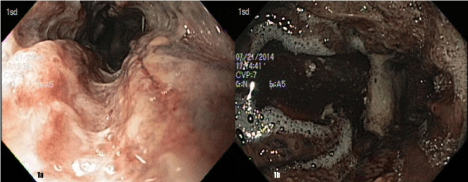
    Figure 1: Esophagogastroduodenoscopy shortly after ingestion of sodium
hypochlorite (bleach). Figure 1a: Proximal esophagus, 1b: Grade 3a injury
of stomach.
He was taken emergently to the OR where a repeat EGD was performed, revealing a 2-mm perforation of his distal esophagus as well as multiple areas of necrosis of the stomach. A left thoracoabdominal incision was performed. Turbid fluid was noted in the lesser sac, and there were multiple small perforations noted in the stomach. The peritoneum and liver were diffusely studded with white implants. A liver biopsy was performed, followed by an esophagogastrectomy with Roux-en-Yesophagojejunostomy for primary reconstruction with feeding jejunostomy.
He was transferred to the ICU and was extubated on the first postoperative day. He was started on tube feeds on postoperative day 3. His tissue culture returned Mycobacterium tuberculosis, and he was started on treatment for this. He was discharged to the inpatient psychiatry unit on postoperative day 44 due to continued requirement for nutritional support and treatment of his tuberculosis. He was ultimately discharged from the inpatient psychiatry unit 3 months postoperatively on risperidone and venlafaxine for major depression without psychotic features. In addition, he was continued on darunavir, abacavir, lamivudine and ritonavir for HIV and dapsone, pyridoxine, and rifabutin for tuberculosis.
Discussion
Clinical presentation
Clinical presentation depends on properties of the chemical agent, quantity ingested, timing, affected tissue, and the intent of ingestion. Our patients represent significant exposure differing agents: patients #1 (pH 12.0), patient #2 (pH 13.0), and patient #3 (pH 12.6). Burns to the lips, mouth, and oropharynx are common; however, the degree of external or visible oropharyngeal injuries does not correlate with injury to the esophagus or stomach [1]. Three serious signs and symptoms of esophageal injury are vomiting, drooling, and stridor [5]. Our three patients presented relatively early after intentional ingestion of alkali. Crain found that 50% of patients with two or more of these serious signs and symptoms had correlating serious esophageal injury on endoscopy [5]. Laryngeal or epiglottic edema leads to stridor, aphonia, hoarseness or dyspnea. Neither of the patients that arrived without airway control demonstrated signs of impending airway compromise. Nonspecific signs include nausea, emesis, hematemesis, dysphagia or odynophagia. Perforation or severe burns may result in acute peritonitis with abdominal pain, rigidity, or rebound tenderness [1]. None of our patients demonstrated signs of acute peritonitis or obvious clinical signs of perforated bowel.
Diagnosis and Staging
Caustic ingestions are relatively uncommon clinical encounters, thus there has been no consensus on the standard of care. Patients presenting with alkali ingestion should undergo standard assessment starting with the airway, noting signs of stridor, laryngeal edema, or dyspnea. Our first patient had his airway controlled before arrival, but the other patients both had evidence of pain and drooling. Neither patients #2 or #3 had stridor, but both had airway control early for diagnostic procedures, and remained intubated afterward for airway edema. Patient #2 was extubated after 5 days, while patient #3 had severe airway edema requiring tracheostomy. Early airway protection is essential as delay may lead to more difficult and traumatic intubation as edema sets in and tissue necrosis. Fiberoptic laryngoscopy may be helpful to directly visualize the airway to prevent additional injury or bleeding as a result of blind intubations. A surgical airway should always be readily available. Once the patient is stabilized, a thorough history regarding the quantity, timing and identity of the ingested substance must be obtained in addition to performing a physical exam [1,6]. The patient should be kept nothing per os and appropriately resuscitated. Vital signs and physical exam should be serially monitored, and laboratory values should be followed to assess for potential acidosis and shock. Initial assessment should include chest and abdominal radiographs (upright when possible) as these are quick modalities to diagnose any possible pneumomediastinum or pneumoperitoneum that may require emergent surgical intervention. Induced vomiting and neutralization of caustic agents should be avoided [1]. Several modalities have been historically used to assess for degree of injury, including barium esophogram or technetiumlabeled sucralfate. Both are limited in the acute setting, and neither have the ability to show extent of injury as well as endoscopy [1]. Endoscopy has also been shown to be superior to CT alone in the assessment of anatomical abnormalities. Those with signs and symptoms of oropharyngeal injury such as drooling, vomiting, pain, or dysphagia should urgently undergo endoscopy to determine if surgical intervention is required, as such signs and symptoms may indicate a higher grade of injury [7]. In the symptomatic patient, it is recommended that endoscopic assessment be performed within 12- 24 hours of ingestion and no more than 48 hours later as endoscopy after 48 hours may increase the chance of esophageal perforation [1,6]. Our patients all had EGD performed on the first hospital day. In two patients (#1 and #2), findings indicated the need for urgent operation. In patient #3, repeat endoscopy was performed on the second day, and indicated progressing of the 3a injury to perforation, necessitating surgery at that time. In a prospective study of 81 patients with corrosive ingestion, Zargar, et al. demonstrated the utility of EGD in the management of corrosive ingestion and further stratified patients into more detailed classifications of esophageal burns (Table 1) [1,8].Our patients all demonstrated early evidence of 3a-3b injuries, indicating severe mucosal damage.
![]()
    Grade
    Degree of Injury


    Grade 0
    Normal


    Grade 1
    Mucosal edema and hyperemia


    Grade 2a
    Superficial ulcers, bleeding, exudates, whitish membranes


    Grade 2b
    Deep or circumferential ulcers


    Grade 3a
    Focal necrosis


    Grade 3b
    Extensive necrosis
Table 1: Degree of Esophageal Injury by Endoscopic Assessment.
Mechanism of injury
In alkali ingestion, esophageal injuries tend to be more prevalent than gastric or duodenal injury. This is hypothesized to be due to the presence of gastric acid and partial neutralization of ingested alkali. Duodenal injury has been documented to occur in only 30% of cases, compared to gastroesophageal in 100% and gastric in 94% [9]. All three of our patients had gastric damage. This is due to the very high pH and large volume of agents used in these cases. Mild duodenal injury was found only in patient #2, who ingested the most alkaline solution in our series.
Ingestion of alkali, especially at pH > 11 or 12, produces liquefaction necrosis, a process that involves break-down of cellular and junctional barriers, causing the epithelium to become permeable to ions and uncharged molecules [10]. This results in deeper injuries as compared to the tissue damage from acids because the coagulation necrosis and eschar formation caused by acid exposure prevents deeper penetration of injury [1]. Liquefaction necrosis can continue over the course of several days, with associated vascular thrombosis, mucosal inflammation, saponification of fats, and denaturation of proteins [1,3]. In the weeks following, the esophageal wall develops granulation tissue and fibrosis due to the epithelial sloughing. It can take up to several months for re-epithelialization to occur [3]. Solid alkali adhere to the mouth and esophagus resulting in proximal burns that spare the stomach, whereas weaker alkali (e.g. detergents) lead to airway injury with associated laryngeal edema or airway compromise. Disk batteries, which contain up to 45% potassium or sodium hydroxide, can become lodged in the esophagus and begin leaking within as little as one hour, which can cause erosion and perforation within 12 hours [1].
Caustic injury may worsen for days after initial ingestion as the deep tissue penetration progresses. Granulation tissue can begin to form after 2-4 days. Necrotic tissue will begin to shed by days 15- 20 as collagen deposition begins to develop in the first few weeks. Between days 5-15, there can be a high risk of injury or perforation during endoscopy, thus care must be taken1. In addition to potential oropharyngeal and upper gastrointestinal injuries, a rare but fatal complication of bleach ingestion is morbidity/mortality secondary to hypernatremia and hyperchloremic metabolic acidosis [11]. Household bleach with 10% sodium hypochlorite content can have a large sodium load that leads to metabolic disturbances and can cause rapid deterioration and cardiac arrest [11,12]. Patient #3 in our series had large volume bleach ingestion, and presented with moderate electrolyte abnormalities. His presentation was soon after ingestion, so that aggressive resuscitation obviated worsening of this condition.
Management
Medical management
While there are no randomized trials to standardize management of esophageal injuries from caustic ingestions, unless there are indications for surgical intervention, the vast majority of caustic injuries can be managed endoscopically and medically. This was not possible in our patients due to the high grade nature of their injuries.
There is generalized agreement that endoscopy is a valuable tool in diagnosis and both early and late management of caustic injuries [13-15], though recent literature has questioned routine use in asymptomatic patients [6]. Patients with grade 0, 1, or 2a injury may escape with no adverse outcome, but those with grade 2b or greater injuries have higher rates of stricture formation and developing systemic complications [9,14]. Under endoscopic guidance, Nasogastric Tubes (NGT) may be placed for grades 2b and 3 injuries to provide nutrition and to evacuate gastric contents1; however blind bedside placement is not recommended as the risk of perforation or inducing vomiting generally outweighs the potential benefit [16]. If safe placement is possible, an NGT may be especially valuable with alkali as these ingestions tend to be viscous fluids, resulting in prolonged mucosal exposure [6]. Endoscopically-placed esophageal stents have shown promise in preventing stricture formation but have an approximately 50% efficacy rate and a relatively high migration rate [17]. Our patients #2 and #3 had NGT placed at the time of surgery.
Other aspects of medical management include pain control and prevention of stricture formation. Antibiotics and steroids have both been proposed and tried in attempts to prevent strictures. Steroids were first shown to decrease granulation tissue and stricture formation in animal models, but retrospective studies of their use in humans do not support their use [18,19]. Literature supporting the use of antibiotics for stricture prevention in the acute setting dates back 60 years and has not since been verified [20]. Antibiotics have been inconsistently applied in clinical series, with their use generally depending on provider and severity of the burn. The use of antibiotics can only be supported in the setting of suspected infection, concurrent steroid treatment, or surgical prophylaxis [6]. Other agents that prevent DNA cross-linking, such as mitomycin, have been suggested to decrease scarring and have shown promising long-term results but need to be used with caution given carcinogenic effects [17,21]. None of these modalities have been studied in prospective randomized controlled trials or extensively verified in case studies and they are currently not routinely used in practice. Our patients required only, and received only, perioperative coverage with antibiotics. None received steroids or other anti-inflammatory agents. One patient (#1) developed dysphagia months after his treatment, but there is no evidence this would have been ameliorated by the use of these agents.
Surgical management
Surgical repair for the effects of caustic injury may be indicated emergently or following failed conservative therapy. In the emergent setting, indications for immediate surgical intervention include signs of peritonitis, presence of pneumoperitoneum or perforation of the esophagus or stomach. If emergency surgery is indicated, most surgeons opt to perform laparotomy; however laparoscopy in skilled hands may be a viable option for more stable patients. More commonly, minimally invasive techniques have been used for patients who require delayed esophagectomy due to strictures [6,22]. Patients requiring surgery may undergo esophagectomy, gastrectomy, or esophagogastrectomy depending on location of perforation or extent of injured areas. Previously, all patients with endoscopy score of 2b or greater were urgently taken to the OR; however, recent studies have shown that up to 15% of pathology specimens from surgical interventions found no full-thickness necrosis [23]. A recent study found that operative management of only those patients with endoscopic grade 3b injuries who also had correlating CT signs of transmural necrosis resulted in no deaths. The study also demonstrated improved outcomes in patients with Zargar 3b injuries without CT evidence of necrosis [24]; however CT alone has poor sensitivity [25].
In most cases, a feeding jejunostomy tube is indicated for enteral feeding access until the patient is healed or undergoes definitive reconstruction [6]. Reconstruction of the neo-esophagus may be performed with a gastric transposition, colonic transposition [26] or with Roux-en-Y jejunal transposition asin two of our patients. Other delayed indications for surgical intervention include bleeding from necrosis or hemodynamic instability as a result of persistent acidosis or burn physiology [6]. Patients who do not require surgery on initial presentation or during an immediately subsequent admission may nonetheless require operative intervention at a later point, often due to stricture formation in the injured esophagus or failed endoscopic therapy.
Delayed reconstruction was performed in patient #1, due to the severity of his injury and the surrounding inflammatory tissue. Both patients #2 and #3 tolerated and did well after immediate reconstruction with roux-en-Y jejunal configuration repair. We suggest immediate repair if it is deemed safe. Also, all our patients had feeding access placed in the jejunum, allowing early initiation of nutritional support.
Complications
Early and late complications of corrosive injury are often related to the anatomical location of the injury or repair. Oropharyngeal injuries can result in tongue fixation, nasopharyngeal reflux, or pharyngeal and laryngeal stenosis. Burns to the larynx lead to loss of airway and strictures that may require tracheostomy or tracheal reconstructive surgeries to repair or bypass. Transmural burns of the esophagus have at least a 20% mortality rate, with perforation complications including tracheoesophageal fistula formation, mediastinitis, pneumonia, sepsis and requirement of esophagogastrectomy. Esophageal strictures can occur as early as 3 weeks after injury or repair in up to 20% of cases. They often occur at regions of natural anatomic narrowing such as at the cricoid cartilage, aortic arch, below the left main stem bronchus or at the esophageal hiatus [1]. Those with Zargar grade 2B or higher injuries have an increased rate of developing strictures14but occurrence is better predicted by increasing CT grade of caustic injuries than by endoscopic grading [27]. Up to 80% of patients experience symptoms of dysphagia by 2 months [1]. The majority of the mortality burden occurs in Zargar grade 3 injury patients [14]. Severe recalcitrant stricture of the esophagus may require esophagectomy with colonic, gastric or Roux-en-Y transpositions as previously described [26]. Injury to and stricture of the esophagus can increase risk of esophageal carcinoma by 1000 fold for as long as 25 years following injury. Gastric perforations result in peritonitis, shock, potentially death, and require emergent exploratory laparotomy [1]. Post-surgical complications include stricture of the anastomosis requiring repeated balloon dilations, as well as anastomosis leak and fistula formation [1,22].
Conclusion
We describe three cases of corrosive ingestions that cause injury to the stomach and esophagus, as well as review of the management of these injuries. There are many possible alkali ingestions; however, easily available household cleaners, such as lye (potassium hydroxide), Liquid Plumr® (sodium hydroxide) and bleach (sodium hypochlorite), are among the most common.
As with any trauma patient, management begins with the airway, which is often compromised due to injuries to the oropharynx. Ingestion injuries may necessitate intubation, at times best completed under direct visualization to prevent further trauma. Chest radiography or other rapid imaging should be performed to assess for pneumoperitoneum or pneumomediastinum. Endoscopy forms the cornerstone of staging. It should be used judiciously and be made readily available within the first 24 hours. Recent expert consensus panel recommends use of CT imaging to complement endoscopy grading and decrease unneeded surgeries [28]. Any sign of viscous perforation or full-thickness necrosis requires emergent surgical intervention. If injuries require esophagectomy, gastrectomy, or esophagogastrectomy, there are multiple surgical techniques available for reconstruction of the new esophagus.
While these are general guidelines, there remain many areas of debate regarding appropriate treatment after ingestion. Use of antibiotics and steroids to prevent stricture formation are not recommended. Strictures remain one of the most common delayed complications of caustic ingestions, often treated endoscopically by dilation and ultimately may require surgical revision. Laparoscopic surgery has become more prevalent and well-practiced in general and may play a greater role in the initial diagnosis of a patient with need for surgical intervention in the future. In their survey of common practices, Kluger has made the first steps towards reaching a consensus on the best diagnosis and treatment pathway for patients with caustic ingestions [6]. This is an area of ongoing research that deserves continued attention.
Our patients represent a series of three severe alkali injuries, all requiring early surgical intervention. We discuss options for diagnosis, management and treatment, and provide a comprehensive framework that describes the clinical considerations in the management in these complex and rare encounters.
References
- Lupa M, Magne J, Guarisco JL, Amedee R. Update on the diagnosis and treatment of caustic ingestion. 2009; 9: 54-59.
- Park KS. Evaluation and management of caustic injuries from ingestion of Acid or alkaline substances. 2014; 47: 301-307.
- Triadefilopolulos G. Caustic ingestion in adults. Up To Date. 2014.
- Riffat F, Cheng A. Pediatric caustic ingestion: 50 consecutive cases and a review of the literature. 2009; 22: 89-94.
- Crain EF, Gershel JC, Mezey AP. Caustic ingestions. Symptoms as predictors of esophageal injury. 1984; 138: 863-865.
- Kluger Y, Ishay OB, Sartelli M, Katz A, Ansaloni L, Gomez CA, et al. Caustic ingestion management: world society of emergency surgery preliminary survey of expert opinion. World J Emerg Surg. 2015; 10: 48.
- Bonnici KS, Wood DM, Dargan PI. Should computerised tomography replace endoscopy in the evaluation of symptomatic ingestion of corrosive substances?. 2014; 52: 911-925.
- Zargar SA, Kochhar R, Mehta S, Mehta SK. The role of fiberoptic endoscopy in the management of corrosive ingestion and modified endoscopic classification of burns. 1991; 37: 165-169.
- Zargar SA, Kochhar R, Nagi B, Mehta S, Mehta SK. Ingestion of strong corrosive alkalis: spectrum of injury to upper gastrointestinal tract and natural history. 1992; 87: 337-341.
- Atug O, Dobrucali A, Orlando RC . Critical pH level of lye (NaOH) for esophageal injury. See comment in PubMed Commons below Dig Dis Sci. 2009; 54: 980-987.
- Ross MP, Spiller HA. Fatal ingestion of sodium hypochlorite bleach with associated hypernatremia and hyperchloremic metabolic acidosis. See comment in PubMed Commons below Vet Hum Toxicol. 1999; 41: 82-86.
- Ward MJ, Routledge PA. Hypernatraemia and hyperchloraemic acidosis after bleach ingestion. 1988; 7: 37-38.
- Poley JW, Steyerberg EW, Kuipers EJ, Dees J, Hartmans R, Tilanus HW, et al. Ingestion of acid and alkaline agents: outcome and prognostic value of early upper endoscopy. 2004; 60: 372-377.
- Abaskharoun RD, Depew WT, Hookey LC. Nonsurgical management of severe esophageal and gastric injury following alkali ingestion. 2007; 21: 757-760.
- Temiz A, Oguzkurt P, Ezer SS, Ince E, Hicsonmez A. Predictability of outcome of caustic ingestion by esophagogastroduodenoscopy in children. 2012; 18: 1098-1103.
- Bonavina L, Chirica M, Skrobic O, Kluger Y, Andreollo NA, Contini S, et al. Foregut caustic injuries: results of the world society of emergency surgery consensus conference. 2015; 10: 44.
- Contini S, Scarpignato C. Caustic injury of the upper gastrointestinal tract: a comprehensive review. 2013; 19: 3918-3930.
- Pelclova D, Navratil T. Do corticosteroids prevent oesophageal stricture after corrosive ingestion? 2005; 24: 125-129.
- Ramasamy K, Gumaste VV. Corrosive ingestion in adults. 2003; 37: 119-124.
- KREY H. On the treatment of corrosive lesions in the oesophagus; an experimental study. 1952; 102: 1-49.
- Berger M, Ure B, Lacher M. Mitomycin C in the therapy of recurrent esophageal strictures: hype or hope? 2012; 22: 109-116.
- Kane TD, Nwomeh BC, Nadler EP. Thoracoscopic-assisted esophagectomy and laparoscopic gastric pull-up for lye injury. 2007; 11: 474-480.
- Chirica M, Resche-Rigon M, Bongrand NM, Zohar S, Halimi B, Gornet JM, et al. Surgery for caustic injuries of the upper gastrointestinal tract. 2012; 256: 994-1001.
- Chirica M, Resche-Rigon M, Pariente B, Fieux F, Sabatier F, Loiseaux F, et al. Computed tomography evaluation of high-grade esophageal necrosis after corrosive ingestion to avoid unnecessary esophagectomy. 2015; 29: 1452-1461.
- Lurie Y, Slotky M, Fischer D, Shreter R, Bentur Y. The role of chest and abdominal computed tomography in assessing the severity of acute corrosive ingestion. Clin Toxicol (Phila). 2013; 51: 834-837.
- Zhou JH, Jiang YG, Wang RW, Lin YD, Gong TQ, Zhao YP, et al. Management of corrosive esophageal burns in 149 cases. 2005; 130: 449-455.
- Ryu HH, Jeung KW, Lee BK, Uhm JH, Park YH, Shin MH, et al. Caustic injury: can CT grading system enable prediction of esophageal stricture?. 2010; 48: 137-142.
- Bonavina L, Chirica M, Skrobic O, Kluger Y, Andreollo NA, Contini S, et al. Foregut caustic injuries: results of the world society of emergency surgery consensus conference. 2015; 10: 44.