Case Report
Austin Surg Case Rep. 2016; 1(2): 1007.
Harvesting Left Lobe Graft from a Living Liver Donor with Extremely Rare Biliary Variation
Aktas H¹, Isik O¹, Gunero S¹, Akan O² and Emiroglu R¹*
¹Department of Surgery, Acibadem Bursa Hospital, Turkey
²Department of Radiology, Acibadem Bursa Hospital, Turkey
*Corresponding author: Emiroglu R, Department of Surgery, Acibadem Bursa Hospital, Turkey
Received: July 14, 2016; Accepted: September 11, 2016; Published: September 14, 2016
Abstract
Purpose: We aimed to emphasize the importance of biliary variations in Living Donor Liver Transplantation (LDLT) by sharing our experience with a living liver donor with extremely rare biliary variation of the left liver lobe.
Case Report: The patient was a thirty-seven year old male who had a right portal venous variation in addition to a left lobe biliary variation. The segment III bile duct was draining to the right posterior bile duct while left hepatic duct was consisted of the segment II and IV bile ducts. A left lobe graft was harvested successfully after detailed preoperative evaluation of the donors’ liver anatomy.
Conclusion: Biliary variations of the liver are not rare and play major role on the postoperative complications associated with biliary anastomoses in LDLT. However, LDLT has to be widely performed in countries with limited cadaveric organ donation. If the only choice is LDLT, preoperative accurate knowledge about the hepatic anatomy of the donor is crucial for better outcomes.
Keywords: Living donor liver transplantation; Biliary variation; Cholangiography
Introduction
Living Donor Liver Transplantation (LDLT) is widely performed in our country because of the limited cadaveric organ donation (76.8% vs. 23.2% of total liver transplantations in 2013). Biliary complications are still one of the main causes of morbidity and mortality in LDLT. Biliary variations of the donor liver play major role on the postoperative complications associated with biliary anastomoses.
It was suggested classifying biliary anatomy into five categories based on the Endoscopic Retrograde Cholangio Pancreatography (ERCP) findings [1]. As said by this classification, Type A1 is the most common biliary anatomy. Variability of the bile ducts in the left lobe of the liver is less common comparing to the right [2]. However, presence of an anatomic variation may be a factor affecting donor safety and increasing the complexity of surgery regardless of the side of hepatectomy. The present paper reports a successful graft harvesting from a living donor with a rare bile duct variation of the left liver lobe.
Case Report
Thirty seven-year-old male patient admitted to our institution to serve as a liver donor for his sister who had fulminant cryptogenic hepatitis. Preoperative Computerized Tomography (CT) of the donor showed a right posterior portal vein branching from the left portal vein, additionally, Magnetic Resonance Cholangio Pancreatography (MRCP) showed that segment III bile duct was draining to the right posterior bile duct, and left hepatic duct was consisted of the segment II and segment IV bile ducts. This type of biliary anatomy was not described in the Huang classification.
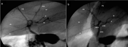
Volumetric imaging study revealed a 23% remnant liver volume after right hepatectomy. Although right hepatectomy was not feasible due to insufficient remnant liver volume, portal venous and biliary variations, a decision was made to use the left liver lobe graft since there was nobody else to serve as a donor for the patient and left lobe volume was satisfactory for the recipient. Intraoperative cholangiography confirmed the preoperative MRCP findings (Figure 1). A left liver lobe graft with one portal vein and two bile ducts was successfully harvested.

    Figure 1:  a-Anterior posterior view of the biliary tree in the intraoperative cholangiography, b- Oblique view of the biliary tree in the intraoperative cholangiography.
(II: Bile duct of segment II, III: Bile duct of segment III, IV: Bile duct of segment IV, CHD: Common Hepatic Duct; LHD: Left Hepatic Duct; RHD: Right Hepatic
Duct; RA: Right Anterior bile duct; RP: Right Posterior bile duct; PS: Posterior Superior bile duct; PI: Posterior Inferior bile duct; AS: Anterior Superior bile duct; AI:
Anterior Inferior bile duct).
Discussion
The normal biliary anatomy exists of the right hepatic duct draining the segments of right liver lobe (V, VI, VII and VIII) with two main branches (right anterior and right posterior ducts) and the left hepatic duct draining segments II, III and IV. The bile duct of caudate lobe drains directly to the right or left hepatic ducts. On the other hand, there is a likelihood of biliary variations up to 40% [3]. To the best of our knowledge, this is the first reported living liver donor who has a segment III bile duct draining to the right posterior bile duct in addition to a left hepatic duct consisted of segment II and IV bile ducts. No published data exist indicating the frequency of this type of biliary anatomy.
It was reported that 54.5% of Anatolian Caucasians have Huang type A1 biliary anatomy [4]. However, utilization of the living liver donors with other Huang types may be unavoidable when emergency transplantation is necessary due to the limited cadaveric donation. The major problem with the grafts harvested from living donors with biliary variation is the high incidence of biliary complications in the recipient due to performing more than one biliary anastomosis.
Despite the use of left liver lobe grafts is recommended by some authors because of the low biliary variation rate [2], there are several living donors reported who have different types of rare left liver lobe biliary duct variants [5]. Preoperative identification of the biliary anatomy allows surgeons to select the optimal graft and determine thorough operative strategy. In our institution, MRCP and intraoperative cholangiography are routinely utilized to evaluate the structure of biliary system of the donors.
In conclusion, biliary variation of the donor liver is not always a contraindication for LDLT. It may be a factor increasing operative complexity of donor hepatectomy and postoperative biliary complications in recipient. Detailed evaluation of the donor liver anatomy may increase donor safety and graft quality in LDLT.
References
- Huang TL, Cheng YF, Chen CL, Chen TY, Lee TY. Variants of the bile ducts: Clinical application in the potential donor of living-related hepatic transplantation. Transplant Proc. 1996; 28: 1669-1670.
- Farias F, Bigolin VA, Cavazzola TL, Filho O, Costa R, Kalil NA. Anatomical study of the intrahepatic biliary ducts. Parameters that guide the surgical approach in transplanting the left lobe of the liver. G Chir. 2013; 34: 210-215.
- Cucchetti A, Peri E, Cescon M, Zanello M, Ercolani G, Zanfi C, et al. Anatomic variations of Intrahepatic bile ducts in a European series and meta-analysis of the literature. J Gastrointest Surg. 2011; 15: 623-630.
- Karakas HM, Celik T, licioglu B. Bile duct anatomy of the anatolian caucasian population: Huang classification revisited. Surg Radiol Anat. 2008; 30: 539- 545.
- Iida T, Kaido T, Yoshizawa A, Yagi S, Hata K, Ogura Y, et al. A rare variation of the biliary tree of relevance to live liver donation. Am J Transplant. 2011; 11: 869-870.