Research Article
Austin J Surg. 2015;2(1): 1046.
Two Phase Derivation / Validation of Clinical Decision Criteria for Finger Radiographs
Kemp CD1, Scholz S3, Subhawong AF2, Abdullah F1 and Rasmussen SK4*
1Department of Emergency Medicine, University of Auckland School of Medicine, New Zealand
2Pittsburgh Mercy Health System, University of Pittsburgh, USA
3Department of Orthopaedic, UPMC St. Margaret Hospital, USA
*Corresponding author: Gregory Luke Larkin, Department of Emergency Medicine, University of Auckland School of Medicine, New Zealand
Received: August 27, 2014; Accepted: January 05, 2015; Published: January 07, 2015
Abstract
Background: A litany of clinical decision rules have been developed to decrease unnecessary emergency department radiography, but there are few clinical guidelines or decision criteria established for the taking of finger radiographs. Objective: To develop criteria for a clinical decision rule for finger radiographs.
Methods: This was a two-phase derivation/validation development of clinical decision rules for finger radiographs. The derivation phase was a retrospective chart review of all ED patients with acute finger and hand injuries. Patients were included based on select ICD-9 codes for finger injuries, age >18 years, and utilization of x-ray to rule out fracture. Demographics, mechanism of injury, physical exam findings, and past medical history were first individually evaluated using a univariate procedure, verifying all assumptions for independence, checking for collinearity, and probability of fracture on radiographs. A logistic model was built using forward and backward elimination, examining assumptions for statistical and clinical reasonableness at each point. Categorical data were compared for association using the Fishers Exact Test. Once derived, the model was validated on a second cohort of ED patients using the same inclusion criteria.
Results: In Phase I (derivation), 394 patients were included representing 186 finger fractures. Of all the demographic, historical and physical exam findings analyzed, only patient gender, location of injury and range of motion were statistically significantly correlated (p<.05) with fracture, predictive of 87% of all finger fractures. Ecchymosis was co-linear with location, and was therefore excluded from further consideration. Mechanism of injury, sensation, deformity, edema, and ecchymosis were not statistically related. Phase II (validation) included 293 finger injuries representing 95 fractures. The overall model from the derivation phase fit the data well.
Conclusion: Current use of finger radiography in the ED is inefficient for identifying fractures. A predictive model incorporating patient gender, location of injury, and range of motio reasonably predicts which patients would benefit from finger radiography. Of these three variables, the most important is decreased range of motion. A larger prospective trial is needed to further validate this model before clinical application.
Keywords: Finger radiographs; Hand trauma; Clinical decision criteria
Introduction
Hand injury is a common Emergency Department (ED) complaint [1], with metacarpal and phalanx fractures accounting for a significant percentage of these injuries [2,3]. The current standard of care obtaining x-rays to rule out fracture is conservative but inefficient. As with most extremity trauma, the incidence of digital fracture is low compared to the number of radiographs obtained [4]. Moreover, finger radiographs do not usually provide additional information that alters patient management.
Studies have shown the overuse of plain films in the evaluation of head, nasal, knee, ankle, lumbar, cervical spine and abdominal complaints [5-12]. Clinical decision rules have been developed in these areas to decrease the number of unnecessary radiographs taken, thereby decreasing patient cost and waiting time without decreasing patient satisfaction or jeopardizing quality of care. No studies, however, have been published examining finger radiograph decision rules. The objective of this study was to establish candidate criteria for a clinical decision rule for finger radiographs.
Methods
Setting
The study was performed at the Mercy Hospital of Pittsburgh, a Level 1 Trauma and tertiary care center with an annual ED census of 41,000. The study was approved by the Mercy Hospital Research and Human Rights Committee.
Design
This was a two-phase derivation/validation development of clinical decision rules for finger radiographs. The derivation phase (Phase I) was a retrospective medical record review of all ED patients with acute finger and hand injuries in a consecutive 26 month period. A multiple logistic regression model was developed, and then validated (Phase II) on a second cohort of ED patients seen with acute finger injuries (same inclusion criteria) who presented to the same ED over an additional 14 months. The presence or absence of fracture on ED x-ray was confirmed by a board-certified radiologist.
Population
Patients were included in the study if they were age 18 years or older, underwent finger radiography to rule out fracture, and had ICD-9 diagnosis codes for finger injuries (fracture [816.10, 816.00, 816.13, 816.03], dislocation [834.00, 834.02, 834.12, 834.10, 834.11], contusion [923.3], sprain/strain [842.10], tendon rupture [842.13, 842.12], deformity [736.21, 755.50, 736.22, 736.20], crush injury [927.3, 927.20], subungal [883.1, 883.0], and avulsion [879.8]).
Procedure
A list of medical records was supplied by Medical Information Systems based on selected ICD-9 coded discharge diagnoses. Medical records were reviewed by trained research assistants for demographic and clinical variables including age, gender, mechanism of injury, location of injury, time of day and day of week, and physical exam findings (deformity, ecchymosis, impaired range of motion at the MCP, PIP, or DIP joints, and tenderness to palpation); past medical history; official radiologic diagnosis, and ED discharge diagnosis.
Statistical analysis
The logistic regression model employed the presence or absence of fracture (as determined by the board certified radiologist) as the dependent variable. All independent variables were individually evaluated using a univariate procedure, verifying all assumptions for independence, checking for interaction and collinearity, and the probability of fracture on radiographs. We then built a logistic model using forward and backward elimination, examining assumptions for statistical and clinical reasonableness at each point to develop a clinical decision rule for the presence of a fracture. Categorical data were compared for association using the Fishers Exact Test, p<.05 level of significance.
Results
Derivation phase (phase I)
While 401 patients met the inclusion criteria, seven records were incomplete or missing either films or essential portions of the ED medical record. Of the 394 remaining pairs of records and radiographs reviewed, 36% were female and 64% were male patients with finger injury. Average age was 33 years (range 18-92). The most common finger injury in this retrospective cohort was contusions/ sprains (n=214; 54%). Fractures accounted for 180 (46%) of the injuries in this ED patient cohort. By gender, a slight majority of the radiographs obtained on male patients (51%) were positive for fracture, but only 37% of the radiographs taken on female patients were positive (p=0.01, Fishers exact). Slightly over half (54%) of the fractures required management beyond simple splinting, particularly those involving the middle and proximal phalanges. There were 41 (10%) dislocations, and only 2 (0.5%) fracture/dislocations. Of those in whom mechanism of injury data were retrievable (n= 388), the predominant mechanism of injury was blunt trauma (n=289; 75%), followed by hyperextension (n= 79; 20%), twisting (n=14; 4%), and crush injury (n= 6; 2%).
Of all the demographic, historical, and physical exam findings analyzed, only patient gender, location of injury, and range of motion were significantly correlated (p<.05) with fracture. When used together, these three variables predict fracture with a sensitivity of 87% and a specificity of 86%. The positive and negative predictive values are 79% and 84%, respectively. Ecchymosis, edema and deformity were highly related with location and were not included in the multivariate analysis. Candidate variables and fracture type are shown in Tables 1 and 2 respectively.
    Variables
    Fracture: Yes/No (%)
      Fracture             No Fracture
      N = 180               n= 214
    P value
      <0.05


    BLUNT
      Mechanism of Injury
      OTHER *
    74

      26
    75

      25

      .469
      (NS)


    YES
      Ecchymosis
      NO
    64

      36
    22

      78

      0.000


    YES
      Sensation Intact
      NO
    98

      2
    99

      1

      0.076
      (NS)


    YES
      Tenderness with Palpation
      NO
    100

      0
    96

      4

      .073
      (NS)


    FULL
      Range of Motion at MCP, PIP,    & DIP
      LIMITED or ABSENT
    30

      70
    76

      24

      0.000


    YES
      Deformity
      NO
    28

      72
    65

      35

      0.000


    YES
      Edema
      NO
    58

      42
    76

      24

      0.0002


    Proximal
      Location
      Middle/Distal
    48

      52
    79

      21

      .004


    Female
      Gender
      Male
    28

      72
    41

      59

      .011


    *Includes hyperextension, crush, twisting,  penetrating,  flexion, abduction, & explosion
Table 1: Candidate Variables Associated with Finger Fracture (Phase I).
    TYPE of FRACTURE (%)


    Prox Phalanx
    Middle
    Distal


    Buckling
    1.1
    0
    0


    Avulsion
    10.3
    4.2
    7.0


    Comminuted
    6.1
    1.6
    9.8


    Transverse
    7.2
    2.2
    9.1


    Tuft
    0
    0
    12.6


    Spiral
    3.5
    0
    0


    Intra-articular
    2.1
    0
    1.4


    Oblique
    4.9
    0
    1


    Nondisplaced
    7.7
    0
    5.6


    Salter II
    1.4
    0
    0


    Impacted
    2.1
    0
    0


    TOTALS
    46%
    8%
    46%
Table 2: Types of Fractures - Phase I.
Validation phase (phase II)
The medical records of 293 finger injuries were reviewed, representing 111 (38%) females and 182 (62%) males. Average age was 36.1 years (range 18-93). The most common finger injury in this retrospective cohort was contusions/sprains (n=176;60%). Fractures accounted for 104 (35.5%)of the injuries in the ED for patients who obtained radiographs. There were 13 (4%) dislocations and only 9 (3%) fracture/dislocations. Of those in whom mechanism of injury data were retrievable (n=260; 89%) the predominant mechanism of injury was blunt trauma (n=145; 56%), followed by crush injury (n=79; 30%), hyperextension (n=16;6%), penetrating trauma (n=11;42%), and twisting (n=9; 4%).
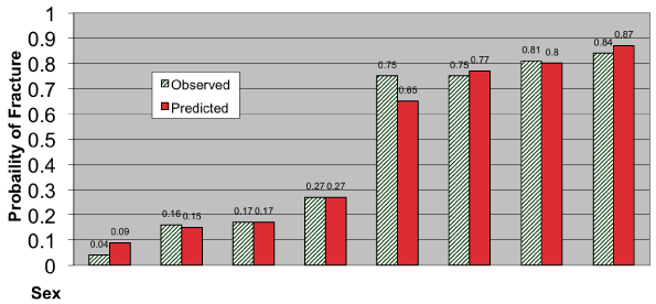
The correlation of finger fractures to clinical data was initially evaluated using Chi-square (p<.05). Of all the demographic, historical, and physical exam findings analyzed, only patient gender, location of injury, and range of motion were significantly correlated with fracture. Ecchymosis was highly related with location of tenderness and was therefore excluded from further consideration. When used together, these three variables fit the data well (Figure 1, p=.892), with diminished range of motion being the most highly predictive clinical criterion. Figure 1 shows the predicted and observed probabilities of the final three variable model, revealing how male gender, distal location, and especially diminished range of motion impact the overall probability of fracture. For any combinations of these three variables, the corresponding probability for fracture may be found on the y-axis of Figure 1.

Figure 1: Predicted & Observed Probabilities of Finger Fractures-Phase II.
Discussion
This study demonstrates that finger radiographs result in a relatively low yield for fractures (46%). This yield can be enhanced significantly by the use of decision criteria studied here: impaired range of motion, male gender, and distal injury, with impaired range of motion being the best discriminator for fracture. Patients without these criteria and especially those who demonstrate full range of motion at the MCP, PIP, and DIP joints are at low risk, and may often be able to be safely treated conservatively and without radiographs. While the criteria established here are not 100% sensitive, they do identify low risk patients (female patients with proximal injury and full range of motion, for example) as having a very low (<10%) risk of fracture. Explicit risk estimates that are independent of other clinical parameters may be generated by applying variations of these three criteria to the nomogram in Figure 1. By predicting the probability of a diagnostic outcome, decision rules/guidelines may help clinicians alter their pretest prior probability of fracture in ways that would otherwise be far less explicit. Several studies have shown clinical decision rules to be highly sensitive in identifying fractures [12,14], but any future clinical application of the three cardinal criteria studied here awaits further validation.
Because of the medical and legal risks associated with practice in the ED, decision rules for ordering extremity radiographs should ideally miss no fractures [15,16]. On the other hand, risk intolerance and the unbridled pursuit of diagnostic certainty is unsustainable in the current healthcare milieu [17]. Many common fractures rarely require different management than nonfractures when the joint space is not involved. For example, the distal phalanx fracture, accounting 15-30% of all hand fractures [3], is managed similar to a nonfracture with splinting, elevation, ice, and pain management.
Many radiographs are ordered because of the perception that patients will be dissatisfied unless they are x-rayed. In a study validating the Ottawa ankle rules, patients were found to be satisfied with care whether or not a radiograph was done [8]. Stiell, et al suggests that physician-patient communication replace the radiograph in the reassurance of patient treatment [8]. Another study demonstrated that one third of patients preferred follow-up to an immediate radiograph in the treatment of an ankle injury [18]. Some physicians may order x-rays because of orthopedic or family practice consultant expectation, a lack confidence in their own clinical judgment, or medical-legal fears [4]. Reducing even a small fraction of the many radiographs obtained in the outpatient setting may lead to improved efficiency, cost savings, increased patient throughput, and decreased exposure to ionizing radiation [19,20]. Approaches such as these described herein may be applied to toe radiographs and possibly other indications as trauma centers and health systems are increasingly asked to do more with less.
Limitations
There are multiple limitations to this study. First, there were many patients who may not have been included in the derivation cohort such as those with occult extremity injury as well as the very young, intoxicated, mentally debilitated, or those with other impaired neurosensoria or distracting injuries. Inconsistent documentation is another possible limitation of this retrospective study; since we were able to include only those records in which documentation was felt to be both legible and complete. This risk for selection bias could not be systematically accounted for in this analysis, however random. Since this study was a retrospective ED trial, patients with upper extremity trauma that did not come to the ED or whom did not receive radiographs were not included. Many patients with minor injury will not seek medical care or are such poor historians (e.g. the chronically disabled) they will never fit into a decision rule, guideline, or algorithm and their injuries cannot be identified by this or any other approach. Although we were unable to verify the accuracy of the information recorded, the validation using a second cohort suggests that these easily evaluable criteria are modestly robust. The use of finger radiography in this institution may not be representative of other hospitals, and further work must include multiple sites to ascertain the external validity of these findings.
Conclusion
Current use of finger radiography in the ED is inefficient for identifying fractures. A predictive model incorporating patient gender, location of injury, and range of motion reasonably predicts which patients would benefit from finger radiography. Of these three variables, the most important discriminator for fracture is decreased range of motion. A larger, multicenter, prospective trial is needed to further validate this model before clinical application.
Acknowledgement
The authors thank Chris Connor for her invaluable assistance with data management and manuscript preparation. This work was supported in part by the Emergency Medical Association of Pittsburgh.
References
- Hossfeld GE, Uehara DT. Acute joint injuries of the hand. Emerg Med Clin North Am. 1993; 11: 781-796.
- Bowman SH, Simon RR. Metacarpal and phalangeal fractures. Emerg Med Clin North Am. 1993; 11: 671-702.
- Gurland M. Carpometacarpal joint injuries of the fingers. Hand Clin. 1992; 8: 733-744.
- Stiell IG, Wells GA, McDowell I, Greenberg GH, McKnight RD, Cwinn AA, et al. Use of radiography in acute knee injuries: need for clinical decision rules. Acad Emerg Med. 1995; 2: 966-973.
- Masters SJ. Evaluation of head trauma: efficacy of skull films. AJR Am J Roentgenol. 1980; 135: 539-547.
- Seaberg DC, Jackson R. Clinical decision rule for knee radiographs. Am J Emerg Med. 1994; 12: 541-543.
- Stiell IG, Greenberg GH, McKnight RD, Nair RC, McDowell I, Reardon M, et al. Decision rules for the use of radiography in acute ankle injuries. Refinement and prospective validation. JAMA. 1993; 269: 1127-1132.
- Stiell IG, McKnight RD, Greenberg GH, McDowell I, Nair RC, Wells GA, et al. Implementation of the Ottawa ankle rules. JAMA. 1994; 271: 827-832.
- Anis AH, Stiell IG, Stewart DG, Laupacis A. Cost-effectiveness analysis of the Ottawa Ankle Rules. Ann Emerg Med. 1995; 26: 422-428.
- Kelen GD, Noji EK, Doris PE. Guidelines for use of lumbar spine radiography. Ann Emerg Med. 1986; 15: 245-251.
- Cadoux CG, White JD, Hedberg MC. High-yield roentgenographic criteria for cervical spine injuries. Ann Emerg Med. 1987; 16: 738-742.
- Hoffman JR, Mower WR, Wolfson AB, Todd KH, Zucker MI. Validity of a set of clinical criteria to rule out injury to the cervical spine in patients with blunt trauma. National Emergency X-Radiography Utilization Study Group. N Engl J Med. 2000; 343: 94-99.
- Greene CS. Indications for plain abdominal radiography in the emergency department. Ann Emerg Med. 1986; 15: 257-260.
- Stiell IG, Wells GA. Methodologic standards for the development of clinical decision rules in emergency medicine. Ann Emerg Med. 1999; 33: 437-447.
- Wasson JH, Sox HC, Neff RK, Goldman L. Clinical prediction rules. Applications and methodological standards. N Engl J Med. 1985; 313: 793-799.
- Laupacis A, Sekar N, Stiell IG. Clinical prediction rules. A review and suggested modifications of methodological standards. JAMA. 1997; 277: 488-494.
- Kassirer JP. Our stubborn quest for diagnostic certainty. A cause of excessive testing. N Engl J Med. 1989; 320: 1489-1491.
- Mower WR, Hoffman JR, Schriger DL. The feasibility of selective radiography inpatients with trauma-induced neck pain. Ann Emerg Med. 1990; 19: 1220-1221.
- Huda W, Bissessur K. Effective dose equivalents, HE, in diagnostic radiology. Med Phys. 1990; 17: 998-1003.
- Committee on the Biological Effects of Ionizing Radiation. Health effects of exposure to low levels of ionizing radiation. BEIR V. Washington DC, National Academy Press. 1990: 281.