Special Article – Pediatric Surgery
Austin J Surg. 2019; 6(3): 1165.
Retroperitoneal Teratoma in a Child with Clinical and Operative Photos
Pan P*
Departments of Pediatric Surgery, Ashish Hospital Jabalpur, India
*Corresponding author: Pradyumna Pan, Departments of Pediatric Surgery, Ashish Hospital, Jabalpur, India
Received: January 24, 2019; Accepted: February 01, 2019; Published: February 08, 2019
Clinical Image
Retroperitoneal teratoma are rare lesions seen in 3.5-4% of all germ cell tumors in children and 1-11% of primary retroperitoneal neoplasms. Germ cell tumors are congenital tumors containing derivatives of all the three germ layers. They are frequently seen in gonads [1]. The may present in extragonadal sites also like mediastinum, sacrococcygeal region and retroperitoneum [2] (Figure 1).
Patients usually present with abdominal distension or a palpable mass. The tumor detected antenatally has a higher incidence of malignancy than those in older children [3]. Ultrasonography and CT are useful tools for evaluation of the lesion. Alfa feto protein is the tumor marker that helps to detect recurrence (Figures 2 & 3).
Complete excision of the tumor offers the best chance of cure [4]. Even large tumors with bilateral involvement of the retroperitoneum can be excised while preserving adjacent organs. Malignancy is uncommon in retroperitoneal teratoma [5]. Prognosis is generally good and curative if the tumor is completely removed.

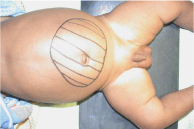
Figure 1: Clinical picture showing extension.

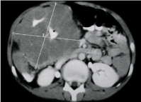
    Figure 2:  CT scan demonstrates a large retroperitoneal heterogeneous
mass.

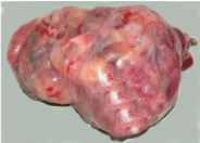
Figure 3: Excised teratoma.
References
- Aldhilan A, Alenezi K, Alamer A, Aldhilan S, Alghofaily K, Alotaibi M. Retroperitoneal teratoma in 4 months old girl: Radiology and pathology correlation. Curr Pediatr Res. 2013; 17: 133-136.
- Schmoll HJ. Extragonadal germ cell tumors Ann Oncol. 2002; 13: 265-272.
- Auge S, Satge D, Sauvage P. Retroperitoneal teratomas in the perinatal period. Review of literature concerning a neonatal immature aggressive teratomas. Ann Pediat. 1993; 40: 613-621.
- Chaudhary A, Misra S, Wakhlu A, Tandon RK, Wakhlu AK. Retroperitoneal teratomas in children. Indian J Pediatr. 2006; 73: 221-223.
- Rattan KN, Kadian YS, Nair VJ, Kaushal V, Duhan N, Aggarwal S. Primary retroperitoneal teratomas in children: a single institution experience. Afr J Paediatr Surg. 2010; 7: 5-8.