
Abstract
Oncoplastic surgery extends the indication of breast conserving surgery without compromising oncological principles and aesthetic outcome. We report a successful level 1 (dual plane undermining) lateral mammoplasty technique entailing a wide resection (20% of breast volume) of a 4cm upper outer quadrant breast lesion in a 61- year-old African woman. Oncoplastic breast conserving surgery is feasible and oncologically safe in a low resource setting if there is ready access to adjuvant radiotherapy for loco-regional control.
Keywords: Breast cancer; Surgery; Conserving; Oncoplastic
Abbreviations
OPS: Oncoplastic Surgery; BCS: Breast Conserving Surgery; IMF: Inframammary Fold; NAC: Nipple Areolar Complex.
Background
Oncoplastic surgery combines conserving surgery for breast cancer and plastic surgery techniques [1-4]. The benefits include improved aesthetic outcome, more accurate tumour resection with better control of tumor margins, high patient satisfaction, and the ability to extend the option of breast conservation to patients who would have traditionally required mastectomy [5-7]. The oncoplastic approach allows symmetrizing surgery which may in addition allow the diagnosis of occult cancers [1,2]. It would allow implant augmentation and simultaneous delivery of intraoperative radiotherapy [1]. The technique used for reconstruction (local tissue rearrangement, reduction mammoplasty or mastopexy, or transfer of local-regional flaps) would depend on tumor location and size, tumor to breast size ratio, and the desire of the patient [1,3,4].
Case Presentation
A 61- year- old African woman presented with a painless, firm mass in the upper outer quadrant of the right breast associated with nipple discharge of clear fluid. A mammogram was not available but ultrasonography suggested breast carcinoma. Fine needle aspiration cytology confirmed ductal carcinoma. Clinical examination revealed a well-looking woman with medium- sized grade 1ptotic breasts. Physical examination of the left breast was normal. There was a firm 4cm mass in the upper outer quadrant of the right breast associated with tethering of the lateral aspect of the areolar skin. The mass had irregular edges and was fixed to adjacent breast tissue but not to underlying muscle or skin (Figure 1). There was no palpable axillary nor supra clavicular lymphadenopathy. Chest and abdominal examination were unremarkable. A full blood count was normal. A chest x-ray and abdominal ultrasound were normal. She was clinically staged as T2, N0, M0 (Table 1). With informed consent the patient’s desired for breast conserving surgery. After periareolar deepitheliasation, a lateral cutaneo-glandular incision was made to the prepectoral fascia with an enbloc resection of the skin overlying the tumour and a tethered lateral aspect of the Nipple- Areolar Complex (NAC). The quadrantectomy then removed the tumour in a wide resection up to the axilla (Figure 2). The tumour was excised with a > 1 cm margin and about 20% volume of breast tissue excised (Figure 3). The breast was then raised at the sub-glandular and subcutaneous planes (dual plane undermining). The gland and skin were reapproximated beginning from the base to produce a higher breast with a narrower base. The NAC was transposed higher and more medially to avoid the inevitable inferolateral retraction exacerbated by adjuvant radiotherapy (Figure 4). The resulting scar was periareolar with lateral radial extension (Figure 5). Symmetrisation was not necessary as there was minimal disparity from the contralateral breast (Figure 6). She made good post- operative recovery with no complications and was discharged on the 3rd postoperative day. Being post-menopausal she was commenced on adjuvant endocrine therapy (tamoxifen 20mg daily) for 5 years. At 10 days follow-up, the wounds were healed with no evidence of nipple and Nipple Areola Complex (NAC) necrosis, seroma, haematoma, infection, wound dehiscence nor fat necrosis (Figure 6). The histological examination reported the receipt of a brown coloured, elastic breast biopsy specimen measuring 8.0 x 6.0 x 3.5 cm on whose surface is attached skin of 4.3 cm. Serial sections revealed a multi-coloured nodule of 4.0 cm diameter. Microscopically the breast tissue showed fibrocystic mastopathy with >50% of specimen occupied by invading duct cells (SBR III). Some in tubular, trabecular or linear pattern. She awaits adjuvant radiotherapy to the tumour bed and remnant breast only, and routine follow-up is planned.
![]()
    T (Tumour)
    N (Nodes)
    M (Metastases)


    TO: impalpable tumour
    No palpable axillary lymph nodes
    M0: No evidences of distant metastases


    T1: <2cm d. No fixation or tethering
    N1: Mobile palpable axillary lymph nodes
    M1: distant metastases or contralateral lymph nodes


    T2: 2-5cm with tethering or nipple retraction
    N2: fixed axillary nodes


    T3: 5-10cm with infiltration, Ulceration or peaud’orange, deep    fixation
    N3: Palpable supraclavicular lymph nodes (M1), oedema of the arm


    T4: >10cm d or infiltration/Ulceration > its diameter
Table 1: TNM (Clinical) staging (Union International contre le Cancer- IUCC).
![]()
    Early    Breast Cancer (t0-2,N0-1,M0)
    Locally    advanced breast cancer (t4, N0-2, M0)
    Late    breast cancer (M1)


    Operable    curable, 50% mortality
    805    mortality, Neoadjuvant therapy to make operable
    Not    curable, inoperable, palliative care, 100%, mortality
Table 2: Significance of clinical staging of Breast cancer.
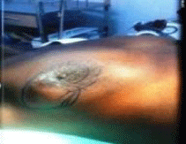
Figure 1: Upper outer tumour (R) breast.
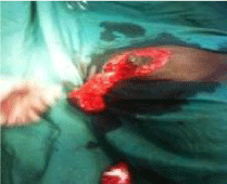
    Figure 2:  After periareolar de-epitheliasation, wide resection (quadrantectomy)
up to axilla.
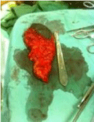
Figure 3: Widely resected specimen (>5 cm diameter) with axillary tail.

Figure 4: NAC transposed higher and more medially.
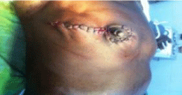
    Figure 5:  Skin closure giving a good result in both form (reduced ptosis) and
volume.
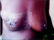
    Figure 6:  10 day’s postoperative breast (periareolar and lateral radial scar
and reduced ptosis of right breast).
Discussion
Local tissue rearrangement is an essential component of many oncoplastic techniques. This case demonstrates how a simple oncoplastic technique may extend the indication of breast conserving surgery without compromising oncological principles and aesthetic outcome. For a medium-sized breast, > 80-100 g tissue removal or 20% volume loss as in this case would leave deformity [4]. Lateral mammoplasty with minimal skin-gland dissection and easy, reliable closure is a simple and reliable intervention. It combines wide tumor excision with supero-medial NAC repositioning on a dermo-glandular pedicle, thereby counteracting the lateral axial scar contraction which is exacerbated by radiotherapy, and breast ptosis [5]. Additionally, the incision may be extended superiorly to access the axilla [1,2]. It is most commonly used in women with moderate-sized breasts, small tumors, grade 1 ptosis and when neoadjuvant chemotherapy has downloaded a large T2 tumour [3-6]. Tumours of the lateral quadrant junction are most frequent (60%) and lateral mammoplasty technique is therefore finding increasing use [4-7]. Medial mammoplasty, used for medial tumors, is almost the mirror image of lateral mammoplasty. Upper pole tumors are more difficult to reconstruct than lower pole tumours given the difficulty of maintaining upper pole breast volume. Tumors of the upper inner quadrant are especially difficult to reconstruct due to the much reduced tissue volume, and relative immobility of the inferomedial breast [1-5]. Preoperative planning affords flexibility in terms of incision design, pedicle selection and establishing expectations of both the patient and the surgeon [1-4]. The most commonly employed oncoplastic technique is the ‘Wise’ (inverted T) pattern with inferior pedicle reduction mammoplasty useful for large or ptotic breasts [1-5]. This technique can also be used for peri-areolar and central tumors [5,7]. Wound complication rate ranges between 5 and 14% [1,2]. The true value of the oncoplastic approach in reducing local recurrence is not known [8,9]. Positive margins have been associated with higher stage, positive nodes, positive lymphovascular invasion, use of neoadjuvant chemotherapy, larger initial “T” stage, positive oestrogen receptor and younger age [10]. These are indeed the high risk groups for breast conserving surgery. Intra-operative assessment of margins using intraoperative frozen section improves local control with consequent low rates of completion mastectomy including patients with tumors > 4 cm in size [1,11]. In addition, excision of multifocal tumors within the same quadrant has been shown to be oncologically safe with the wide margins that can be taken with oncoplastic procedures [12].
Appropriate adjuvant chemotherapy would improve the diseasespecific survival and chance of cure [1]. Neoadjuvant chemotherapy downstages the breast and axilla and enhance surgical loco-regional control (Table 2) [13]. Being a systemic disease, it is increasingly used even in node negative breast cancer patients [14]. Knowledge of the oestrogen (ER), Progesterone (PR) and human epidermal growth factor type 2 receptor (HER2) tumour biomarkers would optimize treatment but may not be available in low-resource settings [15-17]. The anti HER2 agent (trastuzumab) has effectively led to the change in the natural history of the disease with substantial improvement in survival particularly in oestrogen receptor-negative patients [18]. However, endocrine therapy with the cheaper anti-oestrogen (tamoxifen) or the current aromatase inhibitor (anastrazole) has been an acceptable option for the first-line treatment of post-menopausal women with locally-advanced breast cancer or as adjuvant treatment in early breast cancer [19].
Conclusion
Oncoplastic surgery can extend the indication of breast conserving surgery (> 4cm and retroareolar/ central lesions) without compromising oncological principles and aesthesis [10]. It is feasible and oncologically safe in a low resource setting if there is ready access to adjuvant radiotherapy for loco-regional control.
Acknowledgment
I acknowledge the theatre nurse, Mirabel Mbiaka for taking the necessary photographs.
Authors’ Contribution
EPW is the main author, researcher and surgeon, FAE, helped with literature search.
Consent for Publication
Written informed consent was obtained from the patient for publication of the case report and any accompanying images.
References
- De Lorenzi F. Oncoplastic surgery: the evolution of breast cancer treatment. Breast J. 2010; 16: 20-21.
- Rainsbury R. Surgery insight: oncoplastic breast-conserving surgery reconstruction- indications, benefits, choices and outcomes. Nat Clin Pract Oncol. 2007; 4: 657-664.
- Clough KB, Kaufman GJ, Nos C, Buccimazza I, Sarfati IM. Improving breast cancer surgery: a classification and quadrant per quadrant atlas for oncoplastic surgery. Ann Surg Oncol. 2010; 17: 1375-1391.
- Iwuchukwu OC, Harvey JR, Dordea M, Critchler AC, Drew PJ. The role of oncoplastic therapeutic mammoplasty in breast cancer surgery--a review. Surg Oncol. 2012; 21: 133-141.
- Anderson BO, Masetti R, Silverstein MJ. Oncoplastic approaches to partial mastectomy: an overview of volume-displacement techniques. Lancet Oncol. 2005; 6: 145-157.
- Ballester M, Berry M, Couturaud B, Reyal F, Salmon RJ, Fitoussi AD. Lateral mammaplasty reconstruction after surgery for breast cancer. Br J Surg. 2009; 96: 1141-1146.
- Chan SW, Cheung PS, Lam SH. Cosmetic outcome and percentage of breast volume excision in oncoplastic breast conserving surgery. World J Surg. 2010; 34: 1447-1452.
- Losken A, Dugal CS, Styblo TM, Carlson GW. A meta-analysis comparing breast conservation therapy alone to the oncoplastic technique. Ann Plast Surg. 2014; 72: 145-149.
- De Lorenzi F, Hubner G, Rotmensz N, Bagnardi V, Loschi P, Maisonneuve P, et al. Oncological results of oncoplastic breast-conserving surgery: Long term follow-up of a large series at a single institution: A matched-cohort analysis. Eur J Surg Oncol. 2016; 42: 71-77.
- Chang EI, Peled AW, Foster RD, Lin C, Zeidler KR, Ewing CA et al. Evaluating the feasibility of extended partial mastectomy and immediate reduction mammoplasty reconstruction as an alternative to mastectomy. Ann Surg. 2012; 255: 1151-1157.
- Losken A, Pinell-White X, Hart AM, Frietas AM, Carlson GW, Styblo TM. The oncoplastic reduction approach to breast conservation therapy: benefits for margin control. Aesthet Surg J. 2014; 34: 1185-1191.
- Staub G, Fitoussi A, Falcou MC, Salmon RJ. Breast cancer surgery: use of mammaplasty. Results. Series of 298 cases. Ann Chir Plast Esthet. 2008; 53: 124-134.
- Rouzier R, Extra JM, Klijanienko, Falcou MC, Asselain B, Vincent-Salomon A, et al. Incidence and prognostic significance of complete axillary downstaging after primary chemotherapy in breast cancer patients with T1 to T3 tumours and cytologically proven axillary metastatic lymph nodes. J Clin Oncol. 2002; 1304-1310.
- Wolmark N, Wang J, Mamounas F, Bryant J, Fischer B. Preoperative chemotherapy in patients with operable breast cancer: nine- year results from National Surgical Adjuvant Breast and Bowel Project B-18. J Natl Cancer Inst Monogr. 2001; 30: 96-102.
- Rastogi P, Anderson HD, Ber CE, Geyer MS, Kahlenherd A, Robidoux A, et al. Preoperative chemotherapy updates of national surgical adjuvant breast and bowel project protocols B-18 and B- 27. J Clin Oncol. 2008; 26: 778-785.
- Mieog JS, van der Hage JA, van de Velde CJ. Preoperative chemotherapy for women with operable breast cancer. Cochrane Database Syst Rev. 2007; CD005002.
- Anderson BO, Cazap E, El Saghir NS, Yip CH, Khaled HM, Otero IV et al. Optimization of breast cancer management in low- resource and middle resource countries: executive summary of the breast health global initiative con sensus, 2010. Lancet Oncol. 2011; 12: 387-398.
- Baselga J, Bradbury I, Eldmann H, Di Cosimo S, de Azambuja E, Aura C, et al. NeoALTTO study team Lanatinib with trastuzumab for HER2-postive early breast cancer (Neo ALTTO): a randomized open-label, multicenter, phase 3 trial, Lancet. 2012; 379: 633-640.
- Dixon MJ, Leonard RCF. Hormones and chemotherapy. In: JR. Farndon(Ed). Breast and Endocrine Surgery, a Companion to Specialist Surgical Practice. Fourth ed. Elsevier ltd. 2009.