
Case Report
Thromb Haemost Res. 2022; 6(2): 1079.
Is It The Blame of The Occluder? Two Cases of Massive Thrombosis in the Left Atrium in Patients with Bioprosthetic Mitral Valve and the Presence of the Occluder Implanted Percutaneously
Jurzak-Mysliwy N*, Kula W and Sobieszek G
Department of Cardiology, 1st Military Clinical Hospital with the Outpatient Clinic, Lublin, Poland
*Corresponding author: Natalia Jurzak-Mysliwy, Department of Cardiology, 1st Military Clinical Hospital with the Outpatient Clinic, Lublin, Poland
Received: July 18, 2022; Accepted: August 17, 2022; Published: August 24, 2022
Abstract
In the following case report, we present two clinical cases of patients after valvular intervention and percutaneous occluder implantation into the left atrium.
The first patient is a 68-year-old male admitted to the Department of Cardiology because of Non-ST-Elevation Myocardial Infarction (NSTEMI). He had a medical history of paroxysmal atrial fibrillation and implantation of a biological mitral prosthesis due to severe secondary mitral regurgitation. Due to contraindications to the chronic use of anticoagulants, the patient underwent percutaneous closure of the left atrium appendage.
The second patient is an 80-year-old man with a history of permanent atrial fibrillation. 8 years ago, the patient underwent percutaneous atrial septal defect type II (ASD II) closure with and the Amplatz occluder. A year later, the patient underwent Coronary Artery Bypass Graft (CABG) and biological mitral valve implantation due to multivessel coronary disease accompanied by severe mitral regurgitation.
In both patients, Transthoracic Echocardiogram (TTE) showed massive thrombosis in the left atrium.
Introduction
Left Atrial Appendage Closure (LAAC) is a widely used method of preventing stroke in patients with atrial fibrillation who are contraindicated in anticoagulation therapy [1]. Thrombotic events following LAAC are relatively rare. According to a recent studies, Device-Related Thrombosis (DRT) is estimated to account for 1.6% - 16% [2-5]. A similar situation applies to DRT after percutaneous closure of the Atrial Septal Defect (ASD). According to recent metaanalyzes, DRT is estimated at 1% (6)In the study below, we present two clinical cases of patients after valvular intervention and percutaneous implantation of an additional device into the left atrium.
Case Presentation
The first patient is a 68-year-old male. Admitted to the Department of Cardiology due to NSTEMI. The patient’s medical history includes myocardial infarction with ST segment elevation 3 years ago, treated with coronary angioplasty with Drug Eluting Stent implantation (DES). Additionally, the patient suffered from paroxysmal atrial fibrillation, type 2 diabetes, hypertension, obesity and ulcerative colitis.. Due to the presence of significant primary mitral regurgitation, the patient underwent biological mitral prosthesis implantation 3 years ago. One year after cardiac surgery, due to the presence of contraindications to the use of anticoagulants (recurrent gastrointestinal bleeding of unknown etiology), the patient had left atrial appendage closure using the Amplatzer occluder. The coronary angiography showed a multivessel disease. The TTE showed elevated gradients through the mitral bioprosthesis (PG max 30mmHg, PG mean 14mmHg). Additional echostructure was visualized, suggesting the presence of a thrombus in the left atrium. Transesophageal Echocardiogram (TEE) confirmed the presence of a massive thrombus with the largest thickness of 30 mm attached to the implanted occluder. PG mean was assessed at 13mmHg with normal mobility of the bioprosthesis cusps. Additional parameters such as: EOA (effective orifice area) - 1.6 cm2 and EOAi - 0.75 cm2 / m2 prompted us to suspect Prosthesis-Patient Mismatch (PPM).
The second patient is an 80-year-old man with a medical history of permanent atrial fibrillation, hypertension, bladder cancer and chronic obstructive pulmonary disease. 8 years ago, the patient underwent percutaneous closure of ASD II with the Amplatz occluder. A year later, the patient underwent CABG due to multivessel coronary disease, during which a biological mitral valve was implanted due to severe secondary mitral regurgitation. Patient on chronic warfar in therapy with labile International Normalized Ratio (INR) values due to poor adherence. Currently, the patient has been admitted to the Department of Cardiology due to exacerbation of chronic heart failure. TTE showed a reduced ejection fraction up to 30% and the presence of akinesia of the interventricular septum and the anteroseptal wall. Gradients through the mitral prosthesis were proper. The presence of a massive thrombus in the left atrium was visualized. A massive thrombus “papered up” the wall of the left atrium, it was mainly related to the occluder. There was also a thrombus in the left atrium appendage. Additional views showed the presence of a leak around the occluder.
Discussion
Echocardiographic control in patients with biological mitral valve and percutaneous occluder is not currently standardized. The reported cases of massive left atrial thrombosis in patients with mitral bioprosthesis and percutaneous occluder are still missing. It seems rational for patients with risk factors for thrombosis, the presence of biological valve prosthesis, and a percutaneous occluder to undergo routine TTE annually. However, this requires further researches and standardized guidelines. There is still an unanswered question whether patients with implanted biological valve prostheses should be candidates for occluder implantation? The need for research also applies to anticoagulant therapy. Should non-vitamin K antagonist oral anti-coagulation agents (NOACs) be always used, or should Vitamin K Antagonists (VKAs) be used in the case of a patient suspected of PPM?
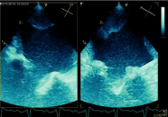
Figure 1: Thrombus on the left atrium.
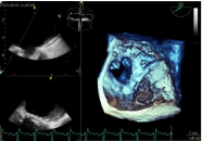
Figure 2: Thrombosis on the left atrial appendage occluder.
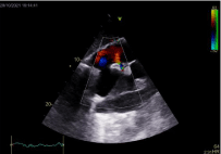
Figure 3: Leakage around the occluder and thrombosis in the left atrium.
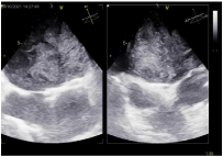
Figure 4: Occluder related thrombosis.
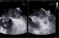
Figure 5: A thrombosis in the left atrium appendage.
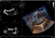
Figure 6: Occluder related thrombosis.
Conclusion
Late device-related thrombosis is very rare. The same applies to patients with a biological mitral prosthesis. In the cases described above, a thrombotic complication was found several years after the cardiac surgery. The common element of these patients was the presence of percutaneous occluders implanted into the left atrium. In both cases, the major mass of the thrombus was on the occluder. Is it a coincidence or is the presence of valve prosthesis and an additional device in the left atrium a “thrombogenic duo”?
References
- Tzikas A Gafoor S, Meerkin D, Freixa X, Cruz-Gonzalez I, Lewalter T, Saw J, et al. Left atrial appendage occlusion with the AMPLATZER Amulet device: an expert consensus step-by-step approach. Euro Intervention. 2016; 11: 1512–1521.
- Lempereur M, Aminian A, Saw J. Rebuttal with regards to “Device-associated thrombus formation after left atrial appendage occlusion: a systematic review of events reported with the Watchman, the Amplatzer Cardiac Plug and the Amulet”. Catheter Cardiovasc Interv. 2018; 92: E216–E217.
- Fauchier L, Cinaud A, Brigadeau F, Lepillier A, Pierre B, Abbey S, et al. Device-related thrombosis after percutaneous left atrial appendage occlusion for atrial fibrillation. J Am Coll Cardiol. 2018; 71: 1528–1536.
- Hildick-Smith D, Landmesser U, Camm AJ, Diener HC, Paul V, Schmidt B, et al. Left atrial appendage occlusion with the Amplatzer™ Amulet™ device: full results of the prospective global observational study. Eur Heart J. 2020; 41: 2894-2901.
- Cochet H, Iriart X, Sridi S, Camaioni C, Corneloup O, Montaudon M, et al. Left atrial appendage patency and device-related thrombus after percutaneous left atrial appendage occlusion: a computed tomography study. Eur Heart J Cardiovasc Imaging. 2018; 19: 1351-1361.
- Abaci A Unlu S, Alsancak Y, Kaya U, Sezenoz B. Short and long term complications of device closure of atrial septal defect and patent foramen ovale: meta-analysis of 28,142 patients from 203 studies. Catheter Cardiovasc Interv. 2013; 82: 1123-1138.