Case Report
Austin J Urol. 2023; 9(1): 1080.
Unclassified Sex Cord/Gonadal Stromal Testis Tumour with a “Pure “Spindle Cell Component: Pathological Features And Review of The Literature
Catherine Cawekazi Cingo¹; Cyril Mnguni²; Meshack Nndweleni Bida³*
¹Registrar, Department of Anatomical Pathology, University of Pretoria and the National Health Laboratory Service, Pretoria, South Africa
²Registrar, Department of Urology, University of Pretoria; Steve Biko Academic Hospital, Pretoria, South Africa
³Department of Anatomical Pathology, University of Pretoria and the National Health Laboratory Service, Pretoria, South Africa
*Corresponding author: Meshack Nndweleni Bida Department of Anatomical Pathology, University of Pretoria and the National Health Laboratory Service, 10 Shingwedzi street; The Wilds; Pretoria; 0167, South Africa. Tel: 012 3542714 Email: meshack.bida@nhls.ac.za
Received: November 21, 2023 Accepted: December 16, 2023 Published: December 22, 2023
Abstract
Background: About 2-5% of adult testicular tumors are classified as sex cord/stromal tumors, which are an uncommon group of tumors of the testis. Occasionally, a small percentage of Sex Cord/stromal Testis Tumors (SCST) that are primarily composed of spindle cells seem to be unclassifiable. There are about six cases reported where spindle cells predominate, four of which are consisting purely of spindle cells. At the morphologic level, a striking resemblance to a fibroma has raised the notion of a possible fibrothecoma, but this is not supported by the immunohistochemistry, and the features are consistent with an unclassified sex cord/stromal tumor with a “pure” spindle cell component.
Description: A 45-year-old man with a firm mass in the lower pole of the left testis for 3 years. The patient had no comorbidities and no features of endocrinopathies. All blood tests were normal. The ultrasound showed a solitary hypoechoic mass with increased blood flow by Doppler scan. A well-circumscribed tumor measuring 30mmx 15mm with no discernible capsule was identified macroscopically. Microscopy showed a well-circumscribed spindle cell lesion with no necrosis, cytological atypia, or mitoses. The tumor cells appeared spindled with eosinophilic cytoplasm, vesicular nuclei, and occasional nucleoli. No nuclear grooves were noted in the tumour cells. No vascular invasion was present. Actin, calretinin, CD99, S100, and inhibin were positively immunoreactive.
Conclusion: The findings were consistent with an Unclassified pure spindle SCST, a rare tumor with a benign course that must be distinguished from several spindle cell neoplasms. There are no biochemical markers for the diagnosis of this tumor, thus requiring meticulous clinical examination, especially if it is small in size.
We report a case of “pure spindle cell sex cord stromal tumour of the testis in a 45 years old man in which the histomorphological as well as the immunohistochemical features are consistent with the diagnosis of unclassified sex cord/stromal testis tumor with “pure” spindle cell component.
Keywords: Unclassified sex cord/gonadal stromal testis tumour with a “pure “spindle cell component; immunohistochemistry
Introduction
Sex cord/stromal tumors of the testis as a group are uncommon, constituting about 2–5% of adult testicular tumors. The majority of patients tend to be children and young adults, constituting about 25% of testicular tumors in infants and children.
There is, however, a wide age range in general, and juvenile granulosa cell tumor is the most common congenital testiculartumor, more common in neonates and infants, associated with ambiguous genitalia or abnormalities of sex chromosomes. Sertoli cell tumors are common before age 1 and often associated with Peutz-Jeghers syndrome and the Carney complex.
Spindle cell tumors, both benign and malignant, are rarely encountered in the testis. The spindle cell morphology of testicular tumors conceptually arouses a variety of tumors, including soft tissue neoplasms. The more common sex cord stromal tumors, such as Leydig cell tumors, Sertoli cell tumors, or granulosa cell tumors, rarely present with spindle cell morphology.
However, cases of conventional sex cord/stromal testis tumors with a spindle cell predominant component that have been reported in the literature are firmly entrenched in the WHO 2016 classification of testicular non-germ cell tumors [1].
These tumors seem to fall into two main groups, viz., biphasic tumors in which the spindle cell component is predominant and intermixed with a conventional sex cord stromal tumor, and one group in which there is a “pure” spindle cell component. Most of these tumors present as a testicular mass, and functional tumors are generally uncommon except for some of those that are intermixed with Leydig cell tumors (unlike ovarian sex cord stromal tumors) or granulosa cell tumors. The tumors often show indolent behavior, with only about 5% showing aggressive behavior. The morphologic prognostic factors include size, mitotic rate, necrosis, cellular atypia, infiltrative borders, and vascular invasion. Clinically, stage is the most important prognostic factor, staged with the TNM, or Children's Oncology Group (COG) staging system, as for germ cell tumors.
Case Report
Clinical Summary
A 45-year-old man was admitted to the Department of Urology of Steve Biko Academic Hospital, Pretoria, South Africa, because of a painless, firm mass in the lower pole of the left testis, which has been present for 3 years.
The patient was otherwise healthy with no underlying systemic disease and, most importantly, no features of endocrinopathies.
The urine analysis, liver enzymes, Prostate-Specific Antigen (PSA), Lactate Dehydrogenase (LDH), serum Alpha-Fetoprotein (AFP), and serum Beta-human Chorionic Gonadotropin (β-hCG) were all normal.
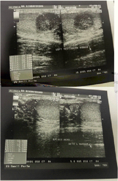
The ultrasound of the scrotum showed a solitary left hypoechoic mass of 13.7mm×17.5mm×14.1mm with increased blood flow detected by Doppler scan (Figure 1). A small hypoechoic nodule in the left epididymis head measuring 6.2x4.1mm was identified. A high left groin node with cortical thickening measuring 4.7mm in diameter was also seen (Figure 2).

Figure 1: (A) The ultrasound of the left testis showing a solitary hypoechoic intratesticular mass; (B) A small hypoechoic nodule in the left epididymis head.

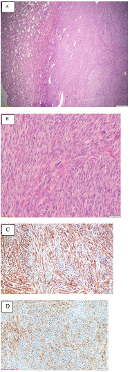
Figure 2: (A) Low magnification showing fascicles of spindle cells with storiform pattern (H&E stain); (B) Representative high-power image with pure spindle cells with bland nuclei and no mitosis (H&E statin). (C) Actin ; (D) Calretinin; (E) S100; (F) Inhibin.
A radical orchiectomy was done, and the specimen was submitted for histologic examination.
Pathological Findings
Macroscopic examination of the specimen showed the testis and spermatic cord weighing 45.7g. On the cut surface, there was a well-circumscribed yellowish tumor present within the testicular gonad measuring 30mm×17mm×15mm with no discernible capsule within the normal surrounding testicular parenchyma. The spermatic cord was normal.
Microscopic examination showed a well-circumscribed spindle cell lesion with no necrosis, cytological atypia, or mitoses present. The tumor cells appeared spindle-shaped with fibrillary eosinophilic cytoplasm, vesicular nuclei, and occasional nucleoli (Figure 2A & 2B). There were no nuclear grooves seen. There was no vascular invasion present.
The immunohistochemical staining was diffusely positive for actin (Figure 2C), calretinin (Figure 2D), and CD99. The tumor was also focally positive for S100 (Figure 2E) and inhibin (Figure 2F). The negative immunohistochemical stains included NAB-STAT6, CD34, bcl-2, Caldesmon, TLE-1, ALk-1, AE1/3, MNF-116, and WT-1.
Discussion
Definition and Classification
Unclassified Sex Cord/gonadal Stromal Testis tumor (SCST) with a pure spindle cell component is a rare testicular tumor that belongs to a wide group of unclassified sex cord-sromal tumors, according to WHO 2016 [1].
Clinical Presentation and Epidemiology
Clinical presentation varies according to the size of the tumor, with some patients reported to have a large, bulky tumor mass with cachexia. Most of the cases reported lack endocrinopathy, such as precocious puberty or gynecomastia, but present as mass lesions in the scrotum. The mass lesions are detected at urologic examination as firm and tense masses, however, with no evidence of abdominal mass or inguinal hernia.
An unclassified sex cord stromal tumor with a pure spindle cell component is rare.
Up to date, including our patient, there are five cases of unclassified sex cord stromal tumors with pure spindle cell morphology documented in the world literature [2].
Pathogenesis
The pathogenesis remains elusive, but suggestions have been made to regard these tumors as a special variant of poorly differentiated granulosa cell tumor, especially given the positive immunoreactivity for inhibin [3]. At the morphologic level, the striking resemblance of fibroma has raised the notion of possible fibrothecoma, but this is not supported by the immunohistochemistry [4,5].
The most likely pathogenetic mechanism will involve testicular somatic stem cells. The germ-line stem cells and somatic stem cells have been found in the testis; however, the somatic stem cells have not been found in the ovary [6].
Biological Behavior
According to Sewell et al. there are no established protocols for surveillance and management of unclassified SCST with a pure spindle cell component [7].
Although this tumor is considered a benign or indolent tumor, it is difficult to predict its clinical behavior based on histological findings alone, given the few cases reported in the literature [2]. A long-term follow-up is recommended. It is also noteworthy that there are too few cases reported, and therefore long-term follow-up studies are lacking. So far, no cases have been reported to recur or metastasize, thus implying the benign course of these tumors.
Even in the presence of high mitotic activity, cellularity, nuclear pleomorphism, and necrosis, there is evidence of no unfavourable outcome [3].
Diagnosis
Given the pure spindle cell morphology of these tumors, a wide range of both benign and malignant differential diagnoses are considered, and the immunohistochemical stains are very useful.
Our case showed pure spindle cell morphology with bland cytologic features, in keeping with the morphologic criteria for an unclassified sex cord/gonadal stromal testis tumor with a pure spindle cell component. The findings resembled those of a fibroma of gonadal origin or testicular myoepithelioma (i.e., ultrastructural evidence of epithelial and myoid differentiation).
A fibroma of gonadal origin may show overlapping immunohistochemical features with SCST, as seen in our case; however, the former is usually negative for inhibin and positive for desmin.
In addition, reticulin stain can also be used to differentiate between fibroma of gonadal origin and unclassified SCST with a pure spindle cell component [8]. The former reveals a rich network enveloping individual cells as opposed to a nested pattern seen in unclassified SCST with a pure spindle cell component [8,9].
Leydig cell tumor immunohistochemical markers were positive; however, co-expression of S100 and smooth muscle actin is typical of an unclassified sex cord/gonadal stromal testis tumor with predominant spindle cell morphology [2,9], as seen in our case. Positive inhibin and CD99 suggest that this tumor is a subset of sex cord-stromal tumors.
Unclassified SCST with a pure spindle component also shares the same immunohistochemical profile as granulosa cell tumors but differs in morphology. Some authors consider these tumors to be variants of a poorly differentiated granulosa cell tumor [5]. Our case, however, did not show nuclei grooves as in some of the previously reported cases.
Sertoli-Leydig cell tumors are negative for smooth muscle actin and may show cytokeratin positivity. In addition, smooth muscle tumors express actin but are S100-negative, and tumors of neural origin do not express actin.
The other differential diagnosis to consider is rete testis carcinoma, which was excluded by the negative cytokeratin markers in our case.
The remaining group of high-grade malignant tumors were excluded on the basis of the absence of atypia, vascular invasion, necrosis, and mitoses.
Concluding Remarks
We have reported the fourth case of unclassified SCST with the pure spindle component of the testicular gonad, a rare tumor with a benign course that must be distinguished from several spindle cell neoplasms in the testis. There are no specific serum biochemical markers for the diagnosis of this tumor, which is detected clinically by palpation of the scrotal mass, thus requiring meticulous clinical examination, especially if it is small in size.
References
- Idrees MT, Ulbright TM, Oliva E, Young RH, Montironi R, Egevad L, et al. The World Health Organization 2016 classification of testicular non-germ cell tumours: a review and update from the International Society of Urological Pathology Testis Consultation Panel. Histopathology. 2017; 70: 513-21.
- Spairani C, Squillaci S, Pitino A, Montefiore F, Fusco W. Unclassified sex cord/gonadal stromal testis tumor with a ”pure” spindle cell component: A case report. Pathologica. 2018; 110: 302-6.
- Tarjàn M, Sarkissov G, Tot T. Unclassified sex cord/gonadal stromal testis tumor with predominance of spindle cells. APMIS. 2006; 114: 465-9.
- Miller MI, Hibshoosh H, Hensle TW. An enormous testicular sex cord/gonadal stromal tumor. J Pediatr Surg. 1996; 31: 980-2.
- Renshaw AA, Gordon M, Corless CL. Immunohistochemistry of unclassified sex cord-stromal tumors of the testis with a predominance of spindle cells. Mod Pathol. 1997; 10: 693-700.
- Liu CF, Barsoum I, Gupta R, Hofmann MC, Yao HHC. Stem cell potential of the mammalian gonad. Front Biosci (Elite Ed). 2009; 1: 510-8.
- Sewell J, Osborne JR, Basto M, Sapre N, Kostos P, Raj M. Unclassified sex cord-stromal tumour of the testis – how to diagnose the unclassified. J Clin Urol. 2018; 11: 372-5.
- Magro G, Gurrera A, Gangemi P, Saita A, Greco P. Incompletely differentiated (unclassified) sex cord/gonadal stromal tumor of the testis with a ”pure” spindle cell component: report of a case with diagnostic and histogenetic considerations. Pathol Res Pract. 2007; 203: 759-62.
- Tarjàn M, Sarkissov G, Tot T. Unclassified sex cord/gonadal stromal testis tumor with predominance of spindle cells. APMIS. 2006; 114: 465-9.