
Case Report
Austin J Vet Sci & Anim Husb. 2015; 2(3): 1017.
Birth Complications and Cesarean Section in a Blacktip Reef Shark (Carcharhinus melanopterus)
Jung-Schroers V*, Hellmann J and Steinhagen D
Fish Disease Research Unit, University of Veterinary Medicine Hannover, Germany
*Corresponding author: Jung-Schroers V, Fish Disease Research Unit, University of Veterinary Medicine Hannover, Bünteweg 17, 30559 Hannover, Germany
Received: November 25, 2015; Accepted: December 16, 2015; Published: December 18, 2015
Abstract
Dystocia occurred in a pregnant blacktip reef shark (Carcharhinus melanopterus) from a public aquarium, where the tail fins of two pups were visible on arrival of the veterinarians. Only one dead pup could be extracted manually, therefore a cesarean section was performed ca. 14 hours later after determination that the swimming performance of the pregnant shark was considerably inhibited and surgery was needed, during which three dead pups were removed. The left uterus showed necrotic areas and signs of severe inflammation and the pups from this uterus were heavier than pups from the right uterus. Two days post operation, the shark was euthanized, because it was immobile. A bacteriological examination of liquid from the body cavity resulted in a septicemia presumably associated with an endotoxemia. In captivity, pregnant sharks are prone to overfeeding which may lead to heavier weights of pups than in natural settings, creating birthing difficulties. Hence, during pregnancy feeding should be controlled, and in the event of dystocia, immediate action is required.
Keywords: Dystocia; Anesthesia; Pup size; Overfeeding
Case Report
The blacktip reef shark (Carcharhinus melanopterus) is a common species in the Indo Pacific, inhabiting shallow reefs and sand-flats of atolls and islands and also occurs in coastal habitats [6,10,14]. Although C. melanopterus is one of the most abundant shark species in the region, the number of individuals has been reduced at many locations due to over fishing [15], and the species is classified as “near threatened” by the International Union for Conservation of Nature. A high proportion of inbreeding among C. melanopterus occurs in French Polynesia which might indicate a reduced population size as well [7]. C. melanopterus also exhibits sexual segregation in habitat use, with females congregating in large numbers inside of lagoons or near atolls, while males use fore-reefs more frequently [7]. Maturation varies geographically, with males maturing at a body length of 95 [4,11] to 111cm [6] Total Length (TL), and females maturing at 110 [15] to 125cm [4] TL. Gestation of C. melanopterus also varies geographically, and ranges from 8-9 months [4] to 16 months [5], and brood sizes ranges from 1 to 5 [6,15], pups with body lengths of 33 and 34cm [1,11] to more than 60cm in TL [14]. In some geographical regions the reproductive cycle is biennial [15], while in other regions it is annual [4,6,12]. As with many shark species, C. melanopterus is often housed in public aquaria, and reproduction is a challenge aquarium staff and attending veterinarian face. As such, understanding how gestation and birthing differ between captive and natural settings is important for conservation purposes by aiding in successful captive rearing.
Here, we present a case study of one female C. melanopterus housed in a public aquarium for seven years with two other females and one male C. melanopterus at a water temperature of 25-26°C. During residency at the aquarium, wounds were present at her pectoral fins, suggesting signs of mating. Over the following month the body silhouette of the female shark of interest became more rounded. Eight months later, in June, the caudal fins of two pups were extending 5-15cm out of the female´s cloaca. After hours of visual evaluation, no birthing progress was recognized and the female shark was transferred to a quarantine tank. By using immense force, the pup which was hanging out of the cloaca by ca. 15cm was removed by manual extraction. The pup did not show any vital signs, like respiration or reflexes, and resuscitation failed. When the veterinarians arrived, the pregnant female shark of interest exhibited an increased buccopharyngeal pumping rate and inconstant swimming behavior, with resting on the bottom of the tank. Because reef sharks require ram ventilation to breath, emergency surgery was conducted. The pregnant shark was placed into a smaller tank, and sedated by slowly adding buffered tricaine methane sulfonate (Tricain, Pharmaq, UK) upto a concentration of 0.05g/l to the water. When control of flight reflex and nictitating eyelid reflex showed a deep sedation, an ultrasound examination was performed and additional pups could be seen inside the uteri. All attempts to remove the pup that remained hanging out of the cloaca failed. Therefore, a cesarean section was conducted. The dose of tricaine methane sulfonate was increased to 0.075g/l. Tricaine methane sulfate has proven as an effective agent for blocking sensory-motor responses and is recommended as singledrug anesthetic for surgical interventions in anamniotes, like fish [13], and therefore no further analgesic substance was administered. Upon anaesthetization, the shark was placed on a vacuum mattress above the tank, and the gills were continuously flushed with water containing 0.05g/l tricaine methane sulfonate to maintain anaesthetization and ensure respiration. The body cavity was opened along the median line, and the right uterus, which was slightly reddened, was opened and two dead pups of 51 and 47cm TL, with body weights of 841 and 557g, respectively, were removed. The left uterus, which was very thin walled, reddened, and showing necrotic areas, was opened, and the dead pup visible from the cloaca was removed from this uterus. The pup from the left uterus was larger (54cm TL) and heavier (963g) than the ones from the right uterus. All pups were arranged with their caudal fins directed toward the posterior of the mother´s body, and all showed first signs of putrefaction in different stages.
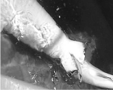
    Figure 1:  Ventral view of the female Carcharhinus melanopterus after manual
extraction of one pup. The tail fin of the second pup can be seen hanging out
of the birth canal. (Picture: Stefan Inselmann, Merlin Entertainments Group).
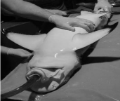
    Figure 2:  Anaesthetized female Carcharhinus melanopterus placed on
a vacuum mattress and intubated with a flexible tube. (Picture: Stefan
Inselmann, Merlin Entertainments Group).
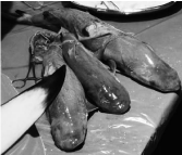
    Figure 3:  Three surgically removed dead Carcharhinus melanopterus pups.
(Picture: Stefan Inselmann, Merlin Entertainments Group).
After removal of necrotic areas, and sampling of uterus tissue and liquid from the body cavity for bacteriological and histological examination, both uteri were closed continuously by a Schmieden´s suture with a polyglycol acid fiber. The body cavity was sutured with single sutures with a polyglycol acid fiber, and the shark received 10mg enrofloxacine and 0.2mg meloxicam per kg body weight by intramuscular (i.m.) injection. After ca. 90min, the female shark was transferred to a quarantine tank without anesthetics and respiration was supported. The shark started swimming independently 90min later.
Each day following the surgery, the female shark was treated with enrofloxacine (10mg/kg) by i.m. injections, however despite the care provided; swimming remained irregular, with a high frequency of resting on the bottom. Two days after the surgery, the female shark could not swim independently, and it was euthanized by adding buffered tricaine methane sulfonate in a concentration of 0.5g/l to the water. The bacteriological examination showed high amounts of Pseudomonas luteola and Lactobacillus spp. Which were sensitive to enrofloxacine according to the antibiogram. Histologically, the most remarkable changes were cell necrosis and severe bleedings in the left uterus, especially directly underneath the epithelium, displacement of the epithelium and an infiltration with high numbers of lymphocytes and granulocytes.
Discussion / Conclusion
This is the first published report of a cesarean section in a blacktip reef shark and probably the first description of such a surgery in sharks in general. The surgery was successful in keeping the pregnant shark alive while anaesthetized for ca. two hours with a very low dose of buffered tricain methan sulfonate. However, because the shark showed a dystocia for ca. 14 hours before anesthesia was started, the time point of the surgery was much too late. Upon opening the body cavity, the left uterus showed signs of necrosis and severe inflammation, which could be followed by a septicemia and an endotoxemia. A total removal of both uteri was desirable, but not possible due to extensive adherence of the uteri to the serous membranes of the body cavity.
After conducting necropsies of the pups, no pathological findings, apart from alterations due to putrefaction were seen macroscopically or histologically. While the pups from the right uterus measured 51 and 47cm TL with a body weight of 841 and 557g respectively, the pups from the left uterus each measured 54cm TL with weights of 950 and 963g. The size of the female shark was 129cm TL with a weight of 11400g after the pups were removed. Median size at birth of C. melanopterus in natural settings differs between geographical locations. In north-eastern Australia, the size at birth is 51 to 67cm TL, and the size of the females is 135 to 160 cm TL [2], at Aldabra Atoll in the Indian Ocean, the size of pups is 45 to 55cm TL and the size of females is maximal at 140cm TL [15], in French Polynesia pups have been observed at 48cm [6] and 34cm TL at Palmyra Atoll [10]. In general blacktip reef sharks from coastal habitats are larger than C. melanopterus from small isolated atolls [2,6 ]. This might be due to a limitation of food resources and the occurrence of other competing species [6,10]. Increased resource availability may therefore not only result in larger maximum sizes but also in increased sizes at birth [10]. In the present case, the body size at birth of the pups from the left uterus were comparatively large in relation to the body size of the female shark, and the suspected diagnosis was a dystocia because of a fetopelvic disproportion. Compared to natural settings, sharks in captivity may feed more frequently, especially while pregnant, because of regular access to larger quantities of food, especially in community tanks. Therefore, overfeeding pregnant sharks in captivity may lead to higher birth weights of pups that inhibit birthing. As such, feeding regulations may need to be adjusted in captivity, especially among pregnant individuals, which will continue to provide valuable information on shark reproduction, of which very little is known for most species.
Acknowledgement
The authors would like to thank Marie Agethen for excellent technical support.
References
- Bonham K. Note on sharks from Rongelap Atoll, Marshall Islands Copeia. 1960; 257.
- Chin A, Simpfendorfer C, Tobin A, Heupel M. Validated age, growth and reproductive biology of Carcharhinus melanopterus, a widely distributed and exploited reef shark. Mar Freshw Res. 2013; 64: 965-975.
- Chin A, Tobin AJ, Heupel MR, Simpfendorfer CA. Population structure and residency patterns of the blacktip reef shark Carcharhinus melanopterus in turbid coastal environments. J Fish Biol. 2013; 8: 1192-1210.
- Lyle JM. Observations on the Biology of Carcharhinus cautus (Whitley), C. melanopterus (Quoy and Gaimard) and C. fitzroyensis (Whitley) from Northern Australia. Aust J Mar Fresh Res. 1987; 38: 701-710.
- Melouk MA. On the development of Carcharhinus melanopterus. Publications of the Marine Biological Station Al Ghardaqa, Egypt. 1957; 9: 229-251.
- Mourier J, Mills SC, Planes S. Population structure, spatial distribution and life-history traits of blacktip reef sharks Carcharhinus melanopterus. J Fish Biol. 2013; 82: 979-993.
- Mourier J, Planes S. Direct genetic evidence for reproductive philopatry and associated fine-scale migrations in female blacktip reef sharks (Carcharhinus melanopterus) in French Polynesia Mol Ecol. 2013; 22: 201-214.
- Neuhaus H, Schroers V, Baumer A, Rosalski A, Kummerfeld N, Meyer K, et al. Surgical excision of a fishhook from the gastrointestinal tract of a freshwater stingray (Potamotrygon falkneri) Praktischer Tierarzt. 2010; 91: 102-110.
- Neuhaus H, Schroers V, Kummerfeld N, Steinhagen D. Diagnosis and therapy of a foreign body obstipation in a freshwater stingray (Potamotrygon itaituba). Kleintierpraxis. 2008; 53: 372-375.
- Papastamatiou YP, Caselle JE, Friedlander AM, Lowe CG. Distribution, size frequency, and sex ratios of blacktip reef sharks Carcharhinus melanopterus at Palmyra Atoll: a predator-dominated ecosystem. J Fish Biol. 2009; 75: 647-654.
- Papastamatiou YP, Lowe CG, Caselle JE, Friedlander AM. Scale-dependent effects of habitat on movements and path structure of reef sharks at a predator-dominated atoll. Ecology. 2009; 90: 996-1008.
- Porcher IF. On the gestation period of the blackfin reef shark, Carcharhinus melanopterus, in waters off Moorea, French Polynesia. Mar Biol. 2005; 146: 1207-1211.
- Ramlochansingh C, Branoner F, Chagnaud BP, Straka H. Efficacy of Tricaine Methanesulfonate (MS-222) as an Anesthetic Agent for Blocking Sensory-Motor Responses in Xenopus laevis Tadpoles. Plos One. 2014; 9: 1-11.
- Speed CW, Meekan MG, Field IC, McMahon CR, Stevens JD, McGregor F, et al. Spatial and temporal movement patterns of a multi-species coastal reef shark aggregation. Mar Ecol Prog Ser. 2011; 429: 261-275.
- Stevens JD. Life-History and Ecology of Sharks at Aldabra Atoll, Indian-Ocean. Proc Roy Soc B. 1984; 222: 79-106.