
Case Report
Austin Alzheimers J Parkinsons Dis. 2023; 6(1): 1037.
Behavioral Variant Frontotemporal Dementia like Syndrome –Atypical Presentation of Intracranial Hypotension
Hiba Khan*; Kasia Gustaw Rothenberg
Lou Ruvo Center for Brain Health, Neurological Institute, Cleveland Clinic, USA
*Corresponding author: Hiba Khan Lou Ruvo Center for Brain Health, Neurological Institute, Cleveland Clinic, 9500 Euclid Avenue, 44195 Cleveland, USA. Tel: 216-894-9673; Fax: 216-636-2645 Email: khanh3@ccf.org
Received: March 01, 2023 Accepted: April 06, 2023 Published: April 13, 2023
Abstract
Spontaneous Intracranial Hypotension (SIH) is an uncommon syndrome which may result from a Cerebrospinal Fluid (CSF) leak. Atypical SIH may present with neurobehavioral symptoms similar to those observed in behavioral variant Frontotemporal Dementia (bvFTD). We present a case of a 55-year-old male with postural headache and behavioral symptoms who was initially diagnosed with bvFTD. On re-evaluation in our clinic, imaging revealed caudal displacement of the midline structures suggestive of Cerebrospinal Fluid (CSF) hypotension. We hypothesized that with unmitigated CSF loss over a long period of time, the patient developed brain sagging clinically resulting in neurobehavioral, frontal lobe dysfunction. After multitude of investigative procedures, a CSF venous fistula originating from a T7 nerve root cyst was found and repaired. Surgical intervention and restoration of CSF volume resulted in resolution of cognitive as well as neurobehavioral symptoms.
Keywords: Behavioral variant frontotemporal dementia; Headache; CSF leak; Spontaneous intracranial hypotension; Sagging brain syndrome
Introduction
Behavioral Variant Frontotemporal Dementia (bvFTD) is a devastating neurodegenerative disease with relatively early onset. The characteristic clinical features of bvFTD include a change in personality and behavior, for example, disinhibition and executive dysfunction, resulting in poor planning, loss of judgment, difficulty with organization and loss of insight. Overeating, particularly of sweets may occur. Other features include apathy, utilization behaviors, and obsessive compulsive-like preservative behaviors. Patients often demonstrate lack of empathy, impaired insight and show little concern for friends or family members. Language deficits occur but may not the presenting feature [10].
Spontaneous Intracranial Hypotension (SIH) may result in symptoms similar to those seen in bvFTD [1,11]. Contrary to bvFTD however, SIH can be successfully treated resulting in reversal of the neuropsychiatric syndrome. SIH commonly manifests as a low-pressure orthostatic headache secondary to a reduction in the CSF pressure, without an apparent explanation. Any breach in the dura mater causes a reduction in CSF volume leading to gravitational traction and downward displacement of the brainstem and cerebellum [9]. Dural defects predispose to the formation of CSF venous fistulas (with unregulated CSF loss) because of communications between the spinal subarachnoid space and dilated paraspinal veins that accompany SIH [2].
SIH has a broad spectrum of clinical and imaging manifestations, and atypical cases have been reported. Atypical clinical presentations of SIH include parkinsonism, ataxia, obtundation, memory deficits and dementia with frontotemporal features [1,8,11]. Symptoms result from prolonged intracranial hypotension and mechanical disruption of frontotemporal anatomy with structural, metabolic or neurodegenerative changes, which can be reversed by restoration of CSF volume [4]. The main MRI finding is a characteristic diffuse pachymeningeal enhancement. Apart from this, MRI may also show caudal displacement of the midline structures of the brain such as sagging of the brain, pituitary enlargement, subdural fluid collection, posterior lobe pituitary hematoma, diffuse dural enhancement of the spinal canal, spinal epidural fluid collection, distension of the spinal epidural venous plexus, and abnormal intensity around the root sleeves [6]. Combination of this characteristic MRI picture and frontotemporal dementia like neurobehavioral syndrome is commonly referred to as Frontotemporal Brain Sagging Syndrome (FTBSS) [12]. FTBSS is defined as an insidious, often progressive decline in behavior and executive functions, hypersomnolence, and orthostatic headaches attributed to cerebrospinal fluid hypovolemia [5,12]. Here by we present a case of a 55 year-old-male diagnosed with bvFTD who in fact was suffering SIH clinically presented as FTBSS which resolved after the source of spinal fluid leak was found and repaired.
Case Report
Initial Case Presentation
The Patient (KW) is a 55 year-old-male who presented with headaches, confusion, and lack of attention. Family reported changes in behavior such as disengagement from surroundings and frequent repetition. Episodes of headaches had been ongoing for at least 14 months which the patient attributed to migraines or allergies. At first, his reported lack of attention was only affecting driving, but soon afterwards he lost two jobs because of cognitive problems. In addition to cognitive changes his behavior changed. Reportedly the patient started laughing and joking inappropriately. These symptoms led to neurological evaluation. He scored 20/30 on Montreal Cognitive Assessment (MoCA) with abnormal performance in: naming, attention, language fluency, abstraction and recall. Imaging was unremarkable and neuropsychological assessment suggestive of frontal as well as temporal lobe dysfunction.
Initial MRI (March 2016): There are a few punctate areas of high signal in the periventricular white matter that are likely related to chronic small vessel ischemic disease. Demyelination cannot be entirely excluded in a young patient. No acute brain parenchymal abnormality.
Initial Neuropsychological assessment (2016): There are prominent deficits in memory (recall and recognition are severely impaired, learning is suppressed). Aspects of executive function (switching/inhibition, errors, rule violations, verbal fluency) are impaired in addition to self-report and wife’s report of presence of significant apathy, disinhibition and executive dysfunction. Attention and processing speed are variable from extremely low to high average. Verbal reasoning, naming, perceptual reasoning, visuospatial functions are intact. Deficits are of moderate severity and suggest involvement of frontal and temporal lobes, concerning for major neurocognitive disorder likely frontotemporal lobar degeneration.
He had an extensive workup including a normal EEG and blood tests, which included, TSH, vitamin B12 level, lead and vitamin D levels, RPR, Lyme disease testing as well an autoimmune and paraneoplastic panel. The results of the above mentioned testing came back within normal range. A1C was in prediabetic range. Thiamine level was low. He was diagnosed with major neurocognitive disorder in the course of frontotemporal lobe degeneration. Treatment with Donepezil and Memantine was initiated and medications were titrated up to the maximum recommended doses.
Re-evaluation at the Center for Brain Health (CBH) Cleveland Clinic
Patient was referred to CBH to be considered for participation in clinical trials. Both the patient and his family were concerned about FTD diagnosis and were seeking re-evaluation. Patient’s cognitive and behavioral symptoms remained the same but in addition to initial clinical presentation he started exhibiting orthostatic headaches, swallowing difficulty and frequent hiccups. On repeat MoCA Patient scored 24/30 with deficits in delayed recall, language and abstraction. Repeated imaging however revealed caudal displacement of the midline brain structures concerning for CSF hypotension.
MRI 2017: Striking caudal displacement of the midline structures suggesting severe CSF hypotension. No significant generalized parenchymal volume loss by visual inspection and quantitative analysis. Hippocampal volumes were at the 53rd percentile when compared to age matched normal controls by quantitative analysis. Mild nonspecific supratentorial white matter disease was reported. Comparison with imaging from 2016 was suggestive of interval progression (Figure 1).
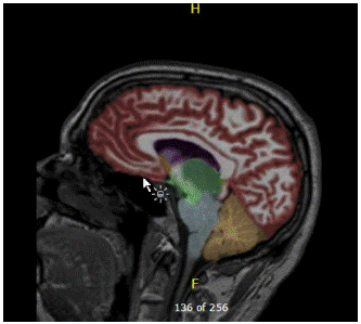
Figure 1: MRI: Caudal displacement of the midline structures suggesting severe CSF hypotension. No significant generalized parenchymal volume loss by visual inspection and quantitative analysis. Hippocampal volumes at the 53rd percentile when compared to age matched normal controls by quantitative analysis. Mild nonspecific supratentorial white matter disease.
Further evaluation by Neurosurgery at the Cleveland Clinic including CSF opening pressure was reported normal and it was suggested that further evaluation was not needed at the time.
He was reevaluated again due to relentless nature of his symptoms. In-hospital imaging did not reveal a CSF leak and it was felt that the appearance of the brainstem was likely due to a congenitally small tentorial incisura.
Repeat MRI (2017): There is effacement of the mesencephalon and pons with narrowing of the dimensions basilar cistern at the superior margin of the tentorium. This is unchanged in appearance when compared to the prior exam. The prepontine cistern as well as the CP angle cisterns and quadrigeminal plate cistern are otherwise normal. The CSF space cephalad to the brain is normal. The overall appearance does not suggest CSF leak. The appearance of the brainstem is likely due to a congenitally small tentorial incisura.
He continued to experience symptoms with increasing severity which led to another, this time more extensive evaluation at an outside facility. CT myelogram failed to localize a specific leak site. Positive pressure combined CT and MR myelogram of the entire spine also failed to localize a leak. This time however treatment in the form of bi-level epidural blood patch (at T10/11 and L2/3) was implemented with significant improvement in all reported symptoms. Headaches nearly resolved within 1 to 2 weeks of receiving blood patch, energy levels improved, dysphagia resolved, and memory was reported to have improved significantly. After 3 to 4 weeks, however, all symptoms recurred. At this point he received another blood patch treatment. Finally, digital subtraction CT myelogram under general anesthesia demonstrated a CSF-venous fistula arising from the right T7 nerve root.
Surgical Intervention
Patient underwent surgical fistula ligation. After surgery subjectively as well as objectively he returned to his premorbid baseline. Neuropsychological testing showed improvement across the majority of cognitive domains compared to earlier assessments. Testing revealed only mild inattention which had a slight influence memory and executive function (digit sequencing, verbal fluency). The Patient remained improved on consecutive follow up appointments
Post – Surgery Neuropsycholgical evaluation: Neuropsychological evaluation revealed a mostly intact cognitive profile with mild inefficiencies (i.e., low average range) in basic auditory working memory, sustained attention, recall of a word-list, and verbal fluency, and borderline performance on a category fluency task. His encoding of a word list was in the borderline range but he demonstrated benefit from repetition (high average learning) and was able to retain and recognize 100% of what he initially encoded. His learning and memory for short stories and simple figures was above average, including superior performance on visual memory acquisition. Performance across all other measures of language, visuospatial functioning, processing speed, and executive functioning ranged from average to high average. He denied clinically significant symptoms of depression or anxiety. On informant measures, his wife indicated the presence of mild agitation and indicated that he has shown minimal impairment in carrying out daily activities. On questionnaires assessing symptoms consistent with frontal systems dysfunction, him and his wife separately reported significant decreases in apathy, disinhibition, and executive dysfunction symptoms since onset of symptoms in 2017. Comparison to the pre-procedure evaluation revealed significant improvements (>2 SD) in all aspects of visual memory, word list recall and recognition, story recall, phonemic verbal fluency, and response inhibition and set-shifting. More subtle improvements (1.5 SD) were seen on measures of abstract verbal reasoning, mental arithmetic, and processing speed. His performance on all other measures remained consistent across evaluations.
Discussion
Most of the cases of intracranial hypotension develop rather quickly and can be related to surgical procedures, and injury to meningeal or parameningeal structures leading to unmitigated CSF loss. Mandal et al (2020) described a case of a marathon runner who presented with postural headache attributable to CSF venous fistulation originating from a lower thoracic nerve root cyst. With unmitigated CSF loss over the following 3 months, the patient became bedbound and developed rapidly progressive behavioral syndrome similar to bvFTD. Ortega et al (2020) published a case of 56-year-old male who developed orthostatic headaches and fatigue after scuba diving. His symptoms included progressive, vertigo, tinnitus, nausea, lack of judgment, inappropriate behavior, memory dysfunction, apathy, tremor, facial dyskinesia, dysarthria, dysphagia and hypersomnolence. Lumbar puncture revealed an opening pressure of 0cm H2O. Magnetic resonance imaging findings included brain sagging, bilateral temporal lobe herniation, and pachymeningeal enhancement. CT myelogram showed a thoracic diverticulum and a CSF-venous leak at the T6-T7 level.
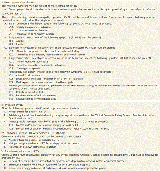
Table 1: International Consensus Criteria for Behavioral Variant Frontotemporal Dementia.
The patient presented in this report had a chronic course of illness with gradually increasing severity of symptoms that were not consistent with the typical presentation of intracranial hypotension. He started experiencing headaches two years before initial evaluation. However, the characteristics of his headaches did not differ from those he had been experiencing throughout his life, which were attributed to migraines, allergies and occluded sinuses. Only when his headaches increased in frequency and became noticeably postural did he start seeking medical attention. Some features of bvFTD-like syndrome started presenting after his headache episodes increased in intensity and severity. The patient might have suffered from symptoms of intracranial hypotension a few years prior to developing the recognizable clinical and radiological features of FTBSS
Prominent cognitive impairment in the course of SIH, although unusual has been reported before [12] analyzed eight patients with radiologically confirmed FTBSS. Every patient had cognitive impairment and daytime somnolence. Behavioral impairment was prominent in some patients. On MRI, all patients had evidence of brain sagging with distortion of the brainstem. CSF opening pressure varied widely but only one patient had low pressure. Daytime somnolence was not reported by our patient. The character of his cognitive dysfunction as well as radiological findings was consistent with FTBSS as defined by Wickling et al (2020). The course of his condition, dominant behavioral features, and sagging brain presentation on MRI and his subsequent response to intervention qualifies his syndrome as atypical [1].
Capizzano et al. (2016) compared clinical and imaging features of spontaneous intracranial hypotension with typical-versus-atypical presentations. Atypical spontaneous intracranial hypotension was a more chronic syndrome compared with classic spontaneous intracranial hypotension, with more severe brain sagging, lower rates of clinical response, and frequent relapses. Patients with atypical spontaneous intracranial hypotension were older than those with classic spontaneous intracranial hypotension and often presented with Parkinsonism, ataxia, hypersomnia, sensorineural deafness, in addition to short term memory loss and behavioral features similar to those of bvFTD. Symptom duration was shorter in classic compared with atypical spontaneous intracranial hypotension. There was no significant difference in dural enhancement, subdural hematomas, or cerebellar tonsil herniation. Patients with atypical spontaneous intracranial hypotension had significantly more elongated anteroposterior midbrain diameter compared to those with classic spontaneous intracranial hypotension and shortened ponto-mammillary distance. Moreover, patients with atypical spontaneous intracranial hypotension were less likely to become symptom-free, regardless of treatment compared with those with classic spontaneous intracranial hypotension.
Even though our patient’s case is consistent with what is described as atypical (use full name or acronym) by Capizzano et al (2016), he responded well to CSF volume restoration after surgical intervention.
Most patients with typical SIH respond well to measures aimed at increasing CSF volume, including hydration with increased salt intake and the administration of caffeine as well as glucocorticoid or mineralocorticoid medications. Autologous epidural blood patches are safe and effective if there is failure in response to conservative management. Success rates are higher if the blood patch is administered directly at the extravasation site, particularly among patients for whom procedures at the lumbar level have been unsuccessful. Injection of fibrin glue as an alternative to blood patch has been used with some effect, and surgical intervention is saved as a last resort when a significant meningeal defect has been located [6].
An autologous epidural patch eased some our patient’s symptoms. Finding and repairing the source of leak seems to be the ultimate solution for symptomatic intracranial hypotension, with a chance for neurobehavioral syndrome to resolve.
Shivent et al (2018) looked into the rate of response to treatment in relatively big group of 29 Patients with SIH and neurobehavioral symptoms. Magnetic resonance imaging showed brain sagging in all patients, CSF opening pressure low in about half of patients, but a spinal CSF leak could not be detected in any. All patients underwent epidural blood patching, but 26 patients eventually underwent one or more surgical procedures. Overall, a good outcome was obtained in majority of patients. Neurobehavioral symptoms in the course of SIH were linked to older age and male gender. The author of the study concluded that neurobehavioral symptoms in SIH are rare and associated with brain sagging and hypersomnolence. Spinal CSF leaks are rarely detected in those cases.
Detection of a source of leak and repair gives a good chance of symptoms reversal. Our Patient benefited from surgical intervention with reversal of both somatic as well as cognitive symptoms. Similarly to our case, the patient presented by Ortega et al (2018) improved postoperatively (T6-T7 laminotomy with dissecting, clipping, and ligating the diverticulum). The follow-up MRIs showed reversion of the sagging index/uncal herniation.
Conclusion
Frontotemporal brain sagging syndrome may mimic bvFTD. It is important to recognize this as a potentially reversible cause of cognitive impairment. As seen in our patient, intracranial hypotension may develop slowly overtime and can lack the emergent presentation of brain herniation. CSF opening pressure varies widely, and thus does not aid in ruling out CSF leak. Neurodegenerative disorders such as frontotemporal dementia are a devastating condition with serious implications on the lives of patients and their caregivers. Every effort should be made to rule out other possibilities, especially reversible causes. Identifying the leak is often very difficult and requires extensive imaging as well as expert consultation. The diagnostic process can be a long and emotionally exhausting journey, but treatment with blood patch can provide temporary relief while workup is continued to identify the leak.
Early referral to tertiary care centers with a group of specialized neurosurgeons should be considered. Clinicians must not hesitate to obtain repeat imaging or a second opinion from a neuroradiologist if there is a clinical suspicion of intracranial hypotension. Lastly, we need to continue educating the medical community to prevent delays in timely diagnosis in order to prevent irreversible changes.
References
- Capizzano AA, Lai L, Kim J, Rizzo M, Gray L, et al. Atypical Presentations of Intracranial Hypotension: Comparison with Classic Spontaneous Intracranial Hypotension. American Journal of Neuroradiology. 2016; 37: 1256-61.
- Fishman RA, Dillon WP. Dural enhancement and cerebral dis-placement secondary to intracranial hypotension. Neurology. 1993; 43: 609–11.
- Mandal AK, Ryatt A, O’Hare K, Missouris CG. Lessons of the month: HLA-B27-associated syndrome and spontaneous intracranial hypotension resulting in behavioral variant frontotemporal dementia. Clinical Medicine (London, England). 2020; 20: 10-11.
- Kent L, Butterworth R, Butler C. Lessons of the month 3: Spontaneous resolution of frontotemporal brain sagging syndrome. Clinical Medicine (London, England). 2019; 19: 336-337.
- Kumar N, Diehn FE, Carr CM, Verdoorn JT, Garza I, et al. Spinal CSF venous fistula: A treatable etiology for CSF leaks in craniospinal hypovolemia. Neurology. 2016; 86: 2310–2.
- Michali-Stolarska M, Bladowska J, Stolarski M, Sąsiadek MJ. Diagnostic Imaging and Clinical Features of Intracranial Hypotension - Review of Literature. Polish Journal of Radiology. 2017; 82: 842-849.
- Ortega-Porcayo LA, Ortega EP, Quiroz-Castro O, Carrillo-Meza RA, Ponce-Gomez JA, et al. Frontotemporal brain sagging syndrome: Craniospinal hypovolemia secondary to aT6-T7 cerebrospinal fluid-venous fistula. Surgical Neurology International. 2020; 11: 250.
- Ozyigit A, Michaelides C, Natsiopoulos K. Spontaneous Intracranial Hypotension Presenting With Frontotemporal Dementia: A Case Report. Frontiers in Neurology. 2018; 9: 673.
- Pabaney AH, Mirza FA, Syed NA, Ahsan H. Spontaneous dural tear leading to intracranial hypotension and tonsillar herniation in Marfan syndrome: a case report. BMC Neurology. 2010; 10: 54.
- Rascovsky K, Hodges JR, Knopman D, Mendez MF, Kramer JH, et al. Sensitivity of revised diagnostic criteria for the behavioral variant of frontotemporal dementia. Brain. 2011; 134: 2456–77.
- Schievink WI, Maya MM, Barnard ZR, Moser FG, Jean-Pierre S, et al. Behavioral Variant Frontotemporal Dementia as a Serious Complication of Spontaneous Intracranial Hypotension. Operative Neurosurgery (Hagerstown). 2018; 15: 505-515.
- Wicklund MR, Mokri B, Drubach DA, Boeve BF, Parisi JE, et al. Frontotemporal brain sagging syndrome: an SIH-like presentation mimicking FTD. Neurology. 2011; 76: 1377-82.