Abstract
Ptotic breast deformity is caused by the involution of the breast parenchyma, leading to a loss of volume with a converse laxity of the skin envelope. Several factors including aging, peripartum enlargement, and postpartum involution may contribute to the diminished elasticity of the breast tissue. This preliminary study included five female patients between June 2015 and July 2016. All patients had medium-size breasts with varying degrees of ptosis. The overlying skin of the marked superior pedicle and the lower segment of the dermoglandular flap were de-epithelialized. Dissection of the pedicle was carried out creating a superior pedicle with the Nipple Areola Complex (NAC). The distal part of this flap was 1 cm in thickness. The lower and lateral segments of the dermoglandular flap were dissected from the medial and lateral pillars of the breast but flap were not separated from the pectoral fascia. Dissection was continued underneath the medial and lateral flaps, as well as deep to the central pedicle to create a pocket. A central dermoglandular flap was prepared. This flap was attached to the pectoral fascia superior lateral medial and inferior side by ten stitches between the second and fifth costal region to create a flap-base width of 10 cm and flap projection of 4-5 cm same the implants. All patients were highly satisfied in terms of size, shape, projection, and natural appearance at the upper pole of the breasts.In conclusion, using this technique, auto augmentation mastopexy with autologous dermoglandular flaps is a novel, but simple technique which can be used in the lifting of small, medium and large-size breasts.
Keywords: Autoaugmentation, breast ptosis, Breast surgery, Breast implant, Mastopexy, Autoaugmentation mastopexy
Introduction
Ptotic breast deformity is caused by the involution of the breast parenchyma, leading to a loss of volume with a converse laxity of the skin envelope. Several factors including aging, peripartum enlargement, and postpartum involution may contribute to the diminished elasticity of the breast tissue. As the breast tissue descends inferiorly with gravity, there is an apparent volume loss in the upper pole and the central breast, while the lower pole becomes fuller and often wider [1,2]. In recent years, mastopexy augmentation using mammary implants has become the most popular technique for ptotic breasts [3-7]. However, mastopexy of small, medium, and large-size breasts is more challenging to plastic surgeons, when the patient seek to lift their ptosed breast, while maintaining the breast size without the use of a breast implant. In such cases, mastopexy combined with auto augmentation can be an alternative method. In the literature, some authors have reported inferior and superior pedicle auto augmentation mastopexy [8-12]. Although central pedicle reduction mammaplasty has been reported previously in the literature [13,14], central pedicle auto augmentation mastopexy is a novel technique. In this study, we aimed to investigate whether central pedicle auto augmentation mastopexy is superior to inferior and superior auto augmentation mastopexy.
Patients and Methods
This preliminary study included five female patients between June 2015 and July 2016. All patients had medium-size breasts with varying degrees of ptosis. The cause of breast ptosis for all patients was postpartum involution changes. They had also moderate ptosis. The patients requested the lifting of their breasts, improvement of the projection, and preserving the size and natural appearance at the upper pole of the breasts. Routine preoperative assessment of the breasts was carried out and included measurement of the degree of ptosis, skin elasticity, and evaluation of the status of the breast parenchyma. Standard preoperative and postoperative images were taken. Marking of the Lejour technique of vertical scar mastopexy was drawn in the standing position. The distance between the nipple and the sternal notch, and the distance between the nipple and the inframammary fold were measured on both sides. Any degree of asymmetry was adjusted in the marking of the new-positioned nipple. All patients were operated under general anesthesia. Antibiotic was given intravenously at the beginning of surgery. A written informed consent was obtained from each patient. Schematic illustration of the surgical plan is revealed in Figure 1.
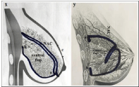
    Figure 1: Schematic illustration of the surgical plan.
x. lateral image of central flap and NAC flap.
y. lateral image of final closure.
Surgical Technique
Ten minutes following the infiltration of 1:500.000 adrenaline/ saline, the overlying skin of the marked superior pedicle and the lower segment of the dermoglandular flap were de-epithelialized Figure 2a. With the use of diathermy connected to a fine Colorado needle, the cutting and dissection of the pedicle was carried out creating a superior pedicle with the Nipple Areola Complex (NAC). The distal part of this flap was 1 cm in thickness. The lower and lateral segments of the dermoglandular flap were dissected from the medial and lateral pillars of the breast but flap were not separated from the pectoral fascia Figure 2b. Dissection was continued underneath the medial and lateral flaps, as well as deep to the central pedicle to create a pocket. A central dermoglandular flap was prepared. This flap was attached to the pectoral fascia superior lateral medial and inferior side by ten stitches between the second and fifth costal region to create a flap-base width of 10 cm and flap projection of 5 cm. The central dermoglandular flap, after attachment by sutures, looks like an implant. Actually, in this technique implant is not used and an implant-like mass is created with autologous tissue Figure 2c. Of note, the term mushroom auto augmentation mastopexy has been coined due to the fact that autologous tissue implanted on the pectoral muscle appears as a mushroom. Medial and lateral flaps were closed after the fixation of the mushroom- or breast implant-shaped tissue on pectoral fascia using permanent sutures. The NAC was sutured to the proposed new site Figure 2d. Only two patients required removal of the submammarian skin. None of the patients had removal of the breast tissue.
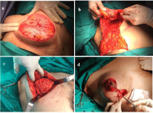
    Figure 2: The intraoperative images of female patient of 35 years-old.
a.Theoverlying skin of the marked superior pedicle and the lower segment of
the dermoglandular flap were deepithelialized.
b. The central segment dermoglandular flap was dissected from the media
land lateral pillars of the breast.
c. Central dermoglandular flap was prepared. The flap was attached to the
pactoral fascia beetween 2. and 5. Costal region (The flap was sutured
superior, lateral, media land inferior side by ten stitches).
d. Final closure.
Results
pOf all patients, the mean Body Mass Index (BMI) was 27.2±3.0 kg/m2 and the mean age was 34 (range: 28 to 42) years. The mean postoperative follow-up was six (range: 3 to 12) months. All patients were highly satisfied in terms of size, shape, projection, and natural appearance at the upper pole of the breasts. Preoperative and postoperative images of the breasts are shown in Figures 3-5. None of the patients had surgery-related complications.
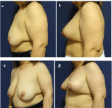
    Figure 3: The oblique and lateral images of female patient of 39 years-old.
a. Preoperative appearances.
b. Postoperative 6. Months appearances
c. Preoperativeappearances.
d. Postoperative 6. Months appearances.
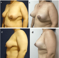
    Figure 4: The oblique and lateral images of female patientof 32 years-old.
a. Preoperative appearances.
b. Postoperative 1. Months appearances.
c. Preoperative appearances.
d. Postoperative 1. Months appearances.
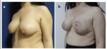
    Figure 5: The oblique images of female patient of 29 years-old.
a. Preoperative appearances.
b. Postoperative 12. Months appearances.
Discussion
Mastopexy of small and medium-size breasts without a decline in size, while improving their projection, and maximizing the fullness of the upper pole, has always been a great challenge for surgeons. In such cases, mastopexy with augmentation of the breast using a mammary implant is reasonable. Followed by others, Gonzales-Ulloa [3-7] introduced the concept of combined augmentation and mastopexy. Over the past few years, there has been an increase in the discussion of augmentation combined with mastopexy in the literature. Fayman [8] published his own technique for auto augmentation. He used inferiorly based dermoglandular flap and transposed it behind the nipple-areolar complex suturing it to the pectoralis fascia. Honig et al. [9] used the same technique and published their satisfactory results. The dermoglandular hammock flap was first described by de la Plaza et al. [10] in 2005. This technique uses a transposition flap to fill the upper and central breast, relocating the lower breast tissue. An upper pedicle dermoglandular flap is raised from the lower pole of the breast and transposed to the upper pole. The flap is fixed like a hammock to the pectoral fascia, and the donor defect, which extends laterally, and is closed by the approximation of the medial and lateral pillars. It makes it possible to augment the upper pole with sagging lower breast tissue and to suspend the whole breast on the pectoral muscle with dermis, improving long-term breast projection and upper pole fullness. In the Gumus modification, the hammock flap design involves a longer and broader flap to reach more easily and effectively to the upper pole through the prepectoral pocket [11]. It also carries more tissue from the lower pole of the breast to the upper pole, increasing the capacity of filling-out both the breast cone behind the NAC and the upper pole of the breast. Through this type of modification, the dermoglandular suspension flap has become a more effective procedure and suitable for all types of ptosis, except for cases of insufficient mammary volumes.
In our study, we performed this new technique in selected patients who had medium-size ptotic breasts and who were unwilling to receive mammary implants. Using this technique, the closure of the medial and lateral pillar flaps was easier, compared to the previously described techniques [8-12]. The application of this method was also simpler, compared to other surgical techniques with sustainable results. As this technique mimics breast implants, high-, mediumand low-profile projections can be easily created as with breast implant. The risk of recurrent ptosis seems to be almost impossible due to use of approximately 10 sutures between the second and fifth ribs. These modifications provide enough permanent central and upper pole fullness and projection and upper pole fullness and breast projection is almost close to the implant results.
In conclusion, using this technique, auto augmentation mastopexy with autologous dermoglandular flaps is a novel, but simple technique which can be used in the lifting of small, medium and large-size breasts.
References
- Regnault P. breast ptosis. Definition and treatment. Clin Plast Surg. 1976; 3: 193-203.
- Wueringer E, Tschabitscher M. New aspects of the topographical anatomy of the mammary gland regarding its neurovascular supply along a regular ligamentous suspension. Eur J Morpho. 2002; 40: 181-189.
- Owsley JQ Jr. Simultaneous mastopexy and augmentation for correction of the small ptotic breast. Ann Plast Surg. 1975; 2: 195.
- Elliott LF. Circumareolar mastopexy with augmentation. Clin Plast Surg. 2002; 29: 337-347.
- Spear SL, Pelletiere CV, Menon N. One-stage augmentation combined with mastopexy: Aesthetic results and patient satisfaction. Aesthetic Plast Surg. 2004; 28: 259-267.
- Gonzales-Ulloa M. Correction of hypotrophy of the breast by exogenous material. Plast Reconstr Surg. 1960; 25: 15.
- Regnault P. The hypoplastic and ptotic breast: A combined operation with prosthetic augmentation. Plast Reconstr Surg. 1966; 37: 31-37.
- Fayman MS. Short Scar Mastopexy with FlapTransposition. Aesth Plast Surg. 1998; 22: 135-141.
- Hönig JF, Frey HP, Hasse FM, Hasselberg J. Auto augmentation Mastopexy with an Inferior-Based Pedicle. Aesth Plast Surg. 2009; 33: 302-307.
- Goés JCS. Periareolar mastopexy: Double skin technique with mesh support. Aesthetic Surg J. 2003; 3: 129-135.
- Gümüs N. A versatile modification of dermo glandular hammock flap for mastopexy: Extended hammock. J Plast Surg Hand Surg. 2013; 47: 252-257.
- Ors S. Auto augmentation mastopexy modification prevents bottoming-out deformity and areola bdistortion: A preliminary report. Aesthetic Plast Surg. 2016; 40: 497-506.
- Balch CR. The central mound technique for reduction mammaplasty. Plast Reconstr Surg. 1981; 67: 305-311
- Hester TR Jr, Bostwick J 3rd, Miller L,Cunningham SJ. Breast reduction utilizing the maximally vascularized central breast pedicle. Plast Reconstr Surg. 1985; 76: 890-900.