Clinical Image
Austin J Anat. 2014;1(2): 1008.
Arrested Pneumatization with Extramedullary Hematopoiesis of the Clivus⁄Sphenoid Simulating Metastatic Carcinoma
Hammer RD* and Butaric LN
Department of Pathology & Anatomical Sciences, University of Missouri, USA
*Corresponding author: Hammer RD, Department of Pathology & Anatomical Sciences, University of Missouri, One Hospital Drive, Columbia, MO 65212, USA.
Received: May 29, 2014; Accepted: May 30, 2014; Published: May 31, 2014
Clinical Image
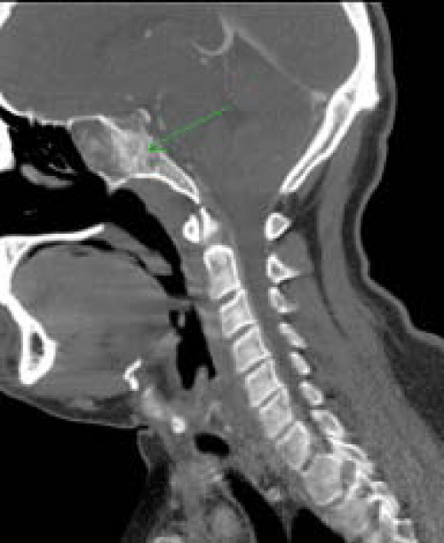
A 37–year–old male presented with bilateral neck masses and lymphadenopathy. The patient was evaluated with ultrasound and CT of the neck. In addition to bilateral masses in the thyroid and multiple enlarged lymph nodes, a destructive lesion involving the clivus and sphenoid sinus was present and initially identified as a metastatic lesion. Subsequent thyroidectomy with neck dissection showed papillary carcinoma with metastatic disease involving multiple bilateral lymph nodes. A biopsy of the bone lesion was performed to r⁄o metastatic disease. The biopsy showed abnormal, curvilinear thin trabeculae that while markedly irregular, lacked osteoclastic activity. The spaces were filled with fat and prominent hypercellular bone marrow, with trilineage hematopoiesis with scattered lymphoid aggregates, some showing reactive follicles. No evidence of metastatic tumor was seen. No underlying hematologic disease was discovered on further analysis. The patient was discharged, with follow–up radiologic studies indicating no metastatic tumors. These findings are consistent with arrested pneumatization and extramedullary hematopoiesis of the cranial base and sphenoidal sinus.
Most of the still developing bones in newborns contain hemapoietic tissue, or red bone marrow, whose main function is erythropoiesis. During relatively well–documented stages of development, adipose tissue within red marrow increases in relative percentage, a process known as red–to–yellow marrow conversion. Among adults, yellow marrow is abundant within medullary cavities of appendicular segments, while red marrow is relatively rare and restricted to the axial skeleton, including some craniofacial bones [1–3]. Although this is the typical adult presentation, yellow marrow can reconvert to red; this is a relatively well–known phenomenon with diseases negatively affecting red–blood–cell function (e.g., leukemia, thalassemia, sickle–cell anemia) [4–8]. This reconversion creates a compensatory site of extra–medullary hematopoiesis. For unclear reasons, even in the absence of pathological evidence as presented here, the red–to–yellow conversion process does not always advance, and red marrow remains into adult–hood.
Within the cranium, this abnormal development is of particular interest because it affects the presentation of normally air–filled structures, confounding clinical interpretations of radiological images [9]. Several studies indicate that pneumatization of the craniofacial bones, such as the paranasal sinuses and mastoid air cells, only occurs after red–to–yellow conversion. If the conversion process does not initiate or complete, pneumatization becomes “arrested,” and the normally aerated area is filled with yellow marrow. More rarely, as reported in the current case, arrested pneumatization results in enduring red marrow, resulting in extramedullary hematopoiesis without accompanying pathologies. Arrested development occurs more frequently the sphenoid sinus, but maxillary [10] and frontal sinus [9] cases have been reported. While this bias toward the sphenoid could relate to lack of recognition inothercranial spaces, there could be an underlying biological cause related to the conversion process itself or sphenoidal anatomy.
In the sphenoid, the red–to–yellow conversion begins anteriorly in the pre–sphenoid, moving posteriorly toward the clivus [3,11–13]. This process can start at 7–months of age, but significant conversion is more common around age 2 [11,13]. As conversion progresses posteriorly, yellow marrow presents in the clivus as early as age 2, more commonly by the age of six (38–48%), and most commonly (90%+) by 15 years–of–age [12]. This anterior–posterior marrow conversion is closely related to pneumatization of the sphenoid body [14]. Unlike the other sinuses, the sphenoid sinus is not yet present in its respective bone at birth [15]. Although pneumatization into the sphenoid may start by 15 months (12% of individuals), it more commonly begins around 43–48 months (85%) [13]– closely approximating the timing of significant yellow conversion. Further pneumatization into the sphenoid occurs with resorptionin the sphenoidal body at age 6, extension into the hypophyseal fossa by age 8, and expansionto the adult form by age 12 [15].
Figure 1: CT shows a lesion in the clivusextending into the sphenoid sinus which is opacified and measures 4.3 cm in maximum dimension. In addition, bilateral masses were present in the thyroid with multiple nodal lesions (not shown). The lesion in the clivus was felt to possibly represent a metastatic lesion.
Although several hypotheses, invoking temperature [16], bone composition (i.e., the amount of trabecular versus cortical bone; [17], and⁄or vasculature changes [13,18], attempt to explain why the red–to–yellow conversion process occurs, relatively little is known about this process and why it sometimes “fails.” These hypotheses are of interest to the current case because each is likely affected by several unique attributes related to the development of the sphenoid sinus and its surrounding bone. Compared to the other craniofacial bones, the sphenoid 1) develops through endochondral, not intramembranous, ossification, 2) joins to the neighboring occiput via a synchrondosis, versus a suture, and 3) is the last of the bones whose paranasal sinuses pneumatize its space. Although beyond the scope of this report, additional studies analyzing the unique vasculature and developmental patterns associated with these specific sphenoidal attributes may provide clues into the conversion process itself and why the sphenoidal sinus shows higher frequencies of arrested development.
References
- Piney A. The Anatomy of the Bone Marrow: With Special Reference to the Distribution of the Red Marrow. Br Med J II. 1922; 2:792-795.
- Kricun ME. Red-yellow marrow conversion: its effect on the location of some solitary bone lesions. Skeletal Radiol. 1985; 14: 10-19.
- Taccone A, Oddone M, Occhi M, Dell’Acqua, A, Ciccone MA. MRI “Road-Map” of Normal Age-Related Bone Marrow. I. Cranial Bone and Spine. Pediat Radiol. 1995; 25:558-595.
- Collins WO, Younis RT, Garcia MT. Extramedullary hematopoiesis of the paranasal sinuses in sickle cell disease. Otolaryngol Head Neck Surg. 2005; 132: 954-956.
- Kulendra K, Butler C, Grant W, Sandison A, Cho G, Patel MC. Unusual clivus lesion demonstrating extramedullary haematopoiesis: case report. J Laryngol Otol. 2009; 123: e15.
- Stamataki S, Behar P, Brodsky L. Extramedullary Hematopoiesis in the Maxillary Sinus. Int J Ped Otorhinolaryngology Extra. 2009; 4: 32-35.
- Sklar M, Rotaru C, Grynspan D, Bromwich M. Radiographic features in a rare case of sphenoid sinus extramedullary hematopoeisis in sickle cell disease. Int J Pediatr Otorhinolaryngol. 2013; 77: 294-297.
- Sohawon D, Lau KK, Lau T, Bowden DK. Extra-medullary haematopoiesis: a pictorial review of its typical and atypical locations. J Med Imaging Radiat Oncol. 2012; 56: 538-544.
- Scuderi AJ, Harnsberger HR, Boyer RS. Pneumatization of the paranasal sinuses: normal features of importance to the accurate interpretation of CT scans and MR images. AJR Am J Roentgenol. 1993; 160: 1101-1104.
- Kuntzler S, Jankowski R. Arrested pneumatization: Witness of paranasal sinuses development? Eur Ann Otorhinolaryngol Head Neck Dis. 2014.
- Aoki S, Dillon WP, Barkovich AJ, Norman D. Marrow conversion before pneumatization of the sphenoid sinus: assessment with MR imaging. Radiology. 1989; 172: 373-375.
- Okada Y, Aoki S, Barkovich AJ, Nishimura K, Norman D, Kjos BO, et al. Cranial bone marrow in children: assessment of normal development with MR imaging. Radiology. 1989; 171: 161-164.
- Szolar D, Preidler K, Ranner G, Braun H, Kern R, Wolf G, et al. Magnetic resonance assessment of age-related development of the sphenoid sinus. Br J Radiol. 1994; 67: 431-435.
- Welker KM, DeLone DR, Lane JI, Gilbertson JR. Arrested Pneumatization of the Skull Base: Imaging Characteristics. Am J Roentgenol. 2008; 190:1691-1696.
- Weiglen AH. Development of the paranasal sinuses. Koppe T, Nagai H, Alt KW, editors. In: The Paranasal Sinuses of Higher Primates: Development, Function, and Evolution. Quintessence Publishing Co, Inc. 1999; 35-50.
- Huggins C, Blocksom BH. Changes in outlying bone marrow accompanying a local increase of temperature within physiological limits. J Exp Med. 1936; 64: 253-274.
- Gurevitch O, Slavin S, Feldman AG. Conversion of red bone marrow into yellow - Cause and mechanisms. Med Hypotheses. 2007; 69: 531-536.
- Yonetsu K, Watanabe M, Nakamura T. Age-related expansion and reduction in aeration of the sphenoid sinus: volume assessment by helical CT scanning. AJNR Am J Neuroradiol. 2000; 21: 179-182.
