
Mini Review
Austin J Anat. 2014;1(2): 1010.
Histomorphology of Metaphysis of Proximal Tibia in Albino Rat
Sarah Ralte*
Department of Anatomy, North Eastern Indira Gandhi Regional Institute of Health and Medical Sciences, India
*Corresponding author: Sarah Ralte, Department of Anatomy, North Eastern Indira Gandhi Regional Institute of Health and Medical Sciences (NEIGRIHMS), Mawdiangdiang, Shillong-793018, Meghalaya, India.
Received:June 01, 2014; Accepted: July 15, 2014; Published: July 18, 2014
Abstract
The metaphysis lies at the distal end of the growth plate, between the epiphysis and diaphysis of bone and is the zone of active growth in a long bone. In this review study, the continuity of metaphysis of proximal tibia in albino rat was studied by light microscope following Hematoxylin and Eosin and Masson’s trichrome stains. The metaphysis has been described as having two distinct regions, the primary and secondary spongiosa, containing mainly osteoblasts and osteoclasts amidst abundant blood vessels. The osteoblasts are the bone forming cells whereas the osteoclasts are the chief cells mediating bone resorption. Hence, the metaphysis is the site of active bone remodelling. Before the fusion of diaphysis and epiphyses, the metaphyses are richly supplied with blood through end arteries forming hair pin bends. This is the common site of osteomyelitis in children. The bone cells are the favourite target site of action of drugs, especially the bisphosphonate class of drugs, which are potent inhibitors of excessive osteoclastic mediated bone resorption.
Keywords: Metaphysis; Primary and Secondary spongiosa; Osteoblast; Osteoclast; Bone remodelling; Tibia; Bisphosphonates
Introduction
The metaphysis is the junctional region of bone lying between the growth plate and the diaphysis. It contains slender calcified cartilage spicules and trabecular bone and is a site of active bone turnover, having large number of osteoblasts, osteoprogenitor cells and osteoclasts amidst highly vascular tissue. The metaphysis is invaded by numerous capillary loops containing osteoblastic cells, which deposit bony matrix on calcified cartilage spicules. The metaphysis is divided into two functionally distinct regions, the primary spongiosa, the area lying adjacent to the growth plate, which is rich in blood capillaries and is the site where primary spongy bone forms, which is characterized by calcified cartilage spicules covered by a thin layer of newly laid bone. The other region lying adjacent to diaphysis is the secondary spongiosa, characterized by interconnecting bony bars of trabeculae. Here, the calcified cartilage spicule is ultimately resorbed by the osteoclast, and is the site of active bone remodelling. Osteoclasts, the giant multinucleated cells, are the bone resorbing cells which resorb the mineralized bone by secreting acids and lysosomal enzymes. Increased activation of osteoclasts results in disruption of normal bone remodelling resulting in increased resorption of the bone [1]. The region of metaphysis is a common site for primary bone tumours and bone infections such as osteomyelitis. The relative predilection of osteosarcoma for the metaphyseal region of long bones in children has been attributed to the rapid bone turnover due to extensive bone remodelling during growth spurts [2]. The effects of large number of drugs have also been studied on the metaphysis of long bone [3-8]. The bisphosphonates are one of the important classes of drugs which act mainly on the bone cell, the osteoclast, and are effective inhibitors of excess osteoclastic mediated bone resorption [9]. The uses of bisphosphonates currently include the prevention and treatment of osteoporosis, osteitis deformans, bone metastasis (with or without hypercalcaemia), multiple myeloma, primary hyperparathyroidism and osteogenesis imperfecta [10- 15].
Histomorphology of Metaphysis by Light Microscope
The histomorphology of metaphysis of proximal end of tibia was studied in young albino rats by light microscope following staining with Hematoxylin and Eosin and Masson’s trichrome. The metaphysis was identified as the area lying adjacent to the lower margin of the epiphyseal growth plate and limited on the sides by the periosteum of the bone. The proximal margin of the metaphysis towards the growth plate was observed to be wider and more or less concavo-convex in comparison to its distal margin, which was narrow and irregular in outline. It was noted that the height of the lateral region was always more than the central region, giving a more or less concave appearance to the distal aspect of the metaphysis (Figure 1).
In Haematoxylin and Eosin stained sections, two distinct regions of the metaphysis were observed. The primary spongiosa, the area lying near the growth cartilage metaphyseal junction (GCMJ), was seen as a network of irregular, fine, thin, longitudinally oriented trabeculae. The trabeculae were connected to each other at places and separated by narrow marrow spaces containing haemopoeitic tissue stained deep purple. The area near the GCMJ was characterized by a scarcity of bone marrow cells when compared to tissue found further from the GCMJ. The trabeculae were composed largely of calcified cartilage spicules covered with a thin layer of bone, the amount of bone increasing as distance from the GCMJ increased. The calcified cartilage spicules were stained darkly eosinophilic while the bony trabeculae appeared light eosinophilic (Figure 1). A large number of capillaries were seen between the calcified cartilage septae. The trabecular surfaces were lined by numerous osteoblasts and occasional osteoclasts. In Masson’s trichrome stain, the bony trabeculae appeared dark blue while the central cores of calcified cartilage were stained light blue colour. At the junction of growth plate and metaphysis, most of the horizontal calcified walls of the lacunae of degenerating hypertrophied cartilage cells were partially eroded and longitudinally oriented calcified walls of lacunae were seen to be invaded by haemopoietic tissue, osteoprogenitor cells, osteoblasts and osteoclasts (Figure 2). The haemopoietic tissues were stained brownish red while the red blood cells took on a bright orange red colour. The secondary spongiosa, the region of metaphysis lying away from the GCMJ, was characterized by large trabeculae composed mainly of bone with occasional central cores of calcified cartilage. The newly formed bone appeared as bars of bony trabeculae of variable width and length. They were interconnected to each other at places and were arranged in a longitudinal direction (Figure 1 & 4). The surfaces of trabeculae were covered with osteoblast, osteoprogenitor cells and osteoclasts. Large marrow spaces containing marrow cells and red blood cells were seen between the trabeculae, the marrow spaces increasing further as the trabeculae of the secondary spongiosa extended towards the diaphyseal region. The occasional osteoprogenitor cells seen in secondary spongiosa were usually located adjacent to the bone surfaces. It was characterized by a poorly visualized cytoplasm. Its nucleus was its most prominent feature, being ovoid to spindle shaped, euchromatic and pale staining in appearance. Under Hematoxylin and Eosin stain, the osteoblasts were seen as a single layer of cells lining the surfaces of trabeculae and appeared cuboidal, polygonal or spindle-shaped cells having a basophilic cytoplasm with an oval, euchromatic nucleus with a single nucleolus were seen lying at one end while a prominent clear area was found at the other end of the cell. The osteoclasts were seen lying between the capillaries and trabeculae composed mainly of calcified cartilage cores. These cells were large, irregular, polymorphus and multi-nucleated with a varying number of closely packed nuclei. The nuclei were randomly placed in the foamy eosinophilic cytoplasm and were found to be round to ovoid, purple stained and usually a single nucleolus were seen in most cells while more than one nucleoli were seen in some cells. The osteoclasts were often seen lying in depressions or pits resorbed from the bone surfaces. The area surrounding the osteoclast closely applied to the trabecular surface was seen as a clear zone at places and fine tooth like extensions projecting from the cytoplasmic membrane were seen (Figure 3). The osteoclasts were found lying against the bone surfaces or sometimes lying freely within the narrow spaces. In Masson’s trichrome stain, the cytoplasm appeared reddish brown while the nuclei were stained brownish black. Oval to spindle shaped osteocytes were seen lying in clear spaces called lacunae embedded within the matrix of the trabeculae (Figure 4).
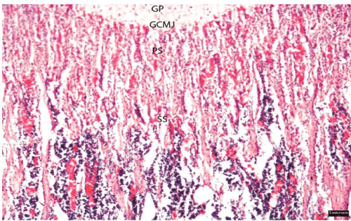
Photomicrograph of a longitudinal section of proximal metaphysis of tibia of rat showing the primary spongiosa (PS) and secondary spongiosa (SS), lying adjacent to the growth cartilage metaphyseal junction (GCMJ; 5 microns bar). Hematoxylin and Eosin (H&E 40X).
Abbreviations: GP-growth plate; GCMJ-growth cartilage metaphyseal junction; PS-primary spongiosa; SS-secondary spongiosa.
Figure 1:Photomicrograph of a longitudinal section of proximal metaphysis of tibia of rat showing the primary spongiosa (PS) and secondary spongiosa (SS), lying adjacent to the growth cartilage metaphyseal junction (GCMJ; 5 microns bar). Hematoxylin and Eosin (H&E 40X).
Abbreviations: GP-growth plate; GCMJ-growth cartilage metaphyseal junction; PS-primary spongiosa; SS-secondary spongiosa.
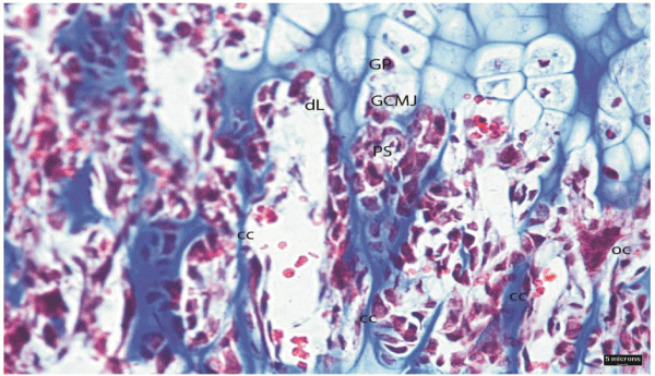
Photomicrograph of primary spongiosa (PS) of proximal metaphysis of rat showing clearly partially eroded, degenerating walls of lacunae (dL) adjacent to the GCMJ invaded by haemopoietic tissue. Pyknotic nuclei of degenerating chondrocytes are also seen in the growth plate (GP). The right margin of photograph shows a distinct multinucleated osteoclast (oc) with 4-5 nuclei. Osteoclast appears reddish brown while the nucleus is stained brownish black (5 microns bar). Masson’s trichrome (200X).
Abbreviations: GP-growth plate; GCMJ-growth cartilage metaphyseal junction; PS-primary spongiosa; cc-calcified cartilage spicule; oc-osteoclast; dL-degenerating lacunae.
Figure 2:Photomicrograph of primary spongiosa (PS) of proximal metaphysis of rat showing clearly partially eroded, degenerating walls of lacunae (dL) adjacent to the GCMJ invaded by haemopoietic tissue. Pyknotic nuclei of degenerating chondrocytes are also seen in the growth plate (GP). The right margin of photograph shows a distinct multinucleated osteoclast (oc) with 4-5 nuclei. Osteoclast appears reddish brown while the nucleus is stained brownish black (5 microns bar). Masson’s trichrome (200X).
Abbreviations: GP-growth plate; GCMJ-growth cartilage metaphyseal junction; PS-primary spongiosa; cc-calcified cartilage spicule; oc-osteoclast; dL-degenerating lacunae.
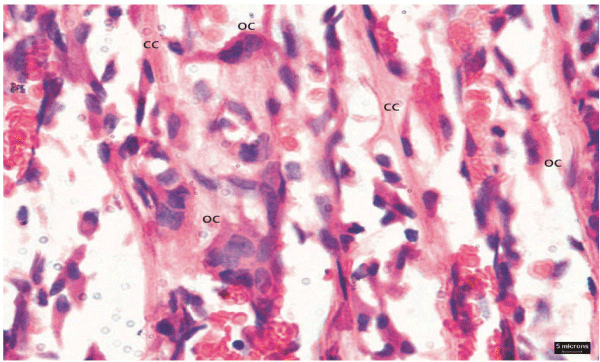
Photomicrograph from proximal tibial metaphysis of rat showing osteoclasts (oc) and trabeculae (trab) mainly composed of calcified cartilage (cc) cores. The osteoclasts show fine tooth like cytoplasmic projections extending towards the trabecular surface. The cytoplasm of osteoclasts is stained darkly eosinophilic with purple nuclei. The osteoblast appears cuboidal or spindle shaped (5 microns bar). Hematoxylin and Eosin (H&E 400X).
Abbreviations: oc-osteoclast; cc-calcified cartilage core.
Figure 3:Photomicrograph from proximal tibial metaphysis of rat showing osteoclasts (oc) and trabeculae (trab) mainly composed of calcified cartilage (cc) cores. The osteoclasts show fine tooth like cytoplasmic projections extending towards the trabecular surface. The cytoplasm of osteoclasts is stained darkly eosinophilic with purple nuclei. The osteoblast appears cuboidal or spindle shaped (5 microns bar). Hematoxylin and Eosin (H&E 400X).
Abbreviations: oc-osteoclast; cc-calcified cartilage core.
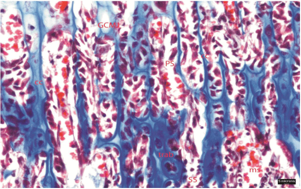
Photomicrograph of secondary spongiosa (SS) of proximal metaphysis of tibia showing areas of calcified cartilage (cc) cores stained light blue covered by bone stained dark blue. The trabeculae (trab) are seen interconnecting with each other at places (5 microns bar). Masson’s trichrome (200X).
Abbreviations: GCMJ-growth cartilage metaphyseal junction; PS-primary spongiosa; SS-secondary spongiosa; cc-calcified cartilage core; trabtrabeculae; ms-medullary space.
Figure 4:Photomicrograph of secondary spongiosa (SS) of proximal metaphysis of tibia showing areas of calcified cartilage (cc) cores stained light blue covered by bone stained dark blue. The trabeculae (trab) are seen interconnecting with each other at places (5 microns bar). Masson’s trichrome (200X).
Abbreviations: GCMJ-growth cartilage metaphyseal junction; PS-primary spongiosa; SS-secondary spongiosa; cc-calcified cartilage core; trabtrabeculae; ms-medullary space.
As early as in 1925, Stump [16] gave one of the earliest qualitative descriptions of the metaphysis of long bone obtained from celloidin embedded long bones of mice, rats, rabbits and sheep. The role of the calcified cartilage in providing longitudinally oriented scaffolding on which osteoblasts would form primary bone was studied. A syncitial cell population termed the ‘osteogenic mesenchyme’, was observed located adjacent to the growth cartilage metaphyseal junction (GCMJ), the point of cartilage lacunar opening and the end of the life of individual chondrocytes. The osteogenic mesenchyme was associated with a relatively high level of multiplication of young connective tissue cells and was found to invade and occupy the spaces which were earlier occupied by chondrocytes. In the osteogenic mesenchyme both osteoblasts and osteoclasts were observed.
Kimmel and Jee [17] studied the bone cell kinetics in the proximal tibial metaphysis in the rat following injection of triatiated thymidine. Animals were sacrificed 1 to 120 hrs. Labelled osteoprogenitor cells and osteoblasts first appeared at 1 hour post-injection within 1 mm of growth cartilage metaphyseal junction (GCMJ), while labelled osteoclast nuclei first appeared at 24 hours post injection within 0.3 mm of GCMJ and was found to be depleted 5 days later, whereas that for the osteoblasts remained. The metaphysis was identified as two regions: primary spongiosa, located within 0.756 mm of the GCMJ with an average age of 4.45 days or less, as a region of high turnover of hard tissue and high numbers of osteoblasts and osteoprogenitor cells. Osteoclasts were found relatively more uniformly distributed through the metaphysis than were osteoblasts and osteoprogenitor cells. The calcified cartilage disappeared at the rate of 0.0359mm2/ day; whereas bone was added at the net rate of 0.0412 mm2/day in the primary spongiosa, the only region of net addition of bone to the metaphysis. The second area composed of tissue located 1.188 mm or further from the GCMJ with an average age of 7 days or more, was a zone of much slower tissue turnover, corresponding to the secondary spongiosa. They observed a net loss of hard tissue, at a rate of 0.0090mm2/day and the rate of loss of calcified cartilage at 0.0014 mm2/day, which was about 20 times slower than the rate of loss found in the primary spongiosa. The lower rate of turnover was correlated with the smaller number of osteoblasts and osteoprogenitor cells located in the secondary spongiosa compared to the primary spongiosa. Most osteoblasts and osteoprogenitor cells were found near the GCMJ where bone was being added, while the osteoclasts were found somewhat more scattered though the metaphysis, being relatively more numerous than osteoblasts in areas characterized by net loss of bone.
In a study on the micro vascular pattern of the metaphysis during bone growth, Aharinejid et al [18] observed that calcified cartilage formed the wall of the cylindrical compartments beneath the hypertrophied chondrocytes of the metaphyseal growth plate. These compartments ran in the bone’s longitudinal axis and contained a single capillary profile. Endothelial cells of these capillaries often showed increased cytoplasmic volume and loose texture of nuclear chromatin. Cast metaphyses by scanning electron microscope showed numerous parallel vascular loops with nodular protrusions 10–12µm in diameter at their tips. The loops had ascending and descending limbs with a luminal diameter of 10–14μm. Small projections 4–5μm in diameter and delicate crests were sometimes found on the tip of the larger nodes. In a 100 × 100µm area, there were 14–17 large nodes. By transmission electron microscope, capillary sprouts were identified at the level beneath the last row of hypertrophied chondrocytes. These capillaries had voluminous endothelial cells rich in free ribosomes and rough endoplasmic reticulum. Endothelial cell nuclei were rounded and showed loose chromatin texture. Endothelial cells were connected by intermediate junctions and there was no basal lamina. Deeper into the metaphysis, arterioles and sinusoids were observed.
As the metaphysis is a site of active bone remodelling containing mainly osteoblasts which are the bone forming cells and osteoclasts, the bone resorbing cells, it is therefore the favourite target area of bone in animal studies for observing the efficacy and potency of various dugs in experimental research and trial studies [19-24]. Animal studies have been conducted in the past to study the effects of various drugs specially bisphosphonates on the metaphysis of growing bones in rats [25, 26] and pigs [27]. As early as 1979, Miller and Jee [25] observed a marked increase in metaphyseal mineralized tissue mass as a result of slowed bone resorption in proximal tibia of rats following short-term administration of clodronate (a first generation bisphosphonate). A dramatic increase in the amount of trabecular bone was seen as compared to controls. Long term treatment of risedronate in ovariectomized induced osteopenic rats resulted in an increase in bone mass in the proximal tibial metaphysis and prevention of further bone loss in ovariectomized rats by depressing the bone resorption and turnover [26]. In pigs, long term administration of pamidronate (a second generation bisphosphonate) has shown a significant increase in trabecular bone volume and density, with a marked decrease in bone resorption [27]. Past studies have shown that bisphosphonates are highly effective in preserving bone mass in estrogen deficient rats [28] and ovariectomized adult rhesus monkey [29, 30]. Pataki et al. [31] observed that zoledronate, a third generation bisphosphonate, was highly effective in accumulating bone mass, preserving the architecture and strength than other bisphosphonates in growing rats. Bisphosphonates have now emerged as a leading therapeutic intervention for the treatment and prevention of skeletal complications of malignancy (skeletal related events) and are the treatment of choice for hypercalcaemia of malignancy, Paget’s disease of bone and postmenopausal osteoporosis [11-15, 32, 33].
References
- Ovalle WK, Nahirney PC. Netter's Essential Histology. 2nd edn. Philadelphia: Elsevier Saunders. 2013.
- De la Roza G. Histology of Bone.
- young mh, crane wa. effect of hydrocortisone on the utilization of tritiated thymidine for skeletal growth in the rat. ann rheum dis. 1964; 23: 163-168.
- Whalen JP, Krook L, MacIntyre I, Nunez EA. Calcitonin, parathyroidectomy and modelling of bones in the growing rat. J Endocrinol. 1975; 66: 207-212.
- Turner RT, Vandersteenhoven JJ, Bell NH. The effects of ovariectomy and 17 beta-estradiol on cortical bone histomorphometry in growing rats. J Bone Miner Res. 1987; 2: 115-122.
- Ma Y, Chen YY, Jee WS, Ke HZ, Ijiri K. Co-treatment of PGE2 and risedronate is better than PGE2 alone in the long-term treatment of ovariectomized-induced osteopenic rats. Bone. 1995; 17: 267S-272S.
- Turan B, Balcik C, Akkas N. Effect of dietary selenium and vitamin E on the biomechanical properties of rabbit bones. Clin Rheumatol. 1997; 16: 441-449.
- Ralte S, Khatri K, Nagar M. Short-term effects of zoledronate on the histomorphology of osteoclast in young albino rats. Ann Anat. 2011; 193: 509-515.
- Fleisch H. Bisphosphonates: mechanisms of action. Endocr Rev. 1998; 19: 80-100.
- Black DM, Delmas PD, Eastell R, Reid IR, Boonen S, Cauley JA, et al. Once-yearly zoledronic acid for treatment of postmenopausal osteoporosis. N Engl J Med. 2007; 356: 1809-1822.
- Wells G, Cranney A, Peterson J, Boucher M, Shea B, Welch V, et al. Risedronate for the primary and secondary prevention of osteoporotic fractures in postmenopausal women. Cochrane Database of Systematic Reviews (1). 2008; CD004523.
- Drake MT, Clarke BL, Khosla S. Bisphosphonates: mechanism of action and role in clinical practice. Mayo Clin Proc. 2008; 83: 1032-1045.
- Russell RG, Watts NB, Ebetino FH, Rogers MJ. Mechanisms of action of bisphosphonates: similarities and differences and their potential influence on clinical efficacy. Osteoporos Int. 2008; 19: 733-759.
- Gnant M, Mlineritsch B, Schippinger W, Luschin-Ebengreuth G, Pöstlberger S, Menzel C, et al. Endocrine therapy plus zoledronic acid in premenopausal breast cancer. N Engl J Med. 2009; 360: 679-691.
- Shapiro JR, Sponsellor PD. Osteogenesis imperfecta: questions and answers. Curr Opin Pediatr. 2009; 21: 709-716.
- Stump CW. The Histogenesis of Bone. J Anat. 1925; 59: 136-154.
- Kimmel DB, Jee WS. A quantitative histologic analysis of the growing long bone metaphysis. Calcif Tissue Int. 1980; 32: 113-122.
- Aharinejad S, Marks SC Jr, Böck P, MacKay CA, Larson EK, Tahamtani A, et al. Microvascular pattern in the metaphysis during bone growth. Anat Rec. 1995; 242: 111-122.
- Halasy-Nagy JM, Rodan GA, Reszka AA. Inhibition of bone resorption by alendronate and risedronate does not require osteoclast apoptosis. Bone. 2001; 29: 553-559.
- Marie PJ. Strontium ranelate: a dual mode of action rebalancing bone turnover in favour of bone formation. Curr Opin Rheumatol. 2006; 18: S11- S15.
- Ke HZ, Jee WS, Mori S, Li XJ, Kimmel DB. Effects of long-term daily administration of prostaglandin-E2 on maintaining elevated proximal tibial metaphyseal cancellous bone mass in male rats. Calcif Tissue Int. 1992; 50: 245-252.
- Jee WS, Tang L, Ke HZ, Setterberg RB, Kimmel DB. Maintaining restored bone with bisphosphonate in the ovariectomized rat skeleton: dynamic histomorphometry of changes in bone mass. Bone. 1993; 14: 493-498.
- Wronski TJ, Yen CF, Qi H, Dann LM. Parathyroid hormone is more effective than estrogen or bisphosphonates for restoration of lost bone mass in ovariectomized rats. Endocrinology. 1993; 132: 823-831.
- Chen HK, Jee WS, Ma YF, Pan Z, McOsker JE, Li XJ. Intermittent treatment of prostaglandin E2 with risedronate is more anabolic than prostaglandin E2 alone in the proximal tibial metaphysis of ovariectomized rats. Bone. 1995; 17: 285S-289S.
- Miller SC, Jee WS. The effect of dichloromethylene diphosphonate, a pyrophosphate analog, on bone and bone cell structure in the growing rat. Anat Rec. 1979; 193: 439-462.
- Ma Y, Chen YY, Jee WS, Ke HZ, Ijiri K. Co-treatment of PGE2 and risedronate is better than PGE2 alone in the long-term treatment of ovariectomized-induced osteopenic rats. Bone. 1995; 17: 267S-272S.
- Vernejoul de, Pointillart A, Bergot C, Bielakoff J, Morieux C, Laval Jeantet AM, et al. Different schedules of administration of pamidronate diphosphonate induce different changes in pig bone remodelling. Calcif Tissue Int. 1987; 40: 160-165.
- Glatt M. The bisphosphonate zoledronate prevents vertebral bone loss in mature estrogen-deficient rats as assessed by micro-computed tomography. Eur Cell Mater. 2001; 1: 18-26.
- Binkley N, Kimmel D, Bruner J, Haffa A, Davidowitz B, Meng C, et al. Zoledronate prevents the development of absolute osteopenia following ovariectomy in adult rhesus monkeys. J Bone Miner Res. 1998; 13: 1775-1782.
- Bare S, Kimmel D, Binkley N. Zoledronate suppresses turnover in cortical bone of the ovariectomized non-human primate. J Bone Miner Res. 1997; 12(Suppl): S473.
- Pataki A, Muller K, Green JR, Ma YF, Li QN, Jee WS. Effects of short term treatment with bisphosphonates, zoledronate and pamidronate on bone: a comparative histomorphometric study on the cancellous bone formed before, during and after treatment. Anat Rec. 1997; 249: 458-468.
- Body JJ, Bartl R, Burckhardt P, Delmas PD, Diel IJ, Fleisch H, et al. Current use of bisphosphonates in oncology. International Bone and Cancer Study Group. J Clin Oncol. 1998; 16: 3890-3899.
- Rossini M, Gatti D, Girardello S, Braga V, James G, Adami S. Effects of two intermittent alendronate regimens in the prevention or treatment of postmenopausal osteoporosis. Bone. 2000; 27: 119-122.