Case Report
Austin J Anat. 2014;1(3): 1011.
Median Nerve Arising from Three Roots
Herath HMLY, Raayiz RM, Rathnayake RMCH, Sandamali SPU, Amaratunga HA and Adikari SB*
Department of Anatomy, University of Peradeniya, Sri Lanka
*Corresponding author: Dr. S B Adikari,Senior Lecturer, Department of Anatomy, Faculty of Medicine, University of Peradeniya, Sri Lanka.
Received:May 20, 2014; Accepted: July 14, 2014; Published: July 18, 2014
Abstract
The brachial plexus is formed by the anterior rami of the lower four cervical and the first thoracic spinal nerves. The median is one of the five major nerves arising from the brachial plexus. It is usually formed by the union of two roots, one from the medial and the other from the lateral cord of the brachial plexus. During routine dissection of the axilla in a male cadaver, it was observed that the median nerves of both sides were formed by three roots. One root arose from the medial cord and the other two from the lateral cord. Knowledge of such variations is useful for doctors in their clinical practice in order to understand the anatomical basis of complicated clinical symptoms and signs.
Keywords: Brachial plexus; Median nerve; Medial root; Lateral root
Case Presentation
During a routine dissection of the axilla at the Department of Anatomy, a variation in the formation of the median nerves was identified. In contrast to the normal pattern, the median nerves of both sides were formed by three roots. One root arose from the medial cord and the other two were from the lateral cord [1]. No other variations in the brachial plexus were identified.
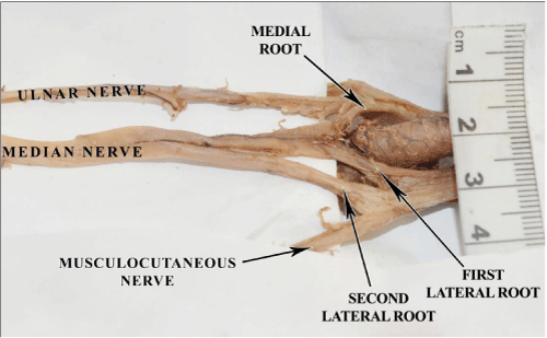
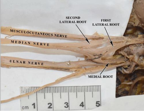
In the left axilla, the medial root was joined by one lateral root to form the main trunk of the median nerve and this was joined by a second lateral root which was given off from the lateral cord about 1cm distal to the first root. The second root was 4cm long and it joined the median nerve 2.5cm distal to the union of the medial root and the first lateral root (Figure 1). In the right axilla the arrangement was similar except the second root joined the median nerve just distal to the union of the medial root and the first lateral root (Figure 2).
Figure 1: Left axilla.
Figure 2: Right axilla.
Discussion
There are many reported cases of variations in the formation of the median nerve and up to four roots can contribute to its formation as opposed to the usual two roots. In a study done on 172 cadavers, Panday and Shukla [2] report that in 7% of cases the medial root received communicating branches from the lateral or posterior cord in the formation of the median nerve. In another study by Choi et al. [3] done on 138 cadavers unusual communications between the musculocutaneous and median nerves were reported in 64 cadavers, 9 bilaterally and 55 unilaterally [4].
The anatomical variations of the musculocutaneous and the median nerve in the arm have been classified into 5 types by Le Minor JM [5].
Type 1: There are no communicating fibres between the musculocutaneous and the median nerves. The musculocutaneous nerve pierces the coracobrachialis muscle and innervates the coracobrachialis, biceps brachii and brachialis muscles.
Type 2: Although some fibres of the medial root of the median nerve unite with the lateral root of the median nerve to form the median nerve, some leave to run within the musculocutaneous nerve and after some distance leave it to join their proper trunk.
Type 3: The lateral root of the median nerve runs into the musculocutaneous nerve and, after some distance, leaves it to join its proper trunk.
Type 4: The fibers of the musculocutaneous nerve unite with the lateral root of the median nerve and, after some distance, emanate from the median nerve.
Type 5: The musculocutaneous nerve is absent. Its fibers run within the median nerve along its course.
The present case describes a Type 2 variation which is the commonest type.
Some authors have described the presence of a small lateral root as the cause for such variations, where the fibers of the median nerve take an unusual course over a short distance along the lateral cord or musculocutaneous nerve to reach the median nerve as additional roots.
Conclusion
Variations of brachial plexus may be responsible for clinical symptoms such as unexplained pain, paresthesia and weakness which may challenge clinicians to arrive at a diagnosis. Nerves with such variations are also vulnerable to damage during routine surgeries on the axilla, radical neck dissections and surgeries in the upper arm.
References
- Gangadhara, Jayanthi V. Variation in the Formation of Right Median Nerve - A Case Report. Anatomica Karnataka. 2011; 5: 24-27.
- Pandey SK and Shukla VK. Anatomical variations of the cords of brachial plexus and the median nerve. Clin. Anat. 2007; 20: 150–156.1
- Choi D, Rodríguez-Niedenführ M, Vázquez T, Parkin I, Sañudo JR. Patterns of connections between the musculocutaneous and median nerves in the axilla and arm. Clin Anat. 2002; 15: 11-17.
- Priti B Khodke, Medha V Ambiye, Seema Khambatta. Unilateral variant median nerve – a case report. Int J Anat Var (IJAV). 2013; 6: 210–212.
- Le Minor JM. A rare variation of the median and musculocutaneous nerves in man. Arch Anat Histol Embryol. 1990; 73: 33–42.

