Mini Review
Austin J Anat. 2024; 11(1): 1112.
Adamantinoma-Like Ewing’s Sarcoma: Misleading Tumor Expressing P40
Imane Boujguenna1,2; Aissam Griche3; Malak El Merrakchi3; Said Ait Benali3; Hanane Rais1
¹Department of Pathological, Anatomy FMPM-UCAM-CHU Mohammed VI Marrakech, Morocco
²Faculty of Medicine and Pharmacy of Guelmim, Ibn Zohr University, Morocco
³Departmen of Neurosurgery, FMPM-UCAM-CHU Mohammed VI Marrakech-Morocco
*Corresponding author: Imane Boujguenna Department of Pathological Anatomy FMPM-UCAM-CHU Mohammed VI Marrakech-Morocco. Tel: 212613243083 Email: imane.boujguenna.fmpg@gmail.com
Received: March 05, 2024 Accepted: April 11, 2024 Published: April 18, 2024
Abstract
Adamantinoma-like Ewing’s sarcoma is a rare variant of the Ewing family of tumors that resembles classic bone adamantinoma. Adamantinoma-like Ewing’s sarcoma shows epithelial differentiation and a more complex immunohistochemical expression profile with keratin immunoreactivity and squamous markers and can resemble a variety of carcinomas.
We report an inhabited case of Ewing’s sarcoma of the vertebral adamantinoma type which mimicked a metastatic localization of squamous cell carcinoma. Like squamous cell carcinomas, this adamantinoma-type Ewing’s sarcoma presents as a basaloid proliferation with a peripheral palisade and a “basaloid” epithelial differentiation demonstrated by cytokeratin AE1 / 3 and p40 positivity. However, unlike most basal cell and epidermoid adenocarcinomas, this tumor presents a high-grade morphology and a tendency to neuroectodermal phenotype with absence of ductal or myoepithelial component and immunoreactivity to CD99. EWSR1 fluorescence in situ hybridization confirmed the presence of a translocation supporting the diagnosis of adamantinoma-like Ewing’s sarcoma.
Keywords: Spinal tumor; p40; CD99; Ewing Adamantinoma
Introduction
The Ewing tumor family is a very rare group of sarcomatous tumors affecting bones and soft tissues. The molecular anomaly (11; 22) (q24; q12), which involves the EWSR1 and FLI-1 genes, is common to these tumors and their variants [1,2]. They usually affect children and young adults. About 5% of Ewing's sarcomas occur in the head and neck and have recently been recognized in the nasosinus tract, parotid gland, thyroid gland and orbit [2].
The adamantinoma variant of Ewing tumors shows histomorphologic and / or immunophenotypic evidence of squamous differentiation. The histologic appearance of this morphologic variant overlaps completely with squamous cell carcinomas of the head and neck region. It may also have a complex immune profile, characterized by diffuse reactivity to CK5 / 6, p40 and p63 [3,4].
Because of these characteristics, the diagnosis of Ewing tumors and their morphological variants always relies on a correlation, notably of morphology, immunohistochemistry (such as CD99 and FLI-1 positivity) and characteristic molecular abnormalities [2]. We report a case of ewing sarcoma of the para-vertebral adamantinoma type with epithelial differentiation.
Medical Observation
This is a 25-year-old young man admitted to the neurosurgery department for rapidly progressive cauda equina syndrome secondary to epiduritis with infiltration of the lumbar vertebrae and infiltration of the adjacent soft parts by a tumor process of starting point unknown. A biopsy was taken, the anatomopathological and immunohistochemical study of which was in favor of a metastatic localization of an epidermoid carcinoma evoked by the strong expression of the anti P40 antibody.
The age and clinical presentation were not in favor of this diagnosis, so the case was sent for review to the pathological anatomy department of MOHAMMED VI University Hospital in Marrakech.
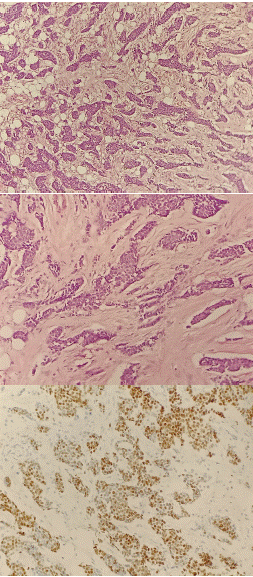
Histological examination showed fibro-fatty and striated muscular tissue largely dissociated by a malignant proliferation made up of lobules carved with rare slits and cords and spans. Tumor cells are arranged in a palisade fashion around the lobules. They are of medium size, provided with anisokaryotic nuclei often increased in volume, hyperchromes, site of mitosis. The mitotic index is estimated at 4 mitoses / 10 fields at high power. The cytoplasm is scanty basophilic. The stroma is hyalinized fibrous punctuated by rare lymphocytes without perinervous sheathing or vascular emboli.
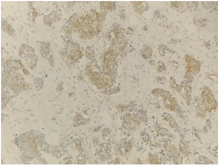
The immunohistochemical study showed a diffuse and moderate positivity of the tumor cells of the broad anti CK antibody, Anti P40 antibody, anti CD99 antibody with an estimated Ki67 proliferation index of 70%. The molecular study objectified the translocation of the EWSR1 gene to 22q12.

Figure 1: P40.

Figure 2: CD99.
The diagnosis was ewing-adamantinoma-like sarcoma.
Discussion
The Ewing sarcoma tumor family has been well known for several decades, but it is only recently that its histological and immunophenotypic spectrum has been fully appreciated. Recent studies have highlighted rare examples with prominent squamous epithelial differentiation, for which the designation “adamantinoma-like” with complex epithelial differentiation has been proposed and represents 0.4% [1,5]. The degree to which these tumors really resemble adamantinomas is admittedly debatable, but this terminology is historical: the term adamantinoma-like ewing's sarcoma has been used since the first reported cases with this phenotype occurred in long bones [1,2,4]. It is only recently that similar cases have been documented in other locations [4,5].
Basaloid or squamous differentiation or the appearance of round cell proliferation should suggest, among other things, this diagnosis and ask the immunohistochemical panel to confirm this diagnosis. Unfortunately, in adamantinoma-like ewing's sarcoma, this diagnostic strategy is likely to mask rather than clarify. Indeed, the histological characteristics (basaloid nests, squamous beads, intraepithelial growth) and immunoprofil (diffuse cytokeratin and p40) of ewing sarcoma of the adamantinoma type strongly suggest a carcinoma, in particular a squamous cell carcinoma. Even the formation of pseudorosettes can easily be misinterpreted as the pseudoglandular spaces of basaloid squamous cell carcinoma. On the other hand, the monomorphic aspect of the cells may point to ewing's sarcoma of the adamantinoma type [1-4,8-10].
The immunohistochemical study shows positivity of anti CD99, FLI1 and P40 antibodies. translocation of the EWSR1 gene to 22q12 can confirm the diagnosis [1].
The main differential diagnoses of adamantinoma-like ewing's sarcoma include midline carcinoma NUT and myoepithelial carcinoma also characterized by squamous differentiation sometimes manifesting as focal keratinization and monomorphic cells. In addition, CD99 expression can rarely be observed in NUT median carcinoma with a positive anti NUT antibody. The latter is negative in adamantinoma-type ewing's sarcoma and myoepithelial carcinoma. The similarities extend to the molecular level, as myoepithelial tumors of soft tissue often harbor rearrangements of EWSR1. Accordingly, for a definitive diagnosis of adamantinoma-like ewing sarcoma, the demonstration of the EWSR1 rearrangement itself it's not enough. The fusion partner gene - typically FLI1 for adamantinoma ewing sarcoma and POU5F1, PBX1, PBX3 or ZNF444 for myoepithelial carcinoma - must be determined to confirm the diagnosis [1-3,6,7].
Conclusion
Adamantinoma-like ewing's sarcoma can occur in any location. The confirmatory diagnosis remains difficult in front of the squamous differentiation, basaloid. Young age. The absence of a history of squamous cell carcinoma. The monomorphic character of the cells and the concomitant positivity of the anti P40 and anti CD99 antibodies should make us think about this diagnosis and push us to look for the EWSRT1 translocation. The anatomo-clinical correlation is the only guarantee of a good diagnostic and management approach.
Author Statements
Conflict of Interest
The authors declare no conflict of interest.
Author’s Responsibilities
Imane.Boujguenna and Fatima.Boukis: drafting of the manuscript
Hanane Rais: correction of the manuscript
Aissam Griche, Malak El Merrakchi, Said Ait Benali: clinical and surgical management of the patient
Chihab bouyaali and Mariem Ouali Idrissi: radiological follow-up of the patient
All authors contributed to the conduct of this work
Acknowledgment
To anyone who has participated in the care of this patient directly or indirectly.
References
- Bridge JA, Fidler ME, Neff JR, Degenhardt J, Wang M, Walker C, et al. Adamantinoma-like Ewing’s sarcoma: genomic confirmation, phenotypic drift. Am J Surg Pathol. 1999; 23: 159–165.
- Barroca H, Souto Moura C, Lopes JM, Lisboa S, Teixeira MR, Damasceno M, et al. PNET with neuroendocrine differentiation of the lung: report of an unusual entity. Int J Surg Pathol. 2014; 22: 427–433.
- Cruz J, Eloy C, Aragues JM, Vinagre J, Sobrinho-Simoes M. Small-cell (basaloid) thyroid carcinoma: a neoplasm with a solid cell nest histogenesis? Int J Surg Pathol. 2011; 19: 620–626.
- Eloy C, Oliveira M, Vieira J, Teixeira MR, Cruz J, Sobrinho-Simoes M. Carcinoma of the thyroid with ewing family tumor elements and favorable prognosis: report of a second case. Int J Surg Pathol. 2014; 22: 260–265.
- Alexiev BA, Tumer Y, Bishop JA. Sinonasal adamantinoma-like Ewing sarcoma: a case report. Pathol Res Pract. 2017; 213: 422–426.
- Bishop JA, Alaggio R, Zhang L, Seethala RR, Antonescu CR. Adamantinoma-like Ewing family tumors of the head and neck: a pitfall in the differential diagnosis of basaloid and myoepithelial carcinomas. Am J Surg Pathol. 2015; 39: 1267–1274.
- Folpe AL, Goldblum JR, Rubin BP, Shehata BM, Liu W, Dei Tos AP, et al. Morphologic and immunophenotypic diversity in Ewing family tumors: a study of 66 genetically confirmed cases. Am J Surg Pathol. 2005; 29: 1025–1033.
- Fujii H, Honoki K, Enomoto Y, Kasai T, Kido A, Amano I, et al. Adamantinoma-like Ewing’s sarcoma with EWS-FLI1 fusion gene: a case report. Virchows Arch. 2006; 449: 579–584.
- Kikuchi Y, Kishimoto T, Ota S, Kambe M, Yonemori Y, Chazono H, et al. Adamantinoma-like Ewing family tumor of soft tissue associated with the vagus nerve: a case report and review of the literature. Am J Surg Pathol. 2013; 37: 772–779.
- Lezcano C, Clarke MR, Zhang L, Antonescu CR, Seethala RR. Adamantinoma-like Ewing sarcoma mimicking basal cell adenocarcinoma of the parotid gland: a case report and review of the literature. Head Neck Pathol. 2015; 9: 280–285.