
Case Report
Austin J Anat. 2015; 2(3): 1041.
An Accessory Left Renal Artery: A Case Report
Gardner S*
Department of Natural and Social Sciences, Bowling Green State University Firelands, USA
*Corresponding author: MGardner, S. Department of Natural and Social Sciences, Bowling Green State University Firelands Campus, Huron, OH USA
Received: October 14, 2015; Accepted: November 12, 2015; Published: November 18, 2015
Abstract
Anatomical variants of the renal vasculature are considered to be normal variants and are extremely common. Variations include accessory renal arteries that enter the hilum of the kidney and aberrant renal arteries that enter the capsule of the kidney. Routine dissection of the abdomen of an 87-year-old female cadaver revealed a left renal accessory artery penetrating the hilum of the kidney. Renal vascular variants are very important to be aware especially for surgical procedures such as kidney transplants or repair of abdominal aortic aneurysms, as well as various diagnostic imaging procedures to ensure patient safety and proper patient diagnosis.
Keywords: Accessory renal artery; Renal vascular variations; Multiple renal arteries
Introduction
The paired renal arteries typically originate from the anterolateral aspect of the abdominal aorta at approximately the level of the L1 and L2 vertebral bodies just below the origin of the superior mesenteric artery crossing the corresponding crus of the diaphragm [1,2]. These two end arteries carry nearly 25% of the cardiac output to the kidneys [1]. Usually there is only one right renal artery and one left renal artery penetrating the hila of each kidney. The right renal artery typically passes behind the inferior vena cava while the left passes posterior to the left renal vein [1,2]. The right renal artery tends to be longer than the left due to the location of the aorta positioned more to the left of the vertebral column and the right renal artery usually passes posterior to the inferior vena cava [1,2]. The renal arteries typically divide into anterior and posterior divisions just prior to entering the hilum of each kidney. Variations of the renal arterial supply usually are described in the literature as “aberrant”, whereby the additional artery enters the kidney directly through the capsule and not the hilum, or the other variation described as “accessory” whereby it branches from the abdominal aorta either superior or inferior to the main renal artery and then enters the hilum as usual [3]. The addition of an extra renal artery or two is one of the most frequent vascular variations within the renal system vasculature [4,5]. A suspected reason for aberrant or accessory arteries could be attributed to embryological development of the lateral mesonephric branches of the dorsal aorta [6,7]. Awareness and clinical knowledge about these renal vascular variations is very important for proper surgical management during procedures such as abdominal aneurysm repair and renal transplants.
Case Presentation
During routine dissection of the posterior abdominal wall of an 87-year old Caucasian female cadaver for the nursing program at Firelands Regional Medical Center in Sandusky, Ohio, it was observed that there was one accessory renal artery branching from the abdominal aorta just below the main renal artery on the left (Figure 1). The left main renal artery branched from the abdominal aorta below the superior mesenteric artery traveling posterior to the left renal vein as it entered the hilum of the left kidney (Figure 1). The accessory renal artery was seen traveling anterior to the left renal vein and ureter upon entering the hilum of the left kidney. The main renal artery on the left was larger in diameter than the accessory renal artery. However, the accessory artery was much more tortuous than the left main renal artery. The venous drainage pattern of the kidneys was normal, however there was a small abdominal aortic aneurysm found just superior to the bifurcation of the abdominal aorta into the right and left common iliac arteries. No other vascular abnormalities were evident in the abdominal vasculature.
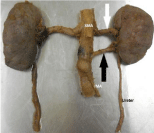
    Figure 1:  Renal artery (white arrow) and accessory renal artery (black
arrow). Also shown are the Superior Mesenteric Artery (SMA) and the Inferior
Mesenteric Artery (IMA).
Discussion
The kidneys are among the most metabolically active organs in the body. They receive approximately twenty-five percent of the cardiac output via two renal arteries [8]. In 70% of the population the kidneys are irrigated by just two renal arteries, a right and a left [5]. Anatomical variations of the renal vasculature (arterial supply and venous drainage) are commonly seen in routine dissection. Recent literature indicates that approximately 30% of the population as having one or more accessory renal arteries [9]. Regarding the arterial terminology of how to describe “extra vessels” some authors state, they are named aberrant, if they enter the renal capsule and not the hilum, or named as accessory if they enter the hilum directly [3,7,11]. The left sided accessory arteries have the potential to cause hydronephrosis as they cross anterior to the ureter obstructing it. It is also known that accessory arteries are “end-arteries”, in that damage or ligation could lead to a decreased blood supply and subsequent infarction. The demand for kidneys for transplants have increased in recent years and the transplant of a kidney with multiple accessory or aberrant arteries is far more difficult than a kidney with only one main renal artery [10,12]. All the accessory renal arteries as well as the main renal artery of the donor kidney must be surgically anastomosed to be certain to completely perfuse the kidney. In light of this trend, it is highly advisable for the patient to undergo arteriography prior to any surgical procedures to reduce post-surgical complications
Conclusion
Complications from an undiscovered aberrant or accessory renal artery may surface during abdominal surgery procedures such as exploratory, aneurysm repair and ureter surgeries. It behooves the surgeon or urologist to spend time familiarizing themselves with various normal variants of the renal vasculature to ensure a positive patient outcome.
References
- Carmine DC. Anatomy, a regional atlas of the human body. Urban & Schwarzenberg. 1987; 214-215.
- Hollinshead, William Henry. "Textbook of anatomy". 1985.
- Graves FT. "The aberrant renal artery". Journal of anatomy. 1956; 90: 553-558.
- Satyapal KS, Haffejee AA, Singh B, Ramsaroop L, Robbs JV, Kalideen JM. "Additional renal arteries incidence and morphometry". Surgical and Radiologic Anatomy. 2001; 23: 33-38.
- Arly BR, Thompson SA, Afifi AK. Catalog of human variation. Urban & Schwarzenberg. 1984.
- Anupma G, Gupta G, Singhla RK. "The accessory renal arteries: a comparative study in vertebrates with its clinical implications". J ClinDiagn Res. 2011; 5: 970-973.
- Felix W. "Mesonephric arteries". Manual of human embryology. 1912; 2: 820-825.
- Saritha S, Jyothi N, Praveen Kumar M, Supriya G. "Cadaveric study of accessory renal arteries and its surgical correlation". 2013; 1: 19-22.
- Olsson O, Wholey M. "Vascular abnormalities in gross anomalies of kidneys". ActaRadiologica (Sweden). 1964; 2: 420-432.
- Ozkan U, Oguzkurt L, Tercan F, Kizilkiliç O, Koç Z, Koca N. "Renal artery origins and variations: angiographic evaluation of 855 consecutive patients". DiagnIntervRadiol. 2006; 12: 183-186.
- Nathan H. "Aberrant renal artery producing developmental anomaly of kidney associated with unusual course of gonadal (ovarian) vessels". The Journal of urology. 1963; 89: 570-572.
- Kacar S, Gurkan A, Akman F, Varýlsuha C, Karaca C, Karaoglan M. "Multiple renal arteries in laparoscopic donor nephrectomy". Annals of Transplantation. 2005; 10: 34-37.