Case Report
Austin J Anat. 2015; 2(3): 1043.
Left Isomerism Sonographic Findings and Post Mortal Autopsy in a Female Foetus
Fröber R¹, Eichhorn K², Schleußner E³ and Schlembach D³*
¹Departmente of Anatomy, Jena University Hospital, Germany
²Center for Prenatal Diagnosis, Gerhart-Hauptmann- Strasse, Germany
³Department of Obstetrics, University of Jena Hospital, Germany
*Corresponding author: Schlembach D, Department of Obstetrics & Gynecology, Prenatal Diagnosis & Fetal Physiology, University of Jena Hospital, Bachstrasse 18, 07743 Jena, Germany
Received: November 25, 2015; Accepted: December 10, 2015; Published: December 16, 2015
Abstract
Background: Left isomerism is a complex malformation syndrome. Important diagnostic pointers like viscera cardiac heterotaxy, complex cardiac anomalies, heart block and interruption of the inferior vena cava provide the prenatal diagnosis. However, although essential for counseling about prognosis, the knowledge about the whole spectrum of associated anomalies in cases of left isomerism is very rare.
Case Presentation: We report a case of a 26-year-old primigravid women presenting at 17+3 weeks of gestation with foetal left isomerism. Following termination of the pregnancy a detailed teratological examination according to the guidelines of the sequential segmental approach was performed, which confirmed the prenatal sonographic findings. Moreover, additional distinct malformations of the cardiovascular, the pulmonal and the gastrointestinal tract were revealed. The main malformation features were: uniatrial-univentricular connection between the left isomeric atrium and a double-outlet right ventricle, interruption of the inferior vena cava combined with juxtaposition of its subhepatic portion and the abdominal aorta, polysplenia, and duodenojejunal malposition with subsequent malrotation of the malfixed bowel.
Conclusion: We demonstrate the importance of an interdisciplinary cooperation of highly skilled sonographer and pathoanatomical experts in teratology. An accurate prenatal diagnosis of left isomerism is feasible. However, the whole dimension of this complex malformation syndrome is usually prenatal not visible. Detailed post mortal autopsy should be recommended to enhance the prenatal diagnosis further with respect to adequate counseling of the parents.
Keywords: Left isomerism; Heterotaxy; Congenital heart disease; Polysplenia; Echocardiography; Autopsy
Background
Heterotaxy syndrome is a rare disease with an incidence of 1-1.5/10,000 live births and high mortality rates [1-3]. This malformation syndrome is defined as the abnormal arrangement of viscera across the left-right axis primarily induced by disorders of the laterality determination during early embryonic development. Its cellular and molecular mechanisms have been extensively investigated in the past decade and the developmental mechanisms of the syndrome have been considerably elucidated [2]. According to the characteristic morphology of the atrial appendages heterotaxy can be divided into “bilateral right-sided” (right isomerism) and “bilateral left-sided” (left isomerism) [4]. Typical findings in case of left isomerism are viscera cardiac heterotaxy, complex cardiac malformations, heart block, and interruption of the inferior vena cava [5]. However, a wide variety of cardiac and extracardiac congenital malformations, with considerable overlap of the anatomical features can occur. In consequence the prenatal differentiation between the two main settings is sometimes delicate. However, it is possible by assessment of specific diagnostic pointers. An intimate knowledge of these markers and the corresponding real anatomy is essential for an exact diagnosis, which is mandatory for adequate parental counseling for planning pregnancy and postnatal management, further diagnostic steps, and also further pregnancies [6]. In this case we report a case of prenatally diagnosed left isomerism illustrated by detailed anatomical pictures.
Case Presentation
Clinical findings
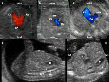
A healthy 26-year-old primigravid woman was referred at 17 + 3 weeks of gestation to a specialist sonographer (K.E.). Ultrasound examination revealed a severe congenital heart defect; brady arrhythmia and heterotaxy syndrome (left isomerism). The sonographic findings are summarized in (Table 1) and (Figure 1). Chromosomal analysis revealed a normal karyotype (46, XX). After detailed counseling, the parents opted for termination of pregnancy at 20 weeks of gestation. Following termination of pregnancy a detailed teratologic examination was performed. The foetal heart has been examined following the guidelines of the sequential segmental analysis [7].
    Fetal Echocardiography
    Additional Sonographic Findings


    complete AVSD
      AV valve insufficiency common atrium
      Persistent brady arrhythmia (58-80 bpm)
    Abdominal situs inversus
      (right-sided spleen and stomach) hyposplenia
      Gall bladder aplasiaI
      VC malposition
Table 1:  Left isomerism-sonographic findings at 17+3 weeks.
AVSD: Atrio Ventricular Septal Defect; IVC: Inferior Vena Cava.

    Figure 1:  Left isomerism -sonographic findings at 17+3weeks.
Complete AVSD (a) with common atrium and AV valve insufficiency, (b) small
preductal aorta, (c) IVC malposition and (d) abdominal situs inversus (e) LV:
Left Ventricle; RV: Right Ventricle; cA: common Atrium; PT: Ulmonal Trunk;
DA: Ductus Arteriosus; pAO: preductalaorta; H: Heart; IVC: Inferior Vena
Cava; AO: Aorta; St: Stomach; S: Spleen
Pathomorphological findings - Cardiovascular
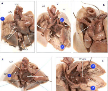
Autopsy confirmed the diagnosis of left isomerism. The heart was left-sided and -orientated. Both atrial appendages were elongated and narrow according the characteristic morphology of left atrial appendages (Figures 2 & 3).

    Figure 2:  Left isomerism - cardiovascular abnormalities found at autopsy.
Precordial venous segment (A): Para aortal course of the interrupted inferior venacava (AZV
and HAZV) and finger like (morphologic left) Right Atrial Appendage (RAA). Atrial segment
(B): Common atrium (A-c) with pulmonary, hepatic vein, and SVC openings; missing coronary
sinus. Atrio ventricular junction and ventricular inflow(C): Common valve and right ventricle,
uniatrial-univentricular connection. Ventricular out flow tract and ventriculo arterial connection
(D): DORV with dextro-transposition of great arteries. Post cardial arterial segment (E): aortic
coarctation (praeductal infund ibularstenos is of the aorticarch).
SVC: Superior Vena Cava; (H) AZV: (hemi) Azygosvein; RAA: Right Atrial Appendage;
A-c/V-c: common Atrium/Ventricle; RPV/LPV: Right/Left Pulmonary Vein; E: Esophagus;
AO: Aorta (descendend); PT: Pulmonal Trunk; HZ-AC: Hepatic Vein Atrial Connection; DA:
Ductusarteriosus

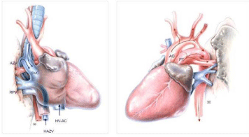
    Figure 3:  Graphic drawings of the distinct cardiovascular anomalies.
Left: precordial venous inflow (right sided view)-paraaortal (H) AZV
continuation of the interrupted IVC and the separate opening of the hepatic
vein.
Right: post cordial arterial outflow (left sided view)-dextro-transposition of the
great arteries and preductal coarctation of the aorticarch.
IVC: Inferior Vena Cava; SVC: Superior Vena Cava; (H) AZV: (hemi)
Azygosvein; RPV: Right Pulmonary Veins; HZ-AC: Hepatic Vein Atrial
Connection; AO: Aorta (ascendend); PT : Pulmonal Trunk
Segmental examination of the heart revealed the following abnormalities: Common atrium with only a thin band of tissue characteristic of a remnant of the atrial septum. The pulmonary veins drained into the central part of the posterior atrial wall. The superior vena cava drained regularly into the right side of the common atrium. The Inferior Vena Cava (IVC) was interrupted, only the hepatic segment entered the atrium inferiorly, whereas the infra hepatic portion of the IVC drained into the hemiazygos-/azygos-system. A large complete Atrioventricular (AV) septal defect with a common AV valve between the common atrium and the right ventricle was present. This uniatrial-univentricular connection between the left isomeric atrium and the bifid single ventricle was ambiguous. At the ventriculoarterial connection a double outlet right ventricle was present accompanied by a dextro-transposition of the great arteries (ascending aorta leaving the heart right-sided and in front of the pulmonary trunk). On the postcardial arterial segment a preductal infundibular stenosis of the aortic arch (aortal coarctation) was present. The morphological cardiac characteristics are summarized in (Table 2).
Segments of the heart
Morphological characteristics
Precordial venous segment
- interrupted IVC(hemiazygos-azygos-continuation)
- PV with atrial connection at posterior wall
Atrial segment
- bilateral left configured appendages
- common atrium
- bilateral left atrium (pectinate muscles confined to the anterior wall)
Atrioventricluar connection
- ambiguous uniatrial-univentricular connection
- common AV valve
Ventricular segment
- rudimentary ventricle septum
- bifid single ventricle
Ventriculoar terial connection
- double outlet right ventricle
- d-transposition of great arteries
Post cardial arterial segment
- aortic coarctation
Table 2:  Post mortal findings of the fetal heart using the segmental approach;
IVC: Inferior Vena Cava; PV: Pulmonary Veins; AV: Atrioventricular.
The interrupted IVC could not be identified in its usual position on the right side of the spine anterolateral to the abdominal aorta. Instead of that both abdominal vessels were found on the left side of the spine. The interruption was located above the left renal vein between the hepatic and the subhepatic portion of the IVC. The hepatic segment had a separate opening on the bottom of the common atrium.
Pathomorphological findings – pulmonal and gastrointestinal
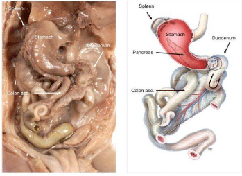
The lungs were bilateral bilobed, morphologic left with two symmetrical hyparterial bronchi. The liver was symmetrically and the extrahepatic biliary structures like the biliary bladder were atretic. The stomach, the pancreatic cauda and multiple splenuli (polysplenia) were found in the right upper abdomen. A malformed and malpositioned duodenum was on the left side of the spine. The location of the duodeno-jejunal junction and the ligament of Treitz were found below the level of the pylorus and right of the midline. Due to the incoherent laterality between the left sided heart axis and the partial situs inversus in the upper abdomen we diagnosed a viscerocardiac heterotaxy; also known as situs ambiguous (Figure 4) [6].

    Figure 4:  Position of the abdominal organs insitus ambiguous with left
isomerism (Anatomical picture (left) and drawing (right)).
Right-sided stomach, spleen and large intestine (small intestine is severed).
Torsion of the duodenum and the following malrotation of the bowel with
malposition of the whole large intestinum on the right side of the lower
abdomen.
Additionally a malpositioned non rotated intestinum and a common mesenterium as a sign of mesenterial malfixation was diagnosed (midgut portion of small bowel was found in the left upper quadrant and the whole large intestinum on the right side and in the midline of the lower abdomen without mesenterial fixation).
Comment
Left isomerism is a laterality disturbance associated with paired left sidedness viscera, malposition of unpaired visceral organs and vessels and multiple small spleens. Congenital heart and gastrointestinal malformations are commonly associated. However, left isomerism syndrome may coexist with very different structurally abnormalities [8]. Despite the progress in prenatal diagnostic imaging some malformations such as bilateral left sided (bilobated) lungs still are not visible prenatally, although the knowledge if these associated malformations is essential for the evaluation of the visceroatrial arrangement.
Complex cardiac lesions are frequently reported in patients with right or left isomerism [5,9]. In cases of left isomerism the associated cardiac anomalies include complete atrioventricular septal defect (68%), complete heart block (38%), right ventricular outflow tract obstruction (35%), double outlet right ventricle (23%), left ventricular outflow tract obstruction (21%) and in about 5% a total anomalous pulmonary vein drainage [3]. Nevertheless, left isomerism syndrome may also coexist with a structural normal heart [10,11]. The sinus node usually has been reported to be absent bilaterally in left isomerism and present bilaterally in right isomerism although exceptions may occur. However, the investigation of Ware et al. indicates that the sinus nodes in hearts with right or left atrial isomerism do not represent morphologically right-sided structures [12].
Our report indicates that even in the foetal heart several prenatal undetectable anomalies are recognized only at a detailed expert teratological examination. Using the segmental approach we detected distinct malformations on each cardiac segment - discordances in the veno-atrial and the atrio-ventricular segment as well as abnormalities in the cono-truncal and the ventriculo-arterial segment.
Interruption of IVC with azygos continuation is another common abnormality observed in cases of left isomerism [8]. This developmental anomaly results in termination of the IVC below the hepatic vein. Systemic venous flow beyond this point is accommodated by the dilated azygos/hemiazygos veins, which eventually 7 empty into the superior vena cava through a dilated azygos arch [13]. The embryonic development of the IVC is very complex, because four different venous precursor segments are involved. The hepatic segment of the IVC develops from the vitelline vein; the three more caudally located segments evolve from paired primitive veins termed as subcardinal vein, supracardinal vein and sub-supracardinal anastomosis. In our case the interruption of the IVC resulted from failure of the right subcardinal-hepatic anastomosis with consequent atrophy of the right-sided primitive veins and subsequent development of the left sided ones. The suprarenal segment was formed by the left instead of the right subcardinal vein and the renal segment by the left instead of the right sub-supracardinal anastomosis. Moreover, the infrarenal segment derived from the left supracardinal vein instead of the right one. Therefore, only, the hepatic segment drained separately into the atrium.
Fetuses with only minor cardiac malformations are at risk postnatal for biliary atresia and for bowel obstruction due to malrotation. In left isomerism a high rate of congenital abnormalities of the foregut, particularly at the level of the hepatobiliary and pancreatic tree are reported [6,8]. It should be noted, that both, the dorsal compound of the pancreas and the spleen develop in the dorsal mesogastrium. We found a mirror-image stomach, a malrotation of the dorsal compound of the pancreas and multiple small spleens in the right upper quadrant. In other studies a high incidence of short or truncated pancreas is reported [8].
Special attention should be directed towards intestinal rotational disorders given the presumed risk of midgut volvulus. While the risk of malrotation in the general population is up to 1%, it may be as high as 70% in cases of heterotaxy although the incidence is not precisely known [8,14]. For patients with situs ambiguous several possible anatomic arrangements of small bowel and colon are reported [14]. We found a duodeno-jejunal malposition and the subsequent malrotation of a malfixed bowel with the caecum in the tight lower quadrant and the transverse colon posterior to the small bowel. The malposition of the duodeno-jejunal junction was combined with ligament of Treitz lying below the level of the pylorus and left of the midline. The suspensory muscle of duodenum, also known as the ligament of Treitz, arised from the right crus of diaphragm as it passed around the esophagus, continued as connective tissue around the stems of the celiac trunk and superior mesenteric artery and inserted into the flexure between the third and fourth portion of the duodenum.
Conclusion
This case report demonstrates the importance of an interdisciplinary cooperation of highly skilled sonographer und pathoanatomical experts in teratology to elucid the real extent of complex multivisceral fetal malformation. Based on this case report, we summarize that: first, the majority of important diagnostic pointers like viscerocardiac heterotaxy, complex cardiac anomalies, heart block and interruption of the IVC provide the accurate prenatal diagnosis of left isomerism and second, a detailed analysis by autopsy is necessary to confirm prenatal and especially echocardiographic findings. It may reveal other minor anomalies, usual undetectable by ultrasound or MRI imaging. Especially the sequential segmental analysis of the foetal heart may complete the malformation pattern, which is necessary for adequate counseling of the parents about additional (genetic) diagnostic steps, and postnatal outcome [6].
References
- Berg C, Geipel A, Smrcek J, Krapp M, Germer U, Kohl T, et al. Prenatal diagnosis of cardiosplenic syndromes: a 10-year experience. Ultrasound Obstet Gynecol. 2003; 22: 451-459.
- Shiraishi I, Ichikawa H. Human heterotaxy syndrome - from molecular genetics to clinical features, management, and prognosis -. Circ J. 2012; 76: 2066-2075.
- Pepes S, Zidere V, Allan LD. Prenatal diagnosis of left atrial isomerism. Heart. 2009; 95: 1974-1977.
- JacobsJP, Anderson RH, Weinberg PM, Walters HL, Tchervenkov CI, DelDuca D, et al. The nomenclature, definition and classification of cardiac structures in the setting of heterotaxy. Cardiol Young. 2007; 1-28.
- Berg C, Geipel A, Kamil D, Knüppel M, Breuer J, Krapp M, et al. The syndrome of left isomerism: sonographic findings and outcome in prenatally diagnosed cases. J Ultrasound Med. 2005; 24: 921-931.
- Gottschalk I, Berg C, Heller R. [Disorders of laterality and heterotaxy in the foetus]. Z Geburtshilfe Neonatol. 2012; 216: 122-131.
- Anderson RH, Shirali G. Sequential segmental analysis. Ann Pediatr Cardiol. 2009; 2: 24-35.
- Tawfik AM, Batouty NM, Zaky MM, Eladalany MA, Elmokadem AH. Polysplenia syndrome: a review of the relationship with viscero-atrial situs and the spectrum of extra-cardiac anomalies. Surg Radiol Anat. 2013; 35: 647-653.
- Berg C, Geipel A, Kamil D, Krapp M, Breuer J, Baschat AA, et al. The syndrome of right isomerism -- prenatal diagnosis and outcome. Ultraschall Med. 2006; 27: 225-233.
- De Paola N, Ermito S, Nahom A, Dinatale A, Pappalardo EM, Carrara S, et al. Prenatal diagnosis of left isomerism with normal heart: a case report. J Prenat Med. 2009; 3: 37-38.
- Dilli A, Gultekin SS, Ayaz UY, Kaplanoglu H, Hekimoglu B. A rare variation of the heterotaxy syndrome. Case Rep Med. 2012; 2012: 840453.
- Ware AL, Miller DV, Porter CB, Edwards WD. Characterization of atrial morphology and sinus node morphology in heterotaxy syndrome: an autopsy-based study of 41 cases (1950-2008). Cardiovasc Pathol. 2012; 21: 421-427.
- Saito T, Watanabe M, Kojima T, Matsumura T, Fujita H, Kiyosue A, et al. Successful blood sampling through azygos continuation with interrupted inferior vena cava. A case report and review of the literature. Int Heart J. 2011; 52: 327-330.
- Papillon S, Goodhue CJ, Zmora O, Sharma SS, Wells WJ, Ford HR, et al. Congenital heart disease and heterotaxy: upper gastrointestinal fluoroscopy can be misleading and surgery in an asymptomatic patient is not beneficial. J Pediatr Surg. 2013; 48: 164-169.