Research Article
Austin J Anat. 2016; 3(1): 1048.
Blood Supply of the Superior Glenohumeral Ligament: A Gross Anatomical and Histological Study
Põldoja E1*, Rahu M2, Kask K2, Bulecza T1, Weyers I3 and Kolts I1
1Department of Anatomy, University of Tartu, Estonia
2Department of Orthopaedics, North Estonia Medical Centre Foundation, Estonia
3Institute of Anatomy, University of Lübeck, Germany
*Corresponding author: Põldoja E, Department of Anatomy, The University of Tartu, Ravila Street 19, Tartu 50411, Estonia
Received: December 27, 2015; Accepted: February 22, 2016; Published: February 26, 2016
Abstract
Background: The purpose of this study was to investigate the blood supply of the superior glenohumeral ligament.The purpose of this study was to investigate the blood supply of the superior glenohumeral ligament.
Method: Thirty two fresh cadaveric shoulder specimens (20M/12F) were used in this study. Arterial injection and dissection were performed in 16 specimens (10M/6F) and in the other 15 specimens (9M/6F) the superior glenohumeral ligament was biopsied to carry out a histological and immunohistochemical analysis. One fixed (1M) specimen was finely dissected to illustrate the glenohumeral joint ligamentous structures.
Results: In the dissection study the superior glenohumeral ligament with the oblique and direct parts was tightly connected with the coracohumeral ligament. In the dissection of injected specimens the superior glenohumeral ligament was found to receive blood supply from the suprascapular, axillary and anterior circumflex humeral arteries in all specimens. A single direct branch of the axillary (so-called “subcoracoid artery”) coursed toward the coracohumeral ligament and bifurcated into two smaller branches that ended in the middle third of the superior glenohumeral ligament. In the histological and immunohistochemical analysis the blood supply of the different thirds of the superior glenohumeral ligament is in correlation with the structural changes from the parallel oriented dense connective with a good vasculature, to fibrocartilage with hypovascular regions of the ligament.
Conclusion: The subcoracoid artery is an important source of blood supply of the rotator interval and the coracohumeral and superior glenohumeral ligaments, in addition to the suprascapular and anterior circumflex arteries.
Keywords: Anatomy; Glenohumeral joint capsule; Rotator interval; Superior glenohumeral ligament; Blood supply; Subcoracoid artery
Introduction
The rotator interval, the area between the anterior border of the supraspinatus tendon superiorly and the superior border of the subscapularis tendon inferiorly, is a surgically important landmark for the repair of subscapularis laxity and supraspinatus tendon tears [1-5]. The coracohumeral and superior glenohumeral ligaments, the anterior joint capsule and the tendon of long head of biceps brachii are encompassed by the rotator interval [2,6-8]. In the literature, hypovascularity of these structures has been suggested to play a key role in the rotator cuff pathology (Table 1) [9-15].
    Study
    N
    Method
    Hypovascularity
      Zone
    Suggested    clinical correlation


    Löhr et al. [9]
    26
      UE
    Histology
    Close to SS tendon
    Degenerative    rotator cuff tears


    Ling et al. [10]
    20
      UE
    Observation
    Articular side of    SS tendon
    SS rupture


    Determe et    l. [11]
    25
      UE
    Dissection
      Histology
    SS tendon
    Rotator cuff    disease


    Katzer et    al. [12]
    8 adult
      6 neonate
    Histology
    SS and IS tendon
      Neonate = adult
    Rotator cuff    rupture


    Andary et    al. [13]
    24
      UE
    Dissection
    Anterior joint    capsule
    Surgery may put    blood supply at risk


    Biberthaler et    al. [14]
    11
      Patients
    Arthroscopy
      Histology
    SS near insertion
    Rotator cuff    lesions


    Cheng et al.    [15]
    20 (E)
      8 (UE)
    Dissection
      Histology
    Long head biceps    brachii
    Weakness and tendon    rupture
Table 1:  Antero-superior view of a dissected right shoulder joint specimen.
The SGHL: Superior Glenohumeral Ligament; O: consists of the “Oblique”;
D: and “Direct” parts. The A (Lateral), B (Middle), C (Medial) on the, SGHL:
Superior Glenohumeral Ligament, correspond to the regions, where the
samples for the light - microscopy and immunohistochemistry were taken.
The coracoid process is cut at its base (cut). The SSP: Supraspinatus; SSC:
Subscapularis Muscles are separated from the shoulder joint capsule and
placed laterally. ISP: Infraspinatus Muscle; MGHL: Middle Glenohumeral
Ligament, TMI: Lesser Tubercle, SGT: Supraglenoidal Tubercle; L: Labrum;
BT: Tendon of Long Head of Biceps Brachii; THL: Transverse Humeral
Ligament; SCHL: Semicircular Humeral Ligament
A zone of hypovascularity has been found laterally in the supraspinatus and infraspinatus tendons in neonates and adults, as well as in the anterior capsule and tendon of long head of biceps brachii, but the blood supply of the superior glenohumeral ligament has not been established. The superior glenohumeral ligament has been clinically identified as an important component of the rotator interval due to its involvement in various pathologies including long head of the biceps tendon rupture, glenohumeral instability, and labral tears [2,15-18]. Although the structural morphology of the superior glenohumeral ligament has been studied by Kolts et al. [8] and Kask et al. [7], no papers were found that examined the blood supply of the superior glenohumeral ligament.
Therefore the purpose of this study was to investigate the blood supply of the superior glenohumeral ligament.
Materials and Methods
Thirty two fresh cadaveric shoulder specimens (age range, 60-84years; 20 male and 12 female) were used in this study. Arterial injection and dissection were performed in 16 fresh specimens (10M/6F) and in the remainig 15 specimens (9M/6F) the superior glenohumeral ligament was biopsied to carry out the histological and Immunohistochemical analysis. One fixed specimen (1M) was finely dissected to illustrate the glenohumeral joint ligamentous structures in which blood supply will be investigated.
The ethics permission was received from Gesetz über das Bestattungsgesetz des Landes Schleswig-Holstein vom 04.02.2005, Abschnitt II, § 9 (Leichenöffnung, anatomisch).
Prior to the dissection, each specimen was injected with 200ml of latex diluted in a 10% aqueous solution stabilized with amonia (0.7% concentration) via the subclavian and the brachial arteries simultaneously. Post injection, the specimens were fixed in an alcohol-formalin-glycerol solution and meticulously dissected using a dissection microscope.
To allow the exposure of the blood vessels the supraspinatus, infraspinatus and teres minor were exposed posteriorly and the pectoralis minor anteriorly by removing the skin and any superficial muscles and tissues. The pectoralis minor was carefully reflected laterally, revealing the neurovascular structures in the axillary sheath. The blood vessels were isolated and the arterial and venous vessels separated, removing axillary fat as needed. The veins, nerves and remaining axillary fat were removed to provide clear visibility of the subclavian and axillary arteries and their branches, as well as the subscapularis. The arteries were traced to the coracohumeral ligament and the surface of the rotator cuff muscles. Arterial supply to the coracohumeral ligament was documented. Next, the coracoid process, coracohumeral ligament and acromion were excised to increase the exposure of the rotator cuff muscles and rotator interval. Each of the four rotator cuff muscles was reflected laterally, taking care not to disrupt the vessels coursing toward the joint capsule. Next, the oblique and direct parts of the superior glenohumeral ligament were identified as they merged to attach to the supraglenoid tubercle and glenoid labrum medially. The oblique part was traced laterally superficial to the intra-articular portion of the tendon of the long head biceps to blend with the semicircular humeral ligament. The direct part was followed to its attachment to the lesser tubercle and the lips of the bicipital groove, where it became continuous with the superior margin of the transverse humeral ligament. All arterial vessels were traced to their termination, until no longer visible under the dissection microscope. Photographs were taken throughout the dissection process and the course and termination of the arterial vessels documented.
Histological and immunohistochemical analysis
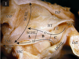
Fifteen specimens that were not injected with latex were dissected as above but for the following identification of the superior glenohumeral ligament, the ligament was excised at its attachment sites and removed from the specimen. Each ligament was biopsied at three locations: A) lateral third, close to the attachment sites of the oblique and direct parts. B) Middle third, through both the oblique and direct parts and, C) medial third, at the common attachment of the oblique and direct parts (Figure 1). Each biopsy was fixed in 4% formalin, and embedded in paraffin. Fourteen consecutive sections, each 5 micrometers thick, were cut from the center of each block using a Microm Type STS microtome (Germany). Four sections were used for the histological analysis and 10 for immunohistochemistry.

    Figure 1:  Antero-superior view of a dissected right shoulder joint specimen.
The SGHL: Superior Glenohumeral Ligament; O: consists of the “Oblique”;
D: and “Direct” parts. The A (Lateral), B (Middle), C (Medial) on the, SGHL:
Superior Glenohumeral Ligament, correspond to the regions, where the
samples for the light - microscopy and immunohistochemistry were taken.
The coracoid process is cut at its base (cut). The SSP: Supraspinatus; SSC:
Subscapularis Muscles are separated from the shoulder joint capsule and
placed laterally. ISP: Infraspinatus Muscle; MGHL: Middle Glenohumeral
Ligament, TMI: Lesser Tubercle, SGT: Supraglenoidal Tubercle; L: Labrum;
BT: Tendon of Long Head of Biceps Brachii; THL: Transverse Humeral
Ligament; SCHL: Semicircular Humeral Ligament.
Four sections from each block were stained using the Trichrome Masson-Goldner method and viewed with a Carl Zeiss Axioplan 2 microscope for the histological assessment of the superior glenohumeral. The orientation of the collagen fiber bundles and the proportion of fibrocytes and chondrocytes were assessed to determine the structural characteristics of the superior glenhumeral ligament at each biopsied site. The number of chondrocytes and fibrocytes was counted manually in the microscopic field of view.
Ten sections from each block were used for immunohistochemistry and processed using the Avidin-Biotin technique to determine if Type IV collagen is found in the basement membranes of the blood vessels. Two of the ten sections were used as negative controls. For immunohistochemical staining, the deparaffinized sections were treated with 0.6% H²O² to inactivate endogenous peroxidase and then were incubated in 5% normal goat serum for 20 minutes to block non-specific binding. Next, the sections were incubated overnight at room temperature in a moist chamber with the primary Mouse Anti-Human Collagen IV antibody (Dako-Clone CIV 22, Code No. M0785, diluted 1:50). A streptABComplex/HRP Duet Mouse/ Rabbit System (DakoCytomation, Glostrup, Denmark) and DAB+ Chromogen (DakoCytomation) kit was used to visualize the primary antiboby. The dehydrated slides were coverslipped with Entellan (Merck, Germany). The washing steps in-between were done in the Tris buffered saline (TBS-pH 7.4). The adjacent sections served as negative controls and were processed using the same protocol without incubation of the primary antibody.
All slides were viewed using a Carl Zeiss Axioplan 2 microscope and photographed with a Sony DXC-950P 3CCD Color photo/video camera.
Results
Dissection of injected specimens
In the dissection study the superior glenohumeral ligament was found to receive blood supply from the suprascapular, direct axillary branch (subcoracoid artery) and anterior circumflex humeral arteries in all specimens.
The branch from the suprascapular artery gives way to the superior transverse scapular ligament. This branch coursed in supraspinatus from below for approximately 3mm before bifurcating into two smaller branches that entered the superior glenohumeral ligament to the medial third and the coracohumeral ligament to the upper surface (Figure 2b).

    Figure 2b:  Antero-superior view of the suprascapular artery branches (***) at
the origin of the CHL: coracohumeral ligament; PC: Coracoid Process; CGL:
Coracoglenoid Ligament; SSP: Supraspinatus Muscle.
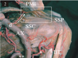
A single direct branch of the axillary, about 1cm distal to the origin of the suprascapular artery, coursed toward the coracohumeral ligament and bifurcated into two smaller branches; these were seen between the fiber bundles. These branches supplied the middle third of the superior glenohumeral ligament with the anterior branch entering the direct part and the posterior branch of the oblique part (Figure 2,2a).

    Figure 2a:  A quadrangular cutout from figure 2.
Blood supply of the CHL: Coracohumeral; SGHL: Superior Glenohumeral
Ligaments; D: by the Direct Branch from the axillary artery (“subcoracoid
artery”). SSC: Subscapularis Muscle; PMI: Pectoralis Minor Muscle.
The anterior circumflex humeral artery has two smaller ascending branches. The ascending branches supplied the subscapularis tendon, the lateral third of the superior glenohumeral ligament and the rotator interval (Figure 2).

    Figure 2:  Latex-injected left shoulder specimen. Overview of the main
arteries and their blood supply regions.
A direct branch (d) arises from the Axillary Artery (AX) and courses under the
coracoid process into the coracohumeral ligament. * - the anterior circumflex
humeral artery; ** - the subscapular artery; SSC: Subscapularis Muscle; SSP:
Supraspinatus Muscle; BT: Tendon of Long Head of Biceps Brachii; PMI:
Pectoralis Minor Muscle
In all the investigated specimens, the direct and oblique parts of the superior Glenohumeral ligament were found.
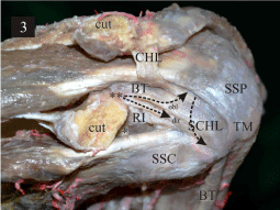
The direct and oblique parts of the superior glenohumeral ligament came from the glenoid labrum and the supraglenoid tubercle. The direct part attached laterally to the lesser tubercle and the lips of the bicipital groove where it became continuous with the superior margin of the transverse humeral ligament. The oblique part ran over the intra-articular part of the tendon of the long head biceps and inserted to the semicircular humeral ligament (Figure 3).The coracohumeral ligament arose from the postero-lateral part of the coracoid process and the coracoglenoid ligament. The coracohumeral and superior glenohumeral ligaments were tightly connected with each other (Figure 2,2a). After the separation from the coracohumeral ligament by sharp dissection, the artery branches became thinner in diameter and were no longer visible in the middle third of the superior glenohumeral ligament (Figure 3).

    Figure 3: Latex-injected left shoulder specimen.
The RI: Rotator Interval; between the tendons of the SSP: Supraspinatus,
SSC: Subscapularis muscles are visualized with the CHL: Coracohumeral
Ligament and the SCHL: Superior Glenohumeral Ligament with the Obl:
Oblique; Dir: Direct (dir) parts. BT: Tendon of long head of biceps brachii ; TM:
Major Tubercle of the Humerus; cut – the coracoid process is cut and moved
posterioly; * - middle glenohumeral ligament; ** - supraglenoidal tubercle.
Histological analysis
The medial and lateral thirds of the superior glenohumeral ligament showed histologically typical characteristics of the fibro cartilaginous tissue with the masked collagen fibres and chonrocytelike cells lying separately, fibrocytes lying in pairs or in rows. The mean ratio of Chondrocytes to Fibrocytes (CC/FC) was approximately similar to the medial and lateral thirds, 1,13 ± 0,28 vs 0,86 ± 0,12 (Figure 4-C,A). The middle third of the ligament showed the typical features of a dense connective tissue with the parallely oriented bundles of collagenfibres and typical fibrocytes. The fibrocytes and chondrocytes which appeared occasionally, dominated in the middle third (Figure 4-B).

    Figure 4:  Light microscopic sections of the lateral (A), middle (B) and medial (C) thirds of the superior glenohumeral ligament. Trichrome Masson-Goldner staining.
The middle third (B) of the superior glenohumeral ligament showed parallel oriented bundles of collagen fibers and typical fibrocytes; both are typical features of
the parallel oriented dense connective tissue. Magnification bar is 50 μm. The lateral (A) and medial (C) third of the superior glenohumeral ligament showed typical
characteristics of the fibrocartilagineous tissue with the masked collagen fibers, chondrocytes lying individually and fibrocytes lying individually, in pairs or in rows.
The magnification bar in the lateral third is 100 μm and in the medial third 50 μm
Immunohistochemistry
The middle third of the superior glenohumeral ligament was richly vascularized, whereas the blood supply of the lateral and medial thirds was less dense (Figure 5-A,B,C). In the medial and lateral thirds of the superior glenohumeral ligament, the reduction in vascularity was accompanied by a relative increase in the fibro cartilaginous tissue in comparison to the higher proportion of the parallel oriented collagen fibres in the middle third (Figure 4,5-A,B,C).

    Figure 5:  Light microscopic sections of the lateral (A), middle (B) and medial (C) thirds of the superior glenohumeral ligament. Immunohistochemical stain for type
Collagen IV.
The middle third (B) of the superior glenohumeral ligament is better vascularized than the medial and lateral parts of the ligament. The blood supply of the lateral
(A) and medial (C) thirds is less dense. The magnification bar is 10 μm in all the thirds
Discussion
Recent decades have brought a new insight into the anatomy of the classically known coracohumeral ligament [19]. The anatomy of the glenohumeral ligaments [7,20] and the “rotator cable” found by Burkhart et al. [21] has been morphologically described as a capsular semicircular humeral ligament [8]. A great progress in understanding the structure of the glenohumeral joint does not fully satisfy the contemporary clinical needs, because the blood supply of the glenohumeral joint capsule and the ligaments have been poorly investigated. The present study concentrated on the blood supply of the coracohumeral and superior glenohumeral ligaments, which are the main structures within the rotator interval - one of the most important regions of the glenohumeral joint.
A tight connection between two ligaments is important for the blood supply of the superior glenohumeral ligament and the rotator interval capsule. The coracohumeral ligament receives blood vessels from two sources – small branches from the suprascapular artery on the superior surface and the direct branch from the axillary artery on the lower surface of the ligament. These two different blood supply sources ensure a good vascularity of the coracohumeral ligament itself and the rotator interval capsule together with the superior glenohumeral ligament. This finding supports the ideas of Golke et al. [22] who suggested, that the vascular supply of the capsular layer of the rotator cuff runs in the direction of the coracohumeralfiber bundles and lays between them.
Our results partly support the findings of Abrassant et al. [23] who investigated the blood supply of the glenoid. The authors found, that the direct branch from the axillary artery and small branches of the suprascapular artery run to the medial part of the anterosuperior joint capsule, but still the region remains poorly vascularized. The similar results of the vasculature of the medial joint capsule are also presented by Andary and Peterson [13].
Determe et al. [11] also found direct branches from the axillary artery (“suprahumeral artery”) to the subscapular muscle. They confirmed that the rotator interval is poorly vascularized by small vessels from the “suprahumeral artery” that is not supported by the present results. The unofficial term “suprahumeral artery” is also used in a glenohumeral joint vasculature study by Löhr and Uhtoff [9] but without any description as to what this term means. According to the present results a strong direct branch from the axillary artery runs under the coracoid process into the coracohumeraland to the superior glenohumeral ligaments. More laterally there are weak and unstable ascending branches to the subscapular muscle that are without any clear reason named as a “suprahumeral artery”. The strong medial direct branch of the axillary artery plays a key role in the blood supply of the coracohumeral and superior glenohumeral ligaments within the rotator interval. It has been mentioned in a study by Garza et al. [24] as a“subcoracoid artery”. Unfortunately, the region of the blood supply of the “subcoracoid artery” was not described and therefore the results are not fully comparable with the results of the present study.
In the previous glenohumeral joint vasculature studies the majority of the investigors have concentrated on the “critical zone” of the supraspinatus tendon [9-12,14]. The vasculature of the joint capsule and the ligaments has generally been the objects of secondary interest, with a few exceptions [25-28]. Therefore, in aninteresting study by Ling et al. [10] the course and branches of the anterior circumflex humeral artery are described in correlation with the supraspinatus tendon vasculature and its role in the vasculature of the rotator interval is undefined.
The histological results of the medial and lateral third of the superior glenohumeral ligament showed features of a typical fibrocartilagineous tissue, and of the middle third,a dense connective tissue. This study produced results which corroborate the findings of a great deal of the previous work of Kask et al. [7]. This finding is in accordance with the grossanatomic results of the some previous studies [10,23]. The immunohistochemical results proved that all three thirds of the superior Glenohumeral ligaments were vascularized. The vascularization of the parallel-oriented dense connective tissue in the middle third of the superior glenohumeral ligament was better than that of a fibrocartilage in the medial and lateral thirds of the ligament. Our findings support the statement of Tillmann and Gehrke in 1995 [29] who suggested that the fibrocrtilagineous tissue is not vascular, but hypovascular.
Conclusion
The purpose of the current study was to determine the blood supply of the superior glenohumeral ligament. This study showed that the superior glenohumeral ligament receives blood supply from the suprascapular, direct branch of the axillary (“subcoracoid artery”), anterior circumflex humeral arteries and the ligament is well vascularized. A better knowledge of the vascular anatomy of the superior capsule has increased significance as arthroscopic and open surgical procedures continue to evolve.
Acknowledgement
The authors thank Raudheiding A, Maynicke J, and Teletzky N, for technical support. The authors wish also to thank the individuals who donate their bodies for the advancement of education and research.
References
- Burkhart SS. A stepwise approach to arthroscopic rotator cuff repair based on biomechanical principles. Arthroscopy. 2000; 16: 82-90.
- Gaskill TR, Braun S, Millett PJ. Multimedia article. The rotator interval: pathology and management. Arthroscopy. 2011; 27: 556-567.
- Lee JC, Guy S, Connell D, Saifuddin A, Lambert S. MRI of the rotator interval of the shoulder. Clin Radiol. 2007; 62: 416-423.
- Lyons RP, Green A. Subscapularis Tendon Tears. J Am AcadOrthop Surgery. 2005; 13: 353-363.
- Millett PJ, van der Meijden OA, Gaskill T. Surgical anatomy of the shoulder. Instr course lect. 2012; 61: 87-95.
- Hunt SA, Kwon YW, Zuckerman JD. The rotator interval: anatomy, pathology, and strategies for treatment. J Am Acad Orthop Surg. 2007; 15: 218-227.
- Kask K, Põldoja E, Lont T, Norit R, Merila M, Busch LC, et al. Anatomy of the superior glenohumeral ligament. J Shoulder Elbow Surg. 2010; 19: 908-916.
- Kolts I, Busch LC, Tomusk H, Raudheiding A, Eller A, Merila M, et al. Macroscopical anatomy of the so-called “rotator interval”. A cadaver study on 19 shoulders joints. Ann Anat. 2002; 184: 9-14.
- Lohr JF, Uhthoff HK. The microvascular pattern of the supraspinatus tendon. Clinical orthopedics and related research.1990; 254: 35-38.
- Ling SC, Chen CF, Wan RX. A study on the vascular supply of the supraspinatus tendon. Surg Radiol Anat. 1990; 12: 161-165.
- Determe D, Rongières M, Kany J, Glasson JM, Bellumore Y, Mansat M, et al. Anatomic study of the tendinous rotator cuff of the shoulder. Surg Radiol Anat. 1996; 18: 195-200.
- Katzer A, Wening JV, Becker-Männich HU, Lorke DE, Jungbluth KH. Die rotatorenmanschettenruptur. Unfallchirurgie.1997; 23: 52-59.
- Andary JL, Petersen SA. The vascular anatomy of the glenohumeral capsule and ligaments: an anatomic study. J Bone Joint Surg Am. 2002; 84-84A: 2258-2265.
- Biberthaler P, Wiedemann E, Nerlich A, Kettler M, Mussack T, Deckelmann S, et al. Microcirculation associated with degenerative rotator cuff lesions. In vivo assessment with orthogonal polarization spectral imaging during arthroscopy of the shoulder. J Bone Joint Surg Am. 2003; 85-85A: 475-480.
- Cheng NM, Pan WR, Vally F, Le Roux CM, Richardson MD. The arterial supply of the long head of biceps tendon: Anatomical study with implications for tendon rupture. Clin Anat. 2010; 23: 683-692.
- Arai R, Mochizuki T, Yamaguchi K, Sugaya H, Kobayashi M, Nakamura T, et al. Functional anatomy of the superior glenohumeral and coracohumeral ligaments and the subscapularis tendonin view of stabilization of the long head of the biceps tendon. J of Shoulder Elbow Surgery. 2010; 19: 58-64.
- Ogul H, Karaca L, Can CH, Pirimoglu B, Tuncer K, Topal M, et al. Anatomy, variants, and pathologies of the superior glenohumeral ligament: magnetic resonance imaging with three-dimensional volumetric interpolated breath-hold examination sequence and conventional magnetic resonance arthrography. Korean J Radiol. 2014; 15: 508-522.
- Schaeffeler C, Waldt S, Holzapfel K, Kirchhoff C, Jungmann PM, Wolf P, et al. Lesions of the biceps pulley: diagnostic accuracy of mr arthrography of the shoulder and evaluation of previously described and new diagnostic signs. Radiology. 2012; 264: 504-512.
- Kolts I, Busch LC, Tomusk H, Arend A, Eller A, Merila M, et al. Anatomy of the coracohumeral and coracoglenoidal ligaments. Ann Anat. 2000; 182: 563-566.
- Kolts I, Busch LC, Tomusk H, Rajavee E, Eller A, Russlies M, et al. Anatomical composition of the anterior shoulder joint capsule. A cadaver study on 12 glenohumeral joints. Ann Anat. 2001; 183: 53-59.
- Burkhart SS, Esch JC, Jolson RS. The rotator crescent and rotator cable: an anatomic description of the shoulder´s “suspension bridge”. Arthroscopy. 1993; 9: 611-616.
- Gohlke F, Essigkrug B, Schmitz F. The pattern of the collagen fiber bundles of the capsule of the glenohumeral joint. J Shoulder Elbow Surg. 1994; 3: 111-128.
- Abrassart S, Stern R, Hoffmeyer P. Arterial supply of the glenoid: an anatomic study. J Shoulder Elbow Surg. 2006; 15: 232-238.
- de la Garza O, Lierse W, Steiner D. Anatomical study of the blood supply in the human shoulder region. Acta Anat (Basel). 1992; 145: 412-415.
- Cooper DE, Arnoczky SP, O'Brien SJ, Warren RF, DiCarlo E, Allen AA. Anatomy, histology, and vascularity of the glenoid labrum. An anatomical study. J Bone Joint Surg Am. 1992; 74: 46-52.
- Fallon J, Blevins FT, Vogel K, Trotter J. Functional morphology of the supraspinatus tendon. J Orthop Res. 2002; 20: 920-926.
- Yepes H, Al-Hibshi A, Tang M, Morris SF, Stanish WD. Vascular anatomy of the subacromial space: a map of bleeding points for the arthroscopic surgeon. J of arthroscopic and related surgery. 2007; 23: 978-984.
- Werner A, Mueller T, Boehm D, Gohlke F. The stabilizing sling for the long head of the biceps tendon in the rotator cuff interval. A histoanatomic study. Am J Sports Med. 2000; 28: 28-31.
- Tillmann B, Gehrke T. Funktionelle anatomie des subakromialen raums. Arthroskopie.1995; 8: 209-217.