Case Presentation
Austin J Anat. 2016; 3(1): 1049.
A Persistent Metopic Suture: A Case Report
Seth Gardner*
Department of Natural and Social Sciences, Bowling Green State University Firelands, USA
*Corresponding author: Seth Gardner, Department of Natural and Social Sciences, Bowling Green State University Firelands, Huron, Ohio, USA
Received: March 08, 2016; Accepted: March 18, 2016; Published: March 22, 2016
Abstract
An adult human skull found in a college osteological collection presented with a persistent metopic suture. The metopic suture or frontal suture is noted to be between the two frontal bones extending from the nasion to the bregma. The metopic suture generally fuses between 1 and 8 years of life. If it remains after that time it is known as metopism. Its presence is a normal variant of the cranial sutures. Its presence may be mistaken for a skull fracture and also may be associated with frontal sinus irregularities.
Keywords: Metopic suture; Cranial sutures; Frontal bone
Introduction
The human frontal bones begin to ossify in the mesenchyme via two ossification centers at approximately eight weeks gestation [1]. The two bones tend to fuse in the midline via the metopic or frontal suture. The term metopic is from Greek meaning “in the middle of the face” [2]. The metopic suture is a dentate-type suture extending from the nasion to the bregma [3]. The fusion of the metopic suture normally begins at the nasion proceeding superiorly and terminates at the anterior fontanelle [4]. The suture is situated almost exactly on the median line of the two frontal bones [2]. It usually will close within the first or second year of life, but it has been reported to take up to seven years to fuse [5]. Racial variations have been reported in the literature [6], as well as complications related to incomplete development of the frontal sinus. When the metopic suture persists into adulthood it is known as “metopism”. It is rare to find this suture in adults and its presence is not considered pathological. However, premature closure of any of the cranial sutures results in a pathology known as craniosynostosis [3].
Case Presentation
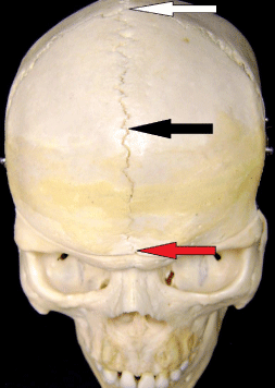
A dry human skull used in the anatomy program at Bowling Green State University Firelands in Huron, Ohio was found to have a persistent metopic suture or metopism. Based upon the size and shape of the piriform aperture as well as the various other anthropometric markings, the skull was suspected to be from a black male of unknown age. The two frontal bones were clearly seen due to the complete metopic suture. The suture extended from the bregma to the nasion as seen in Figure 1.

    Figure 1: The White arrow indicates Bregma. The Black arrow indicates
Metopic Suture. The Red arrow indicates the Nasion.
Discussion
The persistence of the metopic suture is called metopism. This suture disappears by the second or third year of life. It is thought to be a normal variant of the cranial sutures [7]. It forms from the lack of union of the two frontal bones during embryonic development. Del Sol [8] suggested that metopism can be related to abnormal growth of the cranial bones, hydrocephalus, heredity, or atavism. The genetic factor is the one currently accepted by most scientists [9]. Metopism is found in approximately 5% of Asians and 9% of European Caucasians and 1% of Blacks [1,7]. Bergman [7] reported the persistence of the metopic suture in approximately 1-12% of skulls. One author, Agarwal [10] reported the finding of 38.17% in Indian skulls, and Linc [11] observed it in 11% in Czech skulls, and finally Woo [12] reported the finding in 10% in Mongoloid skulls. The data may suggest that metopism is higher in temperate climates than in warmer climates [13].
Berry and Berry [14] reported a 0%-7% incidence associated with ethnicity. Metopism has been found by several investigators as being more prevalent in males than females [15,16]. Castillo reported a world index of incidence of 2.75%. It has also been reported to be perhaps associated with frontal sinus abnormalities but those studies seem flawed [2]. Some authors reported various suspected causes of metopism, including active expression of cytokines during cranial fusion and even resorption of the chondroidal tissue [6]. One author states “that a persistent metopic suture probably occurred in conjunction while refining the ability to walk” [8]. The author further notes that the persistent metopic suture is an adaptation for giving birth to babies with larger brains, and is related to the shift to a rapidly growing brain after birth and even may be related to the expansion of the frontal lobes [8].
Conclusion
The presence of a metopic suture is important from a clinical point of view. It must be included in the differential diagnosis of a suspected skull fracture particularly of the frontal bone. It is not a pathological entity but most certainly should be noted as an incidental finding on an x-ray. The suture is best identified in an A-P view of the skull. This view can help differentiate it from a vertical skull fracture. Neurosurgeons should be aware of the many suture configurations before cranial surgery.
References
- Verma P. International Journal of Anatomical Variations. 2014; 7: 7-9.
- Guerram A, Le Minor JM, Renger S, Bierry G. "Brief communication: The size of the human frontal sinuses in adults presenting complete persistence of the metopic suture". American Journal Of Physical Anthropology. 2014; 154: 621-627.
- Bilgin S, Kantarcı UH, Duymus M, Yildirim CH, Ercakmak B, Orman G, et al. Association between frontal sinus development and persistent metopic suture. Folia Morphol (Warsz). 2013; 72: 306-310.
- Chaoui R, Levaillant JM, Benoit B, Faro C, Wegrzyn P, Nicolaides KH. "Three-dimensional sonographic description of the fetal frontal bones and metopic suture". Ultrasound in obstetrics & gynecology. 2005; 26: 618-621.
- Baaten PJ, Haddad M, Abi-Nader K, Abi-Ghosn A, Al-Kutoubi A, Jurjus AR. Incidence of metopism in the Lebanese population. Clin Anat. 2003; 16: 148-151.
- Vikram S, Padubidri JR, Dutt AR. "A rare case of persistent metopic suture in an elderly individual: Incidental autopsy finding with clinical implications". Archives of Medicine and Health Sciences. 2016; 2: 61.
- Bergman RA, Afifi AK, Miyauchi Ret. "Compendium of human anatomical variation: text, atlas and world literature". Baltimore, Urban and Schwarzenberg. 1988; 41: 282-288.
- Falk D, Zollikoferc CPE, Morimotoc N, de Leónc MSP. "Metopic suture of Taung (Australopithecus africanus) and its implications for hominin brain evolution". Proceedings of the National Academy of Sciences. 2012; 109: 8467-8470.
- Castillo SMA, Oda YJ, Santana GDM. “Metopism in Adult Skulls from Southern Brazil”. International Journal of Morphology. 2006; 24: 61-66.
- Agarwal SK, Malhotra VK, Tewari SP. Incidence of the metopic suture in adult Indian crania. Acta Anat (Basel). 1979; 105: 469-474.
- Linc R, Fleischman J. Incidence of Metropism in the Czech Population and its causes C.R. Ass. Anat. Comptesrendus Del’ Association des Anatomistes’. 1969; 142: 1192-1202.
- Woo JK. Ossification and growth of the human maxilla, premaxilla and palate bone. Anat Rec. 1949; 105: 737-761.
- Anjoo Yadav, Vinod Kumar, Srivastava RK. “Study of Metopic Suture in the Adult Human Skulls of North India”. Journal of the Anatomical Society of India. 2010; 59: 144-244.
- Carolineberry A, Berry RJ. Epigenetic variation in the human cranium. J Anat. 1967; 101: 361-379.
- Murlimanju BV, Prabhu LV, Pai MM, Goveas AA, Dhananjaya KV, Somesh MS. “Median frontal sutures-incidence, morphology and their surgical, radiological importance,” Turkish Neurosurgery. 2011; 21: 489-493.
- Baaten PJ, Haddad M, Abi-Nader K, Abi-Ghosn A, Al-Kutoubi A, Jurjus AR. Incidence of metopism in the Lebanese population. Clin Anat. 2003; 16: 148-151.