Research Article
Austin J Anat. 2016; 3(3): 1057.
Adult Human Tali Calcaneal Articular Facet Patterns of North-West Uttar Pradesh (India) and its Clinical Implication with Fracture
Rehman FU*
Department of Anatomy, Jawahar Lal Nehru Medical College, Aligarh Muslim University, India
*Corresponding author: Rehman FU, Department of Anatomy, Jawahar Lal Nehru Medical College, Aligarh Muslim University, Aligarh, India
Received: September 26, 2016; Accepted: October 18, 2016; Published: October 24, 2016
Abstract
Introduction: Aim of the present study is to find out anatomical presence and percentage of incidence of various patterns of calcaneal articular facets in north-west Uttar Pradesh (India). The prior acquaintance with the anatomical set up of talus and its various articulations holds significance not only in delineating the underlying pathology and fracture fixation but also helps in overall treatment of diseases of the talus bone.
Materials and Methods: Forty human tali adult dry were procured from the bone sets of the department of anatomy, forensic medicine and 1st year MBBS students. Tali were examined individually and were observed for the types of calcaneal articular facets. They were classified into groups, their anatomical setup and percentages of incidence were studied.
Results: In the present study, four patterns of calcaneal articular facets of north-west Uttar-Pradesh adult’s tali were observed and classified. Their percentages of incidence was type-1 (40%=16), type-2 (30%=12), type- 3 (20%=08), type-4 (10%=04). In our study, a majority of calcaneal articular facets of tali shows the 2-facet configuration (Type-1pattern). These findings were compared with the available literature and we find that different type and different dominance of articular facets of tali for calcaneum were present.
Discussion: The present study has revealed that the various types of facets may be due to racial and individual differences and relation of talus and calcaneum with other tarsal bones. This may also be due to gait somato type of the individual and walking habits in plains/ hilly areas. Bruckner in contrast to view of most researchers, argues that the 2-facet configuration (type-1) is more stable than the others types. Severe fractured talus ends up in disruption of articular congruity and/or loss of talar length, alignment, and rotation. Operative treatment to restore hind foot anatomy and mechanics as well as joint congruity requires a detailed knowledge of talar facets and of the subtalar joint. Even small residual-fracture displacement can result in a significant compromise of subtalar, ankle, or talonavicular joint functions. The prior knowledge of tali calcaneal articular facets may be used to place an inter-fragmentary lag screw down the neck of the talus to avoid the sinus tarsi inferiorly, so that the arterial supply of talus in sinus tarsi may not be compromised.
Keywords: Adult human tali; Articulating facets; Fracture talus
Introduction
Morphometric values of talus and calcaneus are important for anatomy, diagnosis of fractures type, corrective orthopedic surgery, kinesiology, physical treatment and rehabilitation. Talus is also the key bone of the longitudinal arch, has a unique structure designed to channel and distribute body weight to the planter arch below [1]. It takes part in the formation of talocrural, subtalar and talocalcaneonavicular joints [2]. Most of the talar body is supplied by branches of the artery of the tarsal canal. The head and neck are supplied by the dorsalis pedis artery and the artery of the tarsal sinus. The posterior part of the talus is supplied by branches of the posterior tibial artery via calcaneal branches that enter through the posterior tubercle. The adequate knowledge of the anatomy of talus is significant not only to the anatomists but also to the orthopedic surgeons as fractures of the talus are quiet common and lead to a vascular necrosis, arthritis and when unrecognized lead to chronic pain and non-union [3]. Talus has three articulating surfaces 1) large oval surface on its most posterior aspect, articulating with sustenticulum tali of calcaneum, 2) a flat surface on its anterolateral surface articulating with upper surface of calcaneum on its anteromedial surface and 3) medial to the above two facets is the third facet articulating with the spring ligament, which is covered by articular cartilage [4]. The body of the talus articulates with posterior facets of the calcaneus, while the head articulates with facet (s) on the anterior third of calcaneus, are clinically referred to as the subtalar joint, where the important movements of inversion and eversion of the foot occur. The integrity of the talus is import for normal function of ankle, subtalar, and transverse tarsal joints. Injuries to the head, neck, or body of the talus can interfere with normal coupled motion of these joints and result in permanent pain, loss of motion, and deformity. Based on the x-ray appearance at time of injury talus fracture is classified in (Type I), an undisplaced vertical fracture of the neck talus, fracture line enters the subtalar joint between the middle and posterior facets. A fracture with clear displacement of even 1 to 2mm is (Type II) fractures rather than (Type I). In (Type II), the fracture line frequently enters a portion of the body and posterior facet of the talus. (Type III) injuries are characterized by a fracture of the neck with displacement of the body of the talus from the subtalar and ankle joints. In (Type IV) injuries, the fracture of the talar neck is associated with dislocation of the body from the ankle and subtalar joints with additional dislocation or subluxation of the head of the talus from the talonavicular joint. Outcomes vary widely and are related to the degree of initial fracture displacement. Pes planus or flatfoot may be congenital or an inherited condition associated with mild subluxation of the subtalar joint [5]. Harris and Beath asserted that the fusion between the talus and the calcaneus was specifically responsible for the peroneal spastic flatfoot [6]. Arora et al. conducted a detailed study on 500 Indian human tali and discovered that there are considerable variations in articular facets on the plantar surface of the head and body of talus [7]. A similar study was conducted by Bilodi [8]. These authors divided talar articular facets into different types, could be due to differences in gait, built and habitat of a person. Therefore, the prior acquaintance with the anatomical set up of talus and its various articulations holds significance not only in delineating the underlying pathology and its treatment but also helps in fracture treatment.
Materials and Methods
Forty human tali adult dry were obtained for study from the bone sets of the department of anatomy, forensic medicine and of MBBS 1st year students. The patterns of articulating facets between the talus and calcaneus have been studied. Types and preponderance of articular facets of talus and calcanium were studied using few parameters such as degree of separation, fusion and shape. They were examined individually and observations were made on types of calcaneal articular facets for tali (their shape and sizes) and were marked with pencil, numbered and photographed. They were classified into four groups and their percentages of incidence were calculated. Later they were well compared and correlated with available literature. Study was used to plan the placement of inter-fragmentry lag screw down the neck of the talus to avoid the sinus tarsi inferiorly, so the arterial supply of talus in sinus tarsi may not be compromised.
Results
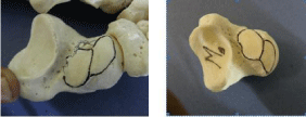
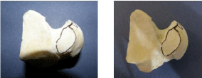
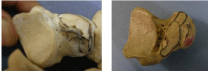
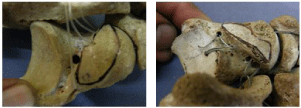
In the present study, four patterns of calcaneal articular facets of north-west Uttar-Pradesh adult’s tali were observed and classified (Type I – IV).
From the above figures it was observed that…
- Type –I (40%) shows two articular facets on their planter surfaces.
- Type –II (30%) shows ridged dividing articular facets into two parts.
- Type –III (20%) shows two articulating facets of different size by non-articulating groove.
- Type –IV (10%) showed a single facet.

Type I: Two articular facets on planter surfaces of the tali.

Type II: Ridge dividing articular facets into two parts.

Type III: Two articulating facets of different size by non-articulating groove.

Type IV: A single facet on planter surface of tali.
Being familiar with the various patterns of facet may help in accurate reduction of fractured tali by open reduction and internal fixation. Fracture stabilization in anatomic reduction can be achieved by screw fixation (With the K-wires placed appropriately and verifying accurate reduction with image intensification, cannulated screws can be placed over the K-wires). Typically lag screws are used to compress and fix the talar neck fractures, to bear early motion which is beneficial for ankle and subtalar joint function. When there is comminution of the medial talar neck, the use of a lag screw may be contraindicated, as it will cause deformity and malunion. Transfixion screws are used to avoid over compression and maintain the correct length of the talus [9,10]. Inserting the screw in two ways (i.e antero-posterior and postero-anterior). Two screws (one medial and one lateral) are inserted from a point just off the articular surface of the head and directed posteriorly into the body. Posterior-toanterior screw placement provides superior mechanical strength compared with insertion from anterior to posterior [11]. Drawbacks of posterior-to-anterior screw fixation include penetration of the subtalar joint or lateral trochlear surface, injury to the flexor hallucis longus tendon, and restriction of ankle plantar-flexion due to screw head impingement. These potential problems can be minimized by placement of smaller-diameter countersunk screws directed along the talar axis. Once the fixation completed image intensification intraoperatively will ensure accurate reduction of all talar facets. The Canale views of the ankle and foot will demonstrate whether or not talar neck reduction and fixation has been achieved satisfactorily.
Discussion
Most researchers view the differences in facet configuration of talus and calcaneum as anatomical variations of no functional significance. Bruckner in contrast, argues that the 2-facet configuration is more stable than the others. The 2-facet configuration is typically associated with a higher angled subtalar joint axis and a sharper intersecting angle, i.e. “Critical angle of Gissane” of the anterior and medial facets [12]. These characteristics, in conjunction with the posterior talocalcaneal facet, cause the talus to sit on an ‘osseous tripod’ and prevent excess motion of the talar head. A 2-facet configuration is more stable, there is less evidence of pathological changes associated with this configuration as compared to unstable joints [13-15]. In the present study majority of tali shows the 2- facet configuration. Different type and similar dominance of articular facets of tali for calcaneum are also present in different foetal studies indicating that they are probably genetically determined, as foetuses are not exposed to post natal influences [16-17]. Severe fracture of talus ends up in disruption of articular congruity and/or loss of talar length, alignment, and rotation. Operative treatment is usually necessary to restore hind foot anatomy and mechanics, as well as joint congruity in the majority of these fractures. Operative treatment of talar neck and body fractures required detailed knowledge of talar facets and of the subtalar joint. Even small residual-fracture displacement (1- 2mm) can result in a significant compromise of subtalar, ankle, or talonavicular joint function. Failure to unite in anatomical alignment results in malunion and posttraumatic arthritis, a malunited fracture drives the foot into a varus (adducted posture) and the patient bears weight along the lateral border of the foot. Corrective osteotomies have been suggested for treating varus malunions due to the resultant deformity and loss of subtalar motion. Osteonecrosis of a significant portion of the weight bearing surface with collapse is the most dreaded complication of talar neck fractures.
The goal of treatment of talar neck fractures is anatomic reduction, which requires attention to proper rotation, length, and angulation of the neck. Biomechanical studies on cadavers have shown why precisely reducing talar neck fractures leads to better outcomes. In one cadaveric study, displacements by as little as 2mm were found to alter the contact characteristics of the subtalar joint, with dorsal and medial or varus displacement causing the greatest change. The weightbearing load pathway changed, and contact stress was decreased in the anterior and middle facets but was more localized in the posterior facet [18]. In another study, varus alignment was created by removing a medially based wedge of bone from the talar neck. This resulted in inability to evert the hindfoot, and the altered foot position was characterized by internal rotation of the calcaneus, heel varus, and forefoot adduction [19]. The altered hindfoot mechanics with a talar neck fracture may be one factor that leads to subtalar posttraumatic arthrosis. For these reasons, open reduction and internal fixation is recommended for displaced fractures.
Conclusion
The present study on human tali and detailed anatomic information will be the baseline for science of anatomy, various orthopedic diagnostic and treatment procedures. The complexity of talar anatomy, its physiological role in functioning of lower limb and variability of fracture pattern affects the outcomes of talus fracture. Therefore, Orthopaedic surgeon should have thorough knowledge of osseous anatomy, vascular supply and modern methods of fixation. The variations in the inferior surface of the talus enable the tali to be classified according to the number and disposition of the articular facets of the talus for calcaneum, being familiar with various pattern may help in correction of various foot deformity, treating the commonly encountered joint instability and screw fixation of fracture talus. Three dimensional computerized imaging techniques may present facet surfaces of talus and calcaneus. So in the talocalcaneal subluxation, dislocation of fractured fragments of talus, coalition and many dysmorphologies, success rate of diagnosis and treatment will increase and talocalcaneal joint implants and prosthesis may be developed.
References
- Hollinshead WH. Ankle and foot. In: Anatomy for surgeons. Med Book Department Harper and Brothers. 1958; 3: 832-834.
- McMinn RMH. Bones of the foot. In: Last’s Anatomy- Regional and Applied. London: Churchill Livingstone. 1993; 231-233.
- Kleiger B. Fractures of the talus. J Bone Joint Surg. 1948; 30: 735-743.
- Ranganathan TS. A Text book of Human Anatomy Chand and Company Ltd- New Delhi. 1993; 233-234.
- Yu-Chi Huang. The relationship between the flexible flatfoot and plantar fasciitis: Ultrasonographic evaluation. Chang Gung Med J. 2004; 27: 443- 447.
- Harris RI, Beath T. Etiology of peroneal spastic flatfoot. J Bone Joint Surg. 1948; 30: 624-633.
- Arora AK, Gupta SC, Gupta CD, Jeysing P. Variations in calcaneal articular facets in Indian tali. Anat Anz. 1979; 146: 377-380.
- Bilodi AK. Study of calcaneal articular facets in human tali. Medical J Kathmandu Univ. 2006; 4: 75-77.
- Fortin PT, Balazsy JE. Talus fractures: evaluation and treatment. J Am Acad Orthop Surg. 2001; 9: 114–127.
- Herscovici D, Anglen JO, Archdeacon M, Cannada L, Scaduto JM. Avoiding complications in the treatment of pronation-external rotation ankle fractures, syndesmotic injuries, and talar neck fractures. J Bone Joint Surg Am. 2008; 90: 898-908.
- Swanson TV, Bray TJ, Holmes GB. Fractures of the talar neck: A mechanical study of fixation. J Bone Joint Surg Am. 1992; 74: 544-551.
- Bruckner J. Journal of Orthopedic and Sports Physical Therapy. 1987; 8: 489- 494.
- Kapandji IA. The Physiology of the Joints, 2, Lower Limb. New York: Churchill Livingstone. 1970.
- Harris NH. The ankle and foot. In Postgraduate Text-book of Clinical Orthopedics. 1983; 840-870.
- Olson TR, Seidel MR. Foot and Ankle. 1983; 3: 322-341.
- Fazal-Ur-Rehman. Study of human fetal tali calcaneal articular facets. International Journal of Development Research. 2014; 4: 1362-1365.
- Banning PSC, Barnett CH. Journal Anat. 1965; 99: 77-76.
- Sangeorzan BJ, Wagner UA, Harrington RM, Tencer AF. Contact characteristics of the subtalar joint: The effect of talar neck misalignment. J Orthop Res. 1992; 10: 544-551.
- Daniels TR, Smith JW, Ross TI. Varus misalignment of the talar neck: Its effect on the position of the foot and on subtalar motion. J Bone Joint Surg Am. 1996; 78: 1559-1567.