
Research Article
Austin J Anat. 2016; 3(3): 1059.
Incidence of Metopism in Adult Skulls from Southeast Brazil
Bernardes FM¹, Giroldo AM¹, Roquette AGD² and Marques KV³*
¹Medical School of Federal University of Uberlandia, Brazil
²Service of Neurosurgery from Clinical Hospital of Federal University of Uberlandia, Brazil
³Department of Surgery, Medical School of Federal University of Uberlandia, Brazil
*Corresponding author: Marques KV, Department of Surgery, Medical School of Federal University of Uberlandia, Pedro Quirino St, Uberlandia, Minas Gerais, Brazil
Received: October 04, 2016; Accepted: December 21, 2016; Published: December 30, 2016
Abstract
Objectives: The aim of this study was to determine the incidence of persistence of metopic suture in human skulls of Brazil Southeastern and compare with literature.
Methods: A collection of 42 human skulls from Nucleus of Morphology was used in order to identify persistent metopic suture. The subjects were catalogued and documented with photographs. Craniometrics points were used in order to obtain dimensions of metopic suture.
Results: An incidence of 4.76% of metopism was obtained in analyzed skulls, which all were classified as complete metopic sutures, with a linear pattern intercalated with serrated and interdigitating pattern.
Conclusion: Although its low incidence, knowing metopic sutures’ anatomy and its correlation to imaging exams is essential to establish different diagnosis in trauma medical care and practice.
Keywords: Frontal bone; Cranial sutures; Anatomy; Neuroanatomy
Introduction
The anatomical characterization of the metopic suture has been well described in the literature with emphasis on its clinical relevance in diagnosis and surgical options for craniosynotosis [1]. The metopic suture (also known as the medial frontal suture) extending from the nasofrontal suture (nasion) superiorly along the midline to bregma runs exactly through the median line of the frontal bone [2]. Its timing of closure is still controversial. Previous studies suggest that the closure of the metopic suture normally starts from birth to 3 months of age and is generally fused completely at the age of one or two years [3-5], by the fourth year of age [6], between the fifth or sixth year [7-9], or by the seventh year [10] by eight years of age [11-13] or by tenth year of age [14]. However, it is widely accepted closing around the age of 2 years olds [13]. A persistent metopic suture in adult skull is relatively rare. The persistence of metopic suture into adulthood is called as metopism [15-16]. The anatomy of the metopic suture has been reported in many populations such as Lebanese [10], Indian [17-19], Anatolian [20], Iranian [21], Nigerian [9]; and Brazilian [22- 24]. Racial variations in the incidence of metopic sutures and shapes have been observed and reported in the literature [25].
Knowing the anatomy patterns of metopic suture in skulls is important for radiologists and neurosurgeons to differ trauma in the frontlines, differing it from frontal bone’s fracture or even sagittal sutures. Beyond that, it is equally important to paleodemography and forensic medicine professionals establish parameters of frontal bone’s fracture and metopic suture. The aim of this study was to determine the persistence of the suture metopic in human skulls of South Eastern Brazilian and compare it with previous literature.
Materials and Methods
A total of 42 human dry skulls were analyzed for persistency of metopic suture, in the Laboratory of Human Anatomy from Medical School of Federal University of Uberlandia, a southeastern institution. Specimens were obtained in accordance with Law 8501 of November 30th, 1992, which rules the use of unclaimed cadavers for teaching and research purposes in medical schools. Data from specimens, including age and gender were not assessed and none of them showed visible abnormalities.
Classification of the metopic suture
A macroscopic observation of shape and main characteristics of the sample was made and documented in records and photographs. Skulls with metopic suture can be divided in three specific types: type one, normal, without metopic suture; type two, complete metopic suture, which extends from the nasion to the bregma uninterruptedly; and type three, with incomplete metopic suture. They can also be divided in groups according to shape and pattern: linear, ‘U’ shaped and ‘V’ shaped) [9]. In (Figure 1) we show different classifications for metopic suture.
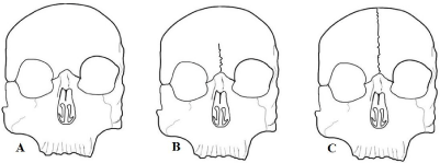
    Figure 1:  Schematic of complete and incomplete metopic suture. (a) Type 1 as normal without any metopic suture; (b) Type 2 with incomplete metopic suture, this
is grouped according to their shape namely linear, ‘U’ shaped and ‘V’ shaped and (c) Type 3, complete metopic suture (extending from the nasion to the bregma
uninterruptedly).
In our present study, two investigators performed a meticulous search for metopic sutures in our sample, and classified it according to pattern and shape. In case of any disagreement during observations, a third investigator was consulted for a final determination. Our method of classification was based on previous descriptions in the literature [26-27].
Morphological analysis and measurements
Morphological analysis was performed in order to classify metopic suture in complete and incomplete pattern. The distance between nasal sutures to coronal suture from midline was measured with measuring tape. Data collected will be compared to findings in previous studies.
Results
Persistent metopic suture was observed in two adult human skulls of our sample, which provides an incidence of 4.76%. Both specimen were classified after morphological assessment in complete sutures, and differ in length (14 and 12 cm, respectively) and pattern. In skull A, a linear sketch in the portion right superior to nasion, which afterword becomes more serrated next to bregma (Figure 2). In skull B, an interdigitating line in portion next to nasion, which becomes linear in middle portion, until its end in bregma with a serrated pattern (Figure 2)? The patter is also shown in different angles and better definition in (Figure 3).
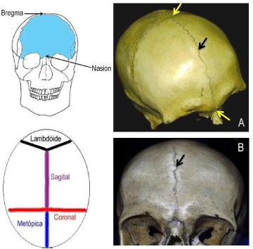
    Figure 2:  Schematic drawing representing the craniometric points (Bregma and nasium). Yellow arrows indicate bregma and nasium; black ones indicate the
metopic suture in adult human skulls.
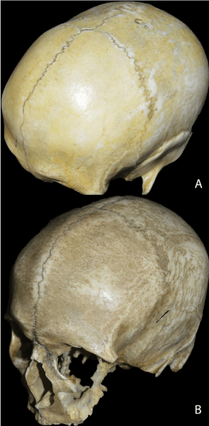
    Figure 3:  Left latero-anterior incidence of human skulls presenting persistence
of metopic suture. The pattern of both samples is showed in more detail.
Discussion
In this presented study, an incidence of 4.76% of persistent metopic suture was observed, which was similar to findings in Brazilian Northeast (4.48%) [24], Asia yellow race and Mongolian [28]. In (Table 1), we present the comparison of incidences among different studies around the world.
![]()
    Slno
    Workers
    Race
    % of the incidence of MS


    1
    Agarwal et al. (1979)
    Indian
    2,66%


    2
    Ajmani et al. (1983)
    Nigerians
    3,40%


    3
    Bergman apud Nayak (2006)
    European Caucasians
    8,70%


    Asians
    5,10%


    Blacks
    1,20%


    Australian
    1,00%


    4
    Berry et al. (1973)
    Nigerian skull
    3,00%


    5
    Bilodi et al. (2003)
    Nepalese
    11,46%


    6
    Breathnach et al. (1958)
    European
    7-10%


    Yellow races
    4-5%


    Africans
    1,00%


    7
    Bryce et al. (1915)
    European
    8,70%


    Scottish
    9,50%


    Mongolism
    5,10%


    Negros
    1,20%


    Australian
    1,00%


    8
    Castilho et al. (2006)
    Southerm Brazil
    7,04%


    9
    Chandrasekaram (2011)
    South Indian
    5,00%


    10
    Das et akm 1973
    Indian (UP)
    3,31%


    11
    Fakhroddin et al. (1967)
    European
    7 to 10%


    Yellow races
    4,50%


    12
    Gupta et al. (2012)
    Indian (U.P)
    5,00%


    13
    Herker et al. (1981)
    Afrikan
    1,00%


    Maharastrian (Miraj)
    3,00%


    14
    Jit & Shah et al. (1948)
    Indian (Panjabi)
    5,00%


    15
    Murlimanju et al. (2010)
    Indian
    1,20%


    16
    Romanes et al. (1972)
    European
    8,00%


    17
    Silva et al. (2013)
    European
    4,48%


    18
    Woo et al. (1949)
    Mongoloid
    10,00%


    Negroids
    2,00%


    19
    Del Sol et al., 1989
    Brazilian
    2,75%


    20
    Sudhakar et al. (2010)
    India Central
    3,95%


    21
    Saheb et al. (2010)
    Indian -South
    3.2%


    22
    Eroglu (2008)
    Anatolium
    3.3 to 14.9%


    23
    Present Study
    South Eastern Brazilian.
    2.00%


    24
    Masih et al. (2013)
    western rajasthan
    6.5%


    25
    Baaten et al. (2003)
    Lebanese
    0.82%


    26
    Chakravarthi & Venumadhav  (2012)
    South Indian
    6.25%


    27
    Silva et al. (2013)
    Adult Brazilian (maceio) northeast region
    7%
Table 1: Comparison of Incidence of metopism in different races as reported by various studies in the last twenty years.
Previous literature described three types of pattern in metopic sutures and its frequency: linear (18.75%), ‘U-shaped” (10%) and “V-shaped” (10%) [28]. Despite that, in our study, only linear pattern was observed, which was similar to several studies. Our suture measurements were similar to found by Castilo et al. (2006), with mean length of 13cm.
In Brazil, previous studies reported different frequencies of metopism in adult human skulls, ranging from 2.5% to 7.4%, showing an important correlation with rationality, with higher incidence in southeast region [22-24]. Comparing to different countries, our incidence was higher than found in India (2.66%) [26] and Nigeria (3.40%) [9] and Australian [29], but lower than Europe (8.7%) [29], Nepal (11.46%) [20], Scotland (9.5%) [31].
Differences in incidence of metopic suture can be linked to geographical differences [32], ethnicity [33] and racial difference [34]. Higher incidence of metopic suture is also associated with temperate zones in globe [35].
The study of sutures’ variation has gained importance in the last years in the area of neurosurgery and radiology. Among several variations, the persistence of metopic suture is an important differential diagnosis with fractures of the frontal bone [36]. In radiographic exams, incomplete metopic sutures can be confused with vertical fractures next to the central line of scalp [23].
It is known that metopic suture usually disappears in childhood by the process of osteogenesis, however, in some cases, it persists like a complete suture extending from nasion to anterior angle of bregma, being denominated metopism. When there is a complete persistence of metopic suture, its part right above the nasion is usually linear, which was also found in our study [9].
Conclusion
This study has identified an incidence of 4.76% persistence of metopic suture in Southeastern Brazilian. The persistence of metopic suture can be associated with regional and racial factors, especially in Brazil. This new study can help us understand the incidence of the metopic suture in sub-populations of Brazil. At the same time, knowledge of its anatomy and correlation to imaging exams is important to differential diagnosis in medical urgency service. Morphometric characteristics, like shape, directions and pattern may be useful for diagnostic and surgical interaction particularly during frontal craniotomy in daily practice of neurosurgeons and help radiologists to prevent mistaken diagnosis for vertical frontal bone fractures in X-ray, CT, or MRI.
Acknowledgment
Our acknowledgments to Miguel Antonio Facury Neto, former titular professor of Human Anatomy from Institute of Biomedical Sciences of Federal University of Uberlandia, by his initiative and support to this study.
References
- Van der Meulen J. Metopic synostosis. Child’s Nervous System. 2012; 28: 1359-1367.
- Standring S, Borley NR, Collins P, Crossman AR, Gatzoulis MA, Healy JC, et al. Anatomy: The Anatomical Basis of Clinical Practice. 2016; 613: 409- 472.
- Ashley-Montagu M. The medio-frontal suture and the problem of metopism in the primates. J Roy Anthrop Inst Great Britain Ireland. 1937; 67: 157-201.
- Keith A. Human embryology and morphology London, Edward Arnold. 1948.
- Manzanares MC, Goret-nicaise M, Dhen A. Metopic sutural closure in the human skull. J Anat. 1998; 161: 203-215.
- Piersol GA. Human Anatomy. Philadelphia: Lippincott. 1996.
- Torgerson J. Developmental, genetic evolutionary meaning of metopic suture. Am J Phys. Anthropol. 9: 193-210; 1951.
- Romanes GJ. Cunningham's Textbook of Anatomy, London: Oxford University Press. 1972.
- Ajmani ML, Mittal RK, Jain SP. Incidence of the metopic suture in adult Nigerian skulls. J Anat. 1983; 137: 177-183.
- Baaten PJJ, Haddad M, Abi-Nader K, Abi-Ghosn A, Al-Kutoubi A, Jurjus AR. Incidence of metopism in the Lebanese population. Clin Anat. 2003; 16: 148–151.
- Warwick R, Williams PL. Gray's Anatomy. London: Longmans. 1980.
- Mathijissen IM, Vaadrager JM, Can Der Meulen JC, Pieterman H, Zonneveld FW, Dreiborg S, et al. The role of bone centers in the pathogenesis of craniosynostosis: an embryologic approach using CT measurements in an isolated craniosynostosis and Apert and Crouzon syndromes. Plast Reconstr Surg. 1996; 98: 17-26.
- Bademci G, Kendi T, Agalar F. Persistent metopic suture can mimic the skull fractures in the emergency setting? Neurocirugia. 2007; 18: 238–240.
- Jit I, Shah MA. Incidence of frontal or metopic suture amongst Punjabi adults. Indian Medical Gazette. 1948; 83: 507.
- Vu HL, Panchal J, Parker EE, Levine NS, Francel P. The timing of physiologic closure of the metopic suture: a review of 159 patients using reconstructed 3D CT scans of the craniofacial region. J Craniofac Surg. 2001; 12: 527–532.
- Gardner S. A Persistent Metopic Suture: A Case Report. Austin J Anat. 2016; 3: 1049.
- Nayak S. Presence of Wormian bone at bregma and paired frontal bone in an Indian skull. Neuroanatomy. 2006; 5: 42-43.
- Mangalgiri AS, Satpathi DK, Razvi R, Naik DC. Study of Metopism in Skulls of Central India. Indian Journal of Forensic Medicine & Toxicology. 2010; 74-76.
- Basha MPA, Sugavasi R. Study of metopic suture in south Indian skulls. Int J Res Med Sci. 2015; 3: 2237-2239.
- Eroglu S. Journal: Clinical Anatomy - CLIN ANATOM. 2008; 12: 471-478.
- Tavassoli MM. Metopism: As an Indicator of Cranial Pathology; A Good Example from Iranian Plateau. Acta Medica Iranica, [S.l.]. 2011; 331-335.
- Del Sol M, Binvignat O, Bolini PDA, Prates JC. Metopismono individuo brasileiro. Rev Paul Med. 1998; 107: 105-107.
- Castilho MAS, Oda JYS, Goncales DM. Metopism in Adult Skulls from Southern Brazil. Int J Morphol. 2006; 24: 61-66.
- Silva IN, Fernandes KJM, Ramalho AJC, Bispo RFM, Rodrigues CFS, Aragao JA. Occurrence of Metopism in Dry Crania of Adult Brazilians. ISRN Anatomy. 2013.
- Vikram S, Padubidri JR, Dutt AR. “A rare case of persistent metopic suture in an elderly individual: Incidental autopsy finding with clinical implications”. Archives of Medicine and Health Sciences. 2016; 2: 61.
- Agarwal SK, Melhotra VK, Tewari SP. Incidence of the metopic suture in adult Indian crania. Acta Anat. 1979; 105: 469-474.
- Chandrasekaran S, Deepti S. A study on metopic suture in adult south Indian skulls. International Journal of Basic Medical Science. 2011: 379-382.
- Chakravarthi KK, Venumadhav N. Morphological study of metopic suture in adult South Indian skulls. Int J Med Health Sci. 2012; 1.
- Nayak S. Presence of Wormian bone at bregma and paired frontal bone in an Indian skull. Neuroanatomy. 2008; 5: 42-43.
- Bilodi AK, Agrawal BK, Mane S, Kumar A. A study of metopic sutures in human skulls. Kathmandu Univ. Med J. 2004; 2: 96-99.
- Bryce TH. Osteology and arthrology. Quain’s Elements of Anatomy. London, Longman Green. 1915; 177.
- Hanihar T, Ishida H. Frequency variations of discrete cranial traits in major human populations. Hypostotic variations J Anat. 2001; 198: 707-725.
- Berry AC. Factors affecting the incidence of non metrical skeletal variations. Journal of Anatomy. 1975; 120: 519-535.
- Masih WF, Gupta S, Saraswat PK, Aggarwal SK. Autopsy Study of Metopic Suture Incidence in Human Skulls in Western Rajasthan. Natl J Med Res. 2013; 3: 63-65.
- Yadav A, Kumar V, Srivastava RK. Study of metopic suture in the adult human skulls of north índia. J Anat Soc India. 2010; 2: 232-236.
- Vikram S, Jagadish RP, Aswini RD. A rare case of persistent metopic suture in an elderly individual: Incidental autopsy finding with clinical implications. Archives of Medicine and Health Sciences. 2014; 2.
- Bergman RA, Afifi AK, Mityauchi R. Compendium of human anatomical variations. Baltimore: Urban and Schwarzenberg, 1989; 197-205.