Research Article
Austin J Anat. 2017; 4(1): 1061.
The Effect of Three-Dimensional Model on Learning of Cardiac Anatomy
Navid S¹, Mehdi Sajjadi S² and Khoshvaghti A³*
¹Department of Anatomical Sciences, Tehran University of Medical Sciences, Iran
²Department of Medical Laboratory Sciences, Birjand University of Medical Sciences, Iran
3Aerospace Medicine Research Center, Aerospace and Subaquatic Medicine Faculty, AJA University of Medical Sciences, Iran
*Corresponding author: Khoshvaghti A, Aerospace Medicine Research Center, Aerospace and Subaquatic Medicine Faculty, AJA University of Medical Sciences, Tehran, Iran
Received: November 04, 2016; Accepted: January 05, 2017; Published: January 09, 2017
Abstract
The aim of this study was to evaluate the effect of a three-dimensional model in learning of cardiac anatomy.
The study was conducted at AJA University of Medical Sciences in 3 phases in 2013:
- Preparation of three-dimensional model image of the heart according with reference to Gray’s Anatomy for Students (2010-1st edition).
- Forming two groups of nursing students; teaching with lecture plus PowerPoint slides to control groups and the first experimental group additional training with heart sheep and the second experimental group additional teaching with three dimensional model.
- Taking similar pre-test and post-test exam and statistical analysis.
- Analysis of pre-test and post-test scores in each group with t-test showed significant differences (p≤0.001). There was a significant difference in the mean of the first experiment with the heart sheep and the second experiment with the three-dimensional model (p= 0.016) but it didn’t cause significant difference in comparison with pre-test scores between groups (p = 0.28). Threedimensional model had positive effect on anatomy learning of cardiac anatomy and it did cause significant difference in comparison with both heart sheep dissection and traditional educational method such as PowerPoint slides.
Keywords: Anatomy; Teaching; Model; Heart
Introduction
The status of anatomical education in modern medical programs is a cornerstone of medicine and its related professions [1]. To serve the public’s needs, teaching approaches are modified in medical schools to educate physicians [2]. Cognitive ability to understand, mentally encode and manipulate Three-Dimensional (3D) visuospatial forms can be determined via spatial visualization ability [3]. Component processes of spatial visualization consist of a visuospatial stimulus encoding, visuospatial image construction from perceptual advice, mental rotation of the image, and changing the visual stimulus to an image in working memory [4]. The ability to relate Two- Dimensional (2D) with 3D representations can be required in some spatial visualization tasks, such as deriving a cross section, which is a 2D section of a 3D object. This ability to derive and interpret the spatial properties of cross-sections is essential in many sciences [5]. There may be good reasons for using these tools to teach human anatomy. First, the traditional teaching method of cadaver dissection may be less useful for some organs, such as the larynx. Among the anatomical structures, some are too small to dissect, such as arytenoid cartilage and some are too delicate, such as the recurrent laryngeal nerve [6,7]. Dissecting these structures is time-consuming and requires practice and dissection skills, which most medical students lack [7]. Second, using these models, the use of cadavers in anatomy medical school curricula can be decreased [8]. Finally, many of the disadvantages of cadaver dissections could be addressed by using 3D educational models [7]. Recent studies have shown that, compared to the traditional methods, 3D models of teaching anatomy are more effective and less stressful when it comes to learning difficult concepts [9-11]. In this study, a 3D model of the heart was created, and its educational value in teaching anatomy was assessed. Moreover, the effect of the 3D model on the learning of cardiac anatomy was studied. Furthermore, the effectiveness of this model was compared with the traditional approach to teaching, in most medical universities in Iran, which involves sheep heart dissection, and PowerPoint slides.
Materials and Methods
The study was conducted in three phases at AJA University of Medical Sciences in 2013. All of the participants were nursing students who had been accepted into the university via the same national exam. Three phases of the study are described below.
Phase 1: Design and preparation of the model
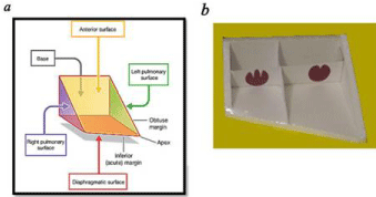
In this phase, a schematic representation of the cardiac section of Gray’s Anatomy for Students [12] was prepared for students (Figure 1a), and an innovative 3D model was designed (Figure 1b).

    Figure 1:  Three-Dimensional (3D) cardiac model (a) schematic diagram of
page 185 of the Gray’s anatomy for students (b) Innovative three-dimensional
model.
The model was created in cube form using affordable facilities and equipment. All surfaces were made of cardboard (thick and thin). Using this model, different parts of the heart were visible including; the surface of diaphragm and left lung, base and apex. To make the cardiac cavities visible, the atrial and ventricular, anterior surfaces of the model were covered with transparent sheets.
Phase 2: Training the experimental groups
In this study, 52 nursing students were selected and randomly divided into two groups. The students had no previous education concerning the relevant subject. The training session was carried out through lectures and PowerPoint slides for both groups. For the first experimental group, an additional training session was carried out using the sheep heart, whereas the 3D model was used for the second experimental group. Moreover, the model structure was explained and each part of the model was educationally described over 15 minutes. It should be noted that both groups were trained by one teacher and the total training time was similar in both groups.
Phase 3: Testing and analysis
The main purpose of study is to evaluate whether our 3D model improves students’learning of 3D anatomical structure cardiac. The 3D model is meant to improve students’ ability to simply recognize and name anatomical structures such as the surface of diaphragm and left lung, base and apex, the atrial, ventricular, and atrial septal, ventricular septal, and anatomic position of the tricuspid and mitral valve, as well as anatomic position of the heart in the thorax between the lungs. Following the Phase 2 training, Identical pre-tests and post-tests were designed to determine the effect of the model on the quality of students’ learning. In this regard, all students were given 12 short-answer questions at the beginning and end of the training session. The two groups were allowed the same amount of time to answer the questions. Exam sheets were corrected by another teacher. Data were collected and analyzed using the SPSS software version 16. The Student T-test was performed to evaluate the differences between groups.
Results
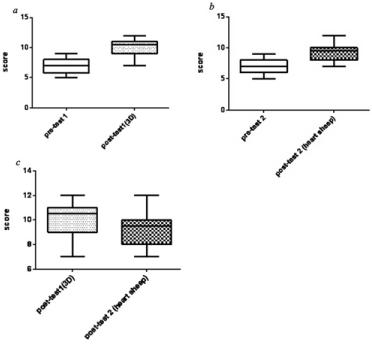
The mean pre-test and post-test scores for the first group were 7.08 ± 1.354 and 9.19 ± 1.327 respectively. Meanwhile, the mean pretest and post-test scores for the second group were respectively 6.69 ± 1.137 and 10.15 ± 1.461. The difference of mean in pre-test and posttest scores in each group with t-test was highly significant (p= 0.00) [Table 1, Figure 2 a,b].
    Group
    Mean Score
    Standard deviation (SD)
    Statically Test
    P-value


    Pre-test 1
    7.08
    1.354
    Paired Samples test
    p=0.00


    Post test 1(3D)
    9.19
    1.327


    Pre-test 2
    6.69
    1.137
    Paired Samples test
    p=0.000


    Post-test 2 (Heart of Sheep)
    10.15
    1.461
Table 1:  Comparison of the Statistical test pre-test and post-test scores in each
group.

    Figure 2:  Box plot of the groups (a) the mean pre-test and post-test scores for
the first group (b) the mean pre-test and post-test scores for the second group
(c) the first experiment with the heart of sheep and the second experiment
with the 3D model.
So practical training (either heart of sheep or 3D model) was useful in both groups.
Considering the mean post-test scores of the groups, there was a significant difference (p= 0.016) [Table 2, Figure 2 c].
    Group
    Mean score
    Statistical test
    P-value


    Pre-test 1
    7.08
    Independent Samples test
    0.28


    Pre-test 2
    6.69


    Post-test 1(3D model)
    9.19
    Independent Samples test
    0.016


    Post-test 2 (Heart of sheep)
    10.15
Table 2: Comparison of the Statistical test between groups.
Thus, 3D model had better effect on students’ learning compared to the sheep’s heart.
With regard to the mean pre-test scores, there were no significant difference between two groups (p = 0.28).
Therefore, the two groups were statistically homogeneous and they were at the same level of intelligence.
Discussion
In this study, we demonstrated that a 3D anatomical model of the heart enhances medical students’ learning of cardiac anatomy. Moreover, a significantly better outcome was obtained for the students who were educated using the 3D model. It is still hard to understand the spatial structure of the heart, and little attention has been given to this concept in previous studies. The negative or positive results of the previous studies may have been partly due to the study design. Due to the small size and delicacy of the middle and inner ear structures and the difficulty in cadaver dissection, Nicholson et al., (2006) created a 3D computer model of the ear. To generate this model, they used high-resolution Magnetic Resonance Imaging (MRI) and performed a randomized controlled trial with medical students as participants. As a result, they proved the efficacy of their model in teaching ear anatomy [7]. However, our 3D of cardiac model differs from the ear model of Nicholson et al. Our model is not expensive and is easy to use without any need to employ unusual equipment or processing steps too. In addition, we imposed a time limit on the participants for completing the tests. In another study, Khoshvaghti et al., (2010) investigated the effects of a 3D model in learning the anatomy of middle ear. They showed that the 3D educational model had positive effects on the learning of middle ear anatomy, however; no significant difference was reported between this method and the traditional educational method [13]. Compared to our study, more participants were included in Khoshvaghti et al.’s research Rubin et al., (1998) made a 3D laryngeal model from MRI and Computed Tomography (CT) imaging of a cadaver larynx. They used several units to create their model. Each unit was designed with individual muscles, cartilage, or ligaments. As an advantage, their model could be viewed on any plane or direction. The model was designed for medical education, but its efficacy for students was not examined. Furthermore, their model was never launched on a Webbased platform [14]. Compared to our study, this research was fully interactive and the greater complexity of the model structure led to a positive effect on the participants’ learning.
In three studies by Garg et al. (2002), a 3D educational computer model of the carpal bones was generated. The importance of this part of the upper limb has been demonstrated in terms of the examination and treatment of the fractures. This area is spatially complex, so the researchers sought to determine its association with the surrounding structures. Their model showed multiple views of the carpal bones via computer-based anatomy software. Garg et al. proved that the “dynamic display of multiple orientations provided by this software” [15-17] may suggest some advantages in medical students’ learning. In comparison, our model was much simpler than this 3D computer model; however, it was effective in the students’ learning increment.
Plank et al. (2009) introduced a method for the 3D individualization of the cardiac models. These models were based on high-resolution 3D anatomical and diffusion tensor MRI [18]. This method may be useful in clinical diagnostics to understand the heart structure for the surgery residents, whereas our 3D model could be helpful for teaching anatomy to medical students. It is also possible to generate this model by using inexpensive and easily available material. Thus, it can be widely used in all universities. Furthermore, the results of this study showed that students were satisfied and accepted this model as a suitable supplement to traditional teaching methods.
Conclusion
In conclusion, this study provides evidence concerning positive effect of the 3D model on the learning of cardiac anatomy. Since this model is less complex than the sheep’s heart, it may facilitate the teaching of cardiac anatomy in the future.
References
- Gilling water TH. The importance of exposure to human material in anatomical education: A philosophical perspective. Anatomical sciences education. 2008; 1: 264-266.
- Gregory JK, Lachman N, Camp CL, Chen LP, Pawlina W. Restructuring a basic science course for core competencies: An example from anatomy teaching. Medical teacher. 2009; 31: 855-861.
- Cohen CA, Hegarty M. Sources of difficulty in imagining cross sections of 3D objects. Proceedings of the Twenty-Ninth Annual Conference of the Cognitive Science Society; Cognitive Science Society Austin TX. 2007.
- Hegarty M, Waller D. Individual differences in spatial abilities. The Cambridge handbook of visuospatial thinking. 2005: 121-69.
- Russell-Gebbett J. Skills and strategies–pupils' approaches to three-dimensional problems in biology. Journal of Biological Education. 1985; 19: 293-298.
- Fritz D, Hu A, Wilson T, Ladak H, Haase P, Fung K. Long-term retention of a 3-dimensional educational computer model of the larynx: a follow-up study. Archives of Otolaryngology–Head & Neck Surgery. 2011; 137: 598-603.
- Nicholson DT, Chalk C, Funnell WRJ, Daniel SJ. Can virtual reality improve anatomy education? A randomised controlled study of a computer‐generated three‐dimensional anatomical ear model. Medical education. 2006; 40: 1081-1087.
- Parker LM. What's wrong with the dead body? Use of the human cadaver in medical education. The Medical journal of Australia. 2002; 176: 74-76.
- Bryner BS, Saddawi‐Konefka D, Gest TR. The impact of interactive, computerized educational modules on preclinical medical education. Anatomical sciences education. 2008; 1: 247-251.
- Davis J, Chryssafidou E, Zamora J, Davies D, Khan K, Coomarasamy A. Computer-based teaching is as good as face to face lecture-based teaching of evidence based medicine: a randomised controlled trial. BMC medical education. 2007; 7: 23.
- Kandasamy T, Fung K. Interactive Internet-based cases for undergraduate otolaryngology education. Otolaryngology--Head and Neck Surgery. 2009; 140: 398-402.
- Drake RL. Gray's anatomy for student: Elsevier Health Sciences. 2010.
- Khoshvaghti A, Ghasemi H. The Effect of Three-Dimensional Model on Anatomy Learning of Middle Ear. Journal of Iranian Anatomical Sciences. 2010; 7: 153-162.
- Rubin JS, Summers P, Harris T. Visualization of the human larynx: a three-dimensional computer modeling tool. Auris Nasus Larynx. 1998; 25: 303-308.
- Garg AX, Norman GR, Eva KW, Spero L, Sharan S. Is there any real virtue of virtual reality? The minor role of multiple orientations in learning anatomy from computers. Academic Medicine. 2002; 77: 97-99.
- Garg A, Norman GR, Spero L, Maheshwari P. Do virtual computer models hinder anatomy learning? Academic Medicine. 1999; 74: 87-89.
- Garg AX, Norman G, Sperotable L. How medical students learn spatial anatomy. The Lancet. 2001; 357: 363-364.
- Plank G, Burton RA, Hales P, Bishop M, Mansoori T, Bernabeu MO, et al. Generation of histo-anatomically representative models of the individual heart: tools and application. Philosophical Transactions of the Royal Society of London A: Mathematical, Physical and Engineering Sciences. 2009; 367: 2257-2292.