
Research Article
Austin J Anat. 2018; 5(1): 1077.
Renal Artery Division Pattern of Black Africans about 50 Cases Treated by Injection-Corrosion Method
Ndiaye Aï¹*, Ongoly M’ Bambo C², Ndiaye AS¹, Gaye M¹, Ndoye AK², Ab Ndiaye¹ and Dia A¹
¹Anatomy and Organogenesis Laboratory, Faculty of Medicine, Cheikh Anta Diop University, Dakar-Senegal
²Urology and Andrology Department, UHC Aristide Le Dantec, Dakar-Senegal
*Corresponding author: Ndiaye Aï, Anatomy and Organogenesis Laboratory, Faculty of Medicine, Cheikh Anta Diop University, Dakar-Senegal
Received: December 01, 2017; Accepted: January 09, 2018; Published: January 16, 2018
Abstract
Conservative renal surgery has undergone significant development in the treatment of kidney tumors. The knowledge of the arterial segmentation of the kidney constitutes a real introduction to this surgery. We have studied the renal artery division pattern by means of the injection-corrosion method applied on 50 kidneys punctured from African melanoderm subjects. The renal artery bifurcated into prepyelic anterior artery and retropyelicposterior artery in 80% of cases. It trifurcated in 20% of cases, in addition to anterior and posterior branches, into an polar branch affecting one of the poles. Secondarily, the anterior branch presented 2 to 5 segmental branches (apical, antero-superior, antero-middle, antéro-inférior and inferior), while the retropelvic branch extended for a segmental branch in the shape of an upper convex arch. These findings will constitute a data base useful to surgeons in order to prepare for a partial nephrectomy.
Keywords: Renal artery; Morphology; Arterial segmentation; Kidney
Introduction
Renal tumor surgery, long summarized in a simple total nephrectomy, is becoming more and more conservative. The indications for partial nephrectomy and lumpectomy are more frequent with diagnostic and therapeutic progress [1]. This state of affairs makes it essential to have a good knowledge of renal arterial segmentation in order to make surgical procedures as safe as possible. The aim of our study is to report the renal artery division mode and to systematize the renal arterial vascularization in the perspective of partial nephrectomy.
Materials and Methods
This study was conducted at the Pathology Anatomy laboratories of the Aristide Le Dantec Hospital, the Grand-Y off General Hospital and the Anatomy and Organogenesis Laboratory of the Sheikh University. Anta Diop from Dakar. We collected 72 kidneys from 36 fresh cadavers of African melanodermal subjects during medicolegal autopsies performed according to Rokitansky’s technique. The pair of kidneys was isolated en bloc with the abdominal aorta, inferior vena cava and the surrounding cellulo-fatty tissue. Then the posterior aspect of the aorta was incised on all its height. After renal catheterization of the ureters and aortic ostia, only 29 kidney pairs irrigated by a single renal artery (80.5% of kidneys removed) were injected with corrosion. We first injected the ureters with resin without dye and then the renal arteries by adding Red Congo to the resin. After 12 hours in the freezer, the kidneys were immersed in 20% hydrochloric acid for 24 h. The molding was obtained by gently rinsing the residue of the corrosion with tap water. Four pairs of poorly corroded kidneys due to improper handling of the resin were excluded from the study.
The parameters studied were:
• The primary division branches of the renal artery,
• The secondary division branches of the renal artery,
• Vascularized territories.
Results
The primary division branches of the renal artery
The renal artery was divided according to 2 modalities:
• The bifurcation into an anterior prepyelic branch and a posterior retropyelic branch, found in 40 cases (80%),
• Trifurcation: in addition to the prepyelic and retropyelic arteries, we had either a superior polar artery (Figure 1) in 3 cases (6%), or a lower polar artery in 4 cases (8%) irrigating respectively the superior pole and the lower pole.
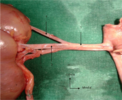
    Figure 1:  Trifurcation of the renal artery.
1: renal artery
2: anterior branch
3: posterior branch
4: superior polar artery
The branches of secondary division of the renal artery
• The prepyelic artery was divided into several segmental branches whose number varied between 2 and 4 (Figure 2a) and more rarely 5 (Figure 3a):
    Figure 2a:  Branches of division of the renal artery: anterior view.
1: anterior branch
2: apical segmental artery
3: antero-superior segmental artery
4: antero-inferior segmental artery
5: inferior segmental artery
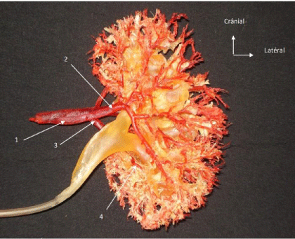
    Figure 2b:  Branches of division of the renal artery: posterior view.
1: posterior branch
2: posterior segmental artery
3: inferior segmental artery
4: posterior branch of the inferior segmental artery
• The apical segmental artery, 36 cases (72%),
• Antero-superior segmental artery, 50 cases (100%),
• The antero-middlesegmental artery, 2 cases (4%),
• The anterior-inferior segmental artery, 44 cases (88%)
• The inferior segmental artery, 46 cases (92%); this lower branch branched into an anterior branch and a posterior branch irrigating respectively the anterior valve and the posterior valve of the inferior pole.
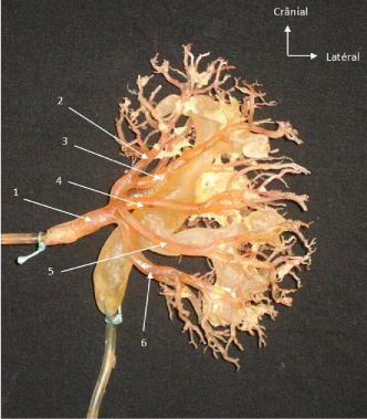
    Figure 3a:  Branches of division of the renal artery: anterior view.
1: anterior branch
2: apical segmental artery
3: antero-superior segmental artery
4: antero-middle segmental artery
5: antero-inferior segmental artery
6: inferior segmental artery
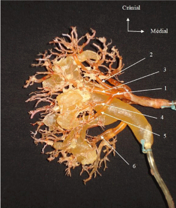
    Figure 3b:  Renal artery division pattern: rear view.
1: posterior artery
2: postero-superior segmental artery
3: superior postero-middle segmental artery
4: inferior postero-middle segmental artery
5: posterior branch of inferior segmental artery
• The retropyelic artery provided fewer segmental branches than the prepyelic artery. In 49 cases (98%), it was prolonged by a posterior segmental artery in the form of a concave arch inwardly pointing downwards and laterally (Figure 2b). From this arcade were born 4 to 5 branches destined mainly for mesorénal segment and superior pole. In one piece, the retropyelic artery gave 3 trunks superimposable to those of the anterior branch except for the inferior branch (Figure 3b).
• The superior polar artery, present in 3 cases (6%), vascularized the superior pole by its secondary branches. Its existence always coincided with the absence of the apical artery, branch of the prepyelic artery.
• The inferior polar artery, present in 4 cases (8%), vascularized the inferior pole by its secondary branches. Its existence always coincided with the absence of the inferior branch of the prepyelic artery.
Systematization
• Despite the differences in the arterial segmentation of each kidney, observations common to the kidneys were noted:
• The absence of anastomosis between the branches of the renal artery with a vascularization of the terminal type;
• The constant existence of the prepyelicanterior and retropyelicposterior arteries, branches of division of the renal artery;
• The existence of a convex avascular plane outside on the frontal plane; it is molded on the lateral edge of the mesorenal segment while being limited inside by the chalices;
• The existence of 2 anterior and posterior parenchymal territories which are separated by the chalices and the avascular plane.
The territorial arterial vascularization was thus:
The upper pole was entirely dependent on the apical artery in 36 cases (72%). If the apical artery did not exist, the superior pole completely depended either on the ramifications of the anterosuperior segmental artery in 7 cases (14%) or on the ramifications of the superior polar artery in 3 cases (6%). In 4 cases (8%), only the anterior valve of the superior pole depended on the antero-superior artery while the posterior valve depended on one of the branches of the retropyelic artery.
As for the mesorenal segment, the variations are less important. The anterior part of the mesorenal segment could be subdivided into 2 antero-superior and antero-inferior territories, each being dependent on the homologous segmental artery. In one case (2%), we had an antero-middle branch and therefore 3 territories in the anterior part of the mesorenal segment. The posterior territory of the mesorenal segment was dependent on a posterior segmental artery; in one case (2%), the retropyelic artery branched into 3 posterior segmental branches: postero-superior, postero-middle, and posteroinferior, dividing the posterior mesorenal segment into 3 territories (Figure 3b).
The lower pole was exclusively dependent on the inferior segmental artery, branch of the posterior artery in 46 cases (92%). This arterygived a branch for the anterior part of the inferior pole and another branch for the posterior part of the pole. The lower polar artery vascularized the entire lower pole in 4 cases (8%).
Discussion
Of all the arteries in the body, the renal arteries are among those with the greatest morphological variety. The purpose of this work is the systematization of arterial segmentation, we deliberately chose to inject and corrode only kidneys with a single renal artery so 80.5% of the pieces removed; kidneys with multiple renal arteries have undergone extensive dissection with skeletalization of the vessels, making difficult a injection-corrosion of quality.
The primary division branches of the renal artery
Although the bifurcation of the two-branched prepyelic and retropyelic renal artery is the divisional mode most commonly found by the majority of authors, we have also noted it in our series (80%), but this finding does not make unanimously. Some authors consider several other modes of division of the renal artery, the most common would be rather the trifurcation [1], this is the case of Ternon [2] and Kyle [3] which also does not find presegmental arteries in 49.3% of the 73 kidneys dissected during his study. In our series, the trifurcation of the renal artery in 3 presegmental branches was of the order of 20%.
• The polar presegmental branches:
They existed in 14% of cases. Several authors classify them in the polar arteries without distinction with the polar arteries arising from the aorta. Thus Sampaio [4] found 14% of polar arteries in his series, Satyapal [5] 10% and Edsman [6] 25% of polar arteries. This confusion could be avoided by clarifying the terminology, for example by retaining the term superior or inferior polar artery for a vessel that would arise from the aorta to penetrate directly to the upper or lower pole of the kidney, the term suprahilar artery or infrahilar for a vessel that would arise from the renal artery and would go directly to one of the kidney pole without passing through the hilum and renal artery any artery arising from the aorta and joining the hilum of the kidney.
The branches of secondary division of the renal artery
• Number of segmental arteries
Three to six segmental arteries are found per unit of kidney with a greater frequency of 5 segmental arteries (44%); exceptionally we found a kidney with 8 segmental arteries (Figures 3a and 3b). Thus, our results are similar to those of Sapte [7]. This author observes the existence of 3 to 6 segmental arteries with a preponderance of 5 segmental arteries per kidney in 42.78% of cases. Sampaio [8] found a number of segmental arteries varying between 4 and 5 with preponderance for 5 arteries found in 61.2% of cases.
• Distribution of segmental arteries
The apical segmental artery is the least common branch: 72% of the cases. The constant segmental branches are the anterosuperior segmental branch and the posterior segmental branch. Despite some disparities between our results and those reported in the literature, some commonalities are notable. Indeed, the majority of authors consider the posterior segmental artery to be a constant branch. Sampaio [8] during his work by the injection-corrosion method on 49 kidneys reports this constancy of the posterior segmental artery, Sapte [7] finds this same regularity on the 194 kidneys he studied with the same method than Sampaio, as well as Fine and Keen [9]. On the other hand, in his dissection work, Kyle [3] only found the posterior segmental branch in 75.3% of cases.
Systematization
The renal arterial ramifications noted that the segmentation of Graves [10] is still relevant. However, the apical segment did not always constitute a homogeneous vascular territory. Indeed in 8% of the cases of our series, it could be split into 2 parts anterior and posterior depending respectively on the prepyelic artery and the retropyelic artery. The avascular line of Hyrtl and Brodel is also a reality found on our molds and is therefore an incision area to be preferred.
Conclusion
The renal artery has many variations in its distribution. This study highlights the most common variations and is a database for conservative renal surgery but also the need to enrich the terminology of renal vessels.
References
- Chrétien Y, Méjean A, Cazalaa JB, Dufour B. Les techniques de chirurgie conservatrice pour tumeur du rein. Progrès en Urologie. 2000; 10: 134-141.
- Ternon Y. Anatomie chirurgicale rénale : base d’une segmentation artérielle du rein. J Chir. 1959; 78: 517-533.
- Kyle WJ, Sam BB, Belani J, Ames CD, Hruby G, Landman J. Extra renal vascular of kidney: Assessment of variations and their relevance to partial nephrectomy. J Urol. 2005; 66: 985-989.
- Sampaio FJB, Passos M. Renal arteries: Anatomic study for chirurgical and radiological practice. Surg Radiol Anat. 1992; 14: 113-117.
- Satyapal KS, Haffejee AA, Singh B, Ramsaroop L, Robbs JV, Kalideen JM. Additional renal arteries incidence and morphometry. Surg Radiol Anat. 2001; 23: 33-38.
- Edsman G. Accessory vessels of the kidney and their diagnosis in hydonephrosis. Acta Radiol. 1954; 42: 26-32.
- Sapte E, Bordei P. Anatomical considerations on the renal arterial segmentation. Rev Med Chir Soc Med Nat Iasi. 2005; 109: 597- 602.
- Sampaio FJ, Schiavini JL, Favorito LA. Proportional analysis of the kidney arterial segments. Urol Res. 1993; 21: 371-374.
- Fine H, Keen EN. Arteries of the human kidney. J Anat. 1966; 100: 881-889.
- Graves FT. The anatomy of the intrarenal arteries and its application to segmental resection of the kidneys. Br J Surg. 1954; 42: 132-139.