Special Article - Angiography
Austin J Anat. 2018; 5(1): 1078.
A Rare Case of Triple Coronary-Cameral Fistula Draining Into Left Ventricle in a Patient with Cocaine-Induced Myocardial Infarction
Patel H*, Hosein K and Shamoon F
Department of Internal Medicine, Division of Cardiology, New York Medical College, Saint Joseph’s Regional Medical Center, USA
*Corresponding author: Hiten R Patel, Department of Internal Medicine, Division of Cardiology, New York Medical College, Saint Joseph’s Regional Medical Center, New Jersy, USA
Received: December 22, 2017; Accepted: January 09, 2018; Published: January 16, 2018
Abstract
Coronary Cameral Fistula (CCF) is rare entity wherein there is an abnormal congenital or acquired anomalous connection between a coronary artery and a cardiac chamber. The right coronary artery is the usual origin of the communication, draining in the right sided chambers of the heart in 90% of cases while the connection between the left coronary arteries and the Left Ventricle (LV) occurs in only 10% of CCF. A CCF originating from all 3 major coronary arteries that terminates into the LV is an extremely rare phenomenon. Acquired CCF can occur secondary to cardiac surgery, trauma or after myocardial infarction. Here, we report a very rare case of triple CCF draining into LV in a patient with prior cocaine induced myocardial infarction and discuss the literature review.
Keywords: Triple; Coronary-cameral fistula; CCF; Left ventricle; Cocaine; Myocardial infarction
Introduction
Coronary Cameral Fistula (CCF) is an abnormal congenital or acquired anomalous connection between a coronary artery and a cardiac chamber. CCF is a rare entity reported in ~0.08% to 0.3% of unselected patients undergoing diagnostic coronary angiography. Acquired cases can be secondary to cardiac surgery, trauma or after myocardial infarction. 90% of CCF involves the right coronary artery with a fistula draining in the right sided chambers of the heart, while the connection between the left coronary arteries and the Left Ventricle (LV) occurs in only 10% of cases. Here we report and discuss a very rare case of a patient with prior cocaine induced myocardial infarction, who was found to have multiple small CCFs originating from all the 3 major coronary arteries and terminating into the LV cavity.
Case Presentation
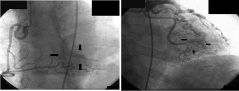
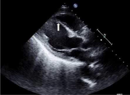
We describe case of a 67-year-old male patient with a history of tobacco and cocaine abuse who presented to the Emergency Department with shortness of breath occurring both at rest and with exertion. Vital signs showed HR of 110bpm, BP 118/70mmHg, respiratory rate of 20/min, O2 saturation of 96% on 2L of oxygen. Physical examination was remarkable for S3 gallop with mild rales over b/l lung. EKG showed sinus tachycardia with significant ST Elevation in leads V2-V5 with significant Q waves in V2-V4. Serial cardiac biomarkers were minimally elevated at 0.9ng/ml. Coronary angiography showed normal epicardial coronary arteries; however, during selective angiography of all 3 major coronary arteries, there was extensive draining of the contrast agent into the Left Ventricular (LV) cavity through many small, diffuse fistulae, resulting in complete LV contrast opacification (Figure 1). Hence, we diagnosed coronary cameral fistula involving all the 3 coronary arteries with each of them draining into LV. The shunt didn’t appear to be large and patient also didn’t complain of any exertional angina, so there was no coronary steal phenomenon and no acute intervention was deemed necessary at this time. Transthoracic echocardiogram revealed decreased LV ejection fraction of 35-40% with a thin akinetic septal and anteroseptal wall (Figure 2). His presentation was consistent with acute systolic heart failure due to prior MI and ischemic cardiomyopathy. He was discharged after improvement with a follow up in 1 week for electrophysiologic studies and possible intra cardiac defibrillator placement.

    Figure 1:  Panel A - Coronary angiography shows no lesions in epicardial
coronary arteries; but there are multiple CCF arising from diagonal artery and
obtuse marginal artery draining into the LV cavity (black arrows).
Panel B - Coronary angiography shows no lesion in right coronary artery but
there are multiple CCF arising from right posterior descending artery again
draining into the LV cavity (black arrow).

    Figure 2:  Transthoracic echocardiogram in parasternal long axis view
showing thin septal and antero-septal wall (arrow pointing to inter ventricular
septum) suggestive of old myocardial infarction.
LA = left atrium, LV = left ventricle, RV = right ventricle
Discussion
Coronary Cameral Fistula (CCF) is an abnormal congenital or acquired anomalous connection between a coronary artery and a cardiac chamber [1]. The incidence of coronary anomalies is estimated to be between 0.6 - 1.5% of patients undergoing invasive cardiovascular imaging [2] but CCF is a rare entity reported in ~0.08% to 0.3% of unselected patients undergoing diagnostic coronary angiography [3]. The right coronary artery is the usual origin of the communication, draining in the right sided chambers of the heart in 90% of cases. The connection between the left coronary arteries and the left ventricle occurs in only 10% of CCF, whereas a CCF originating from all 3 major coronary arteries that terminates into the left ventricle is an extremely rare phenomenon [4].
The clinical presentation depends on factors such as the age of patient, amount of shunting, and possible development of cardiac ischemia. Acquired CCF can be after cardiac surgery [5] or trauma [6] or after myocardial infarction [7,8]. Our case is unusual, not only because of the rareness of the particular type of CCF, but also because the patient does not have symptoms despite the presence of multiple and significant left ventricular communications. Such sizable communications would be expected to cause symptoms of increased left ventricular end-diastolic pressure and myocardial ischemia as a result of arterio-arterial shunt and the myocardial blood flow–stealing effect distal to the site of the CCF connection. Our case could have had congenital CCF and the use of cocaine made him more susceptible for myocardial ischemia and infarction, by worsening stealing effect in the coronaries. Or, our patient might have developed these fistulas after sustaining the myocardial infarction [8]. In any case, our patient is angina free likely because his myocardium is already infracted.
In general, the majority of adult patients are asymptomatic and the lesion is detected on physical examination as a murmur, classically described as soft and continuous and that tends to be crescendo decrescendo in both systole and diastole, but louder during diastole [9]. If symptomatic, chest pain is usually the most common presenting complaint among patients. While coronary angiography remains the gold standard for diagnosis; magnetic resonance imaging and multidetector computed tomography are attractive and safe alternative methods especially in kids to diagnose congenial CCF [10].
When considering treatment it is generally accepted that all symptomatic patients should undergo closure of medium to large coronary artery fistulae [11]. Asymptomatic or small coronary fistulae do not need to be treated; however, if these patients develop endocarditis or a long-term follow-up is not feasible, they should be treated [11]. Although therapy in significant asymptomatic CCF is still controversial, beta blockers have been tried and our patient was already on it for his ischemic cardiomyopathy.
Conclusion
Through our rare case of triple CCF draining in to LV cavity, authors hypothesize that cocaine could make a patient with congenital CCF more susceptible for myocardial infarction or it may cause an acquired CCF secondary to myocardial infarction.
References
- Challoumas D, Agamemnon, P, Inetzi DA. Coronary Arteriovenous Fistulae: A Review. Int J Angiol. 2014; 23: 1-10.
- H Patel, K Nemani, P Shah. Native and aberrant left anterior descending artery arising from left and right coronary sinus respectively: a very rare congenital coronary anomaly. Minerva Cardioangiol. 2017; 65: 94-96.
- Vavuranakis M, Bush CA, Boudoulas H. Coronary artery fistulas in adults: incidence, angiographic characteristics, natural history. Cathet Cardiovasc Diagn. 1995; 35: 116-120.
- Fragakis N, Giazitzoglou E, Katritsis. A Case of Coronary-Cameral Fistulae Involving All Three Major Coronary Arteries. Circulation. 2015; 131: e380-e381.
- Sgalambro A, Olivotto I, Rossi A. Prevalence and clinical significance of acquired left coronary artery fistulas after surgical myomectomy in patients with hypertrophic cardiomyopathy. J Thorac Cardiovasc Surg. 2010; 140: 1046-1052.
- Lowe JE, Adams DH, Cummings RG. The natural history and recommended management of patients with traumatic coronary artery fistulas. Ann Thorac Surg. 1983; 36: 295-305.
- H Patel, Y Sundermurthy, S Bhutani. A rare case of Contusio Cordis: Fist fight leading to an Acute Myocardial Infarction due to Left Anterior Descending artery dissection. J Cardiovasc Med Cardiol. 2017; 4: 026-028.
- Ryan C, Gertz EW. Fistula from coronary arteries to left ventricle after myocardial infarction. Br Heart J. 1977; 39: 1147-1149.
- Haller JA, Little JA. Diagnosis and surgical correction of congenital coronary artery-coronary sinus fistula. Circulation. 1963; 27: 939-942.
- Schumacher G, Roithmaier A, Lorenz HP. Congenital coronary artery fistula in infancy and childhood: diagnostic and therapeutic aspects. Thorac Cardio vasc Surg. 1997; 45: 287-294.
- Mangukia CV. Coronary artery fistula. Ann Thorac Surg. 2012; 93: 2084- 2092.