Case Presentation
Austin J Anat. 2018; 5(1): 1079.
Anastomosed Triplicated Branches of the Superior Cerebellar Arteries: A Rare Case of Cerebral Arterial Plexus
Nagawa E¹*, Aremu AM¹ and Kiryowa H²
¹Department of Human Anatomy, Islamic University in Uganda, Uganda
²Department of Human Anatomy, Makerere University College of Health Sciences, Uganda
*Corresponding author: Nagawa E, Department of Human Anatomy, Islamic University in Uganda, PO Box 2555 Uganda
Received: January 22, 2018; Accepted: February 09, 2018; Published: February 16, 2018
Abstract
Triplications of the superior cerebellar arteries are rare cases but when present are culminating factors to thromboembolism and arterisclerosis leading to infarction of areas supplied. We present a rare case of arterial plexus, where there were not only triplicated branches of the superior cerebellar but these anastomosed forming a single branch that later bifurcated normally lateral to the cerebral peduncles. This kind of formation is attributed to embryological malfusions of primitive neural arteries towards the basilar trunk. Anastomosis can be clinically important in neurovascular surgery ligation therefore caution should be taken not to damage the forming branches.
Keywords: Triplication; Anastomosis; Superior cerebellar artery
Case Presentation
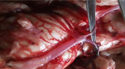
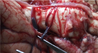
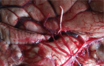
During a postmortem procedure carried out at the Kampala City Council Authority (KCCA) mortuary, the basal arterial complex showed a unique pattern of branching of the superior cerebellar arteries in a 30 year old male cadaver. The terminal end of the basilar artery branched into seven arteries. i.e. two posterior cerebral arteries, three right and two left superior cerebellar arteries (Figure 1). The arterial plexus was formed in such a way that the two branches (branches A and B) of the Superior Cerebellar (SCA) arose from the right side of the basilar artery. One artery (branch B) bifurcated 1.33mm from the main trunk and its upper branch anastomosed with the previous branch A forming a single branch C. (Figure 2). The formed branch C later bifurcated into two branches lateral to the cerebral peduncles (Figure 3). Branch C appeared engorged with a diameter of 2.24mm compared to its forming arteries (1.3mm and 1.86mm). There were no perforating branches from the arteries.

    Figure 1:  Heptafurcation of the basilar artery.
*The black asterics show the seven branches at the basilar artery terminal.
The 2 Posterior cerebral arteries are picked by the forceps.

    Figure 2:  Superior cerebellar arteries.
*Branches A and B- Anastomosing branches. Branch C- formed single
branch.

    Figure 3:  Branches of the formed branch C.
*The arrows point to the cut branches of the branches of the formed branch C.
Discussion
Variations of the SCAs have been observed in their origin, patterns of branching with other anomalies being fenestrations, tortuosity. Triplication cases have been reported by various studies though rarely compared to duplications which are either unilateral or bilateral. Padmavathi reported 23.3% bilateral duplication and 2% triplications [1]; Wankhede reported 17.5% unilateral duplications and 2.5% bilateral duplications [2] while Harrigan reported 26% bilateral duplications and 2% triplications [3]. Literature reviewed shows that, there are more single and duplicated SCA than triplicated cases among the populations studied. Most triplicated SCA were unilateral with a single branch of the SCA on the contralateral side. In this case the unique right triplication co-existed with a duplicated left SCA forming a unique plexus. In cases where the SCA branches were either duplicated or triplicated, there were also variant origins [4]. Some arteries arose from the Posterior Cerebral Artery (PCA) varying from the case presented where all branches arose normally from the basilar trunk.
The SCAs are in close proximity with the oculomotor nerve which separates it from the PCA, the trochlear and the trigeminal nerves [5,6]. The impingement on the oculomotor nerve depends on the angle between the PCA and the SCAs. Triplication of the SCA is most likely to reduce this angle limiting space occupied by both nerve and arteries especially in anastomosed state. This could lead to ocular complications in cases where the oculomotor and trochlear nerves are compromised.
We report an anastomosed kind of triplication where two branches of the triplicate join to continue as a single artery and later dividing into two caudal and rostra branches lateral to the cerebral peduncles [7]. The unison of these branches could be as a result of failed fusion of the primitive neural SCAs towards the basilar trunk to form a single primary branch [5]. The anastomosed kind of triplication can be viewed as advantageous and disadvantageous to the bearer. Anastomosis of arteries usually provides an alternative pathway for blood supply in case one artery is to be ligated for neurovascular surgery [8] or in case of blockage by plaques. The plexus kind of arterial anastomosis is a culminating factor to atherosclerosis, thrombosis that compromise blood flow leading to infarctions. Duplicated and triplicated branches usually contain perforating branches therefore caution should be taken during neurovascular surgery especially at the clivus and the cerebellopontine angle [9].
Conclusion
Although triplication of the SCA is a rare case, their presence is a culminating factor to thromboembolism, arterioscelorosis and atheromas. In situations where the triplicated branches anastomose with one another, care should be taken not to damage the alternative branches during neurovascular surgeries. Triplicated branches are more likely to cause compression of the oculomotor nerve than in duplicated or single branch cases.
References
- Padmavathi G. Study of the variations of superior cerebellar artery in human cadavers. Int J Res Med Sci. 2014; 2: 699-703.
- Wankhede HA, Nimje D, Hosmni P. Variations in the branches of basilar artery in adult human cadavers. Asian Pac. J Health Sci. 2015; 2: 161-166.
- Harrigan MR, Deveikis JP, Ardelt AA. Handbook of Cerebrovascular Disease and Neurointerventional Technique. Humana Press, Springer Science+Business Media. Essential Neurovascular anatomy. 2009; 10: 59- 61.
- Dodevski A, Lazarova DT, Zhivadinovik J, Lazareska M, Stojovska- Jovanovska E. Morphological characteristics of the superior cerebellar artery. Contributions Section of Medical Sciences. 2015; 36.
- Padget D. The development of the cranial arteries in the human embryo. Contrib Embryol Carnegie Inst. 1948; 32: 205–261.
- Pai BS, Varma RG, Kulkarni RN, Nirmala S, Manjunath L, Rakshith S. Microsurgical anatomy of the posterior circulation. Neurology India. 2007; 55: 31.
- Hardy DG, Peace DA, Rhoton AL. Microsurgical anatomy of the superior cerebellar artery. Neurosurgery. 1980; 6: 10-28.
- Vare AM, Bansal PC. Arterial pattern at the base of the human brain. J Anat Soc India. 1970; 19: 71–79.
- Dagcinar A, Kaya AH, Aydin ME, Kopuz C, Senel A, Demir MT, et al. The superior cerebellar artery: anatomic study with review. Neurosurgery Quarterly. 2007; 17: 235-240.