
Research Article
Austin J Anat. 2021; 8(1): 1096.
Morphometric Study of Frontal Horn of Lateral Ventricle of Brain and Its Correlation with Age, Gender and Side among Adults in the University of Gondar Comprehensive Specialized Hospital, Gondar, Northwest Ethiopia, 2019
Agegnehu A¹*, Tenaw B², Gebrewold Y³ and Jemberie M²
¹Department of Biomedical Science, Debre Tabor University, Ethiopia
²Department of Human Anatomy, University of Gondar, Ethiopia
³Department of Radiology, University of Gondar, Ethiopia
*Corresponding author: Assefa Agegnehu, Department of Biomedical Science, College of Health Science, Debre Tabor University, Debre Tabor, Ethiopia
Received: February 26, 2021; Accepted: March 29, 2021; Published: April 05, 2021
Abstract
Background: Knowledge of the frontal horn size is necessary for the initial and precise analysis of Ventriculomegaly. Therefore, having a baseline reference value of the frontal horn size will be beneficial in a huge vary of medical pathologies. The purpose of this study was to determine the frontal horn of lateral ventricle size and its correlation with age among adult patients in the University of Gondar Comprehensive Specialized Hospital, Northwest Ethiopia, 2019.
Methods: Hospital-based cross-sectional study design was conducted on 169 patients in the age group of 20-79 years in the University of Gondar Comprehensive Specialized Hospital. The study participants were assessed by way of structured questionnaires and checklists. Radiological residents were performed computerized tomography measurements of the frontal horn of lateral ventricle size. The data were entered using EPI INFO version 7 and analyzed by SPSS version 20. By Pearson’s product moment correlation coefficients, the correlations of frontal horn size with associated factors were evaluated. A paired t-test (between left and right side), independent t-test (between male and female) ANOVA (mean the difference between age groups) were performed. The p-value of less than 0.05was considered being significant.
Results: The mean right and left frontal horn length, width, and frontal horn tips diameter were 27.57 (±3.72) mm, 28.47 (±3.77) mm, and 4.90 (±1.54) mm, 5.08 (±1.58) mm, and 30.36 (±3.04) mm, respectively. In relation to gender, the measurement of the frontal horn is greater in males as compared to females.
Conclusion: In total, 169 patients satisfied the inclusion criteria. This study found a direct relationship between the frontal horn of lateral ventricle size with the age of both male and female adult participants. In all measurements, the frontal horn of the lateral ventricle size of male participants was greater than that of female participants.
Keywords: Frontal horns of lateral ventricles; Computed tomography; Interventricular foramen
Abbreviations
ANOVA: Analysis of variance; CT: Computed Tomography; GE: General Electric; EPI INFO: Epidemiological information; SPSS: Statistical Package for the Social Sciences
Introduction
Ventriculomegaly is a clinically important finding that associates with a number of pathological conditions [1]. In particular, enlargement of the frontal horn of lateral ventricles has been observed in alcoholism, normal aging, depression, dementia, and schizophrenia [2]. However, there is lack of evidence to date from available literature and in radiology practice of the ranges of the sizes of cerebral ventricles for the adult Ethiopians, because currently used reference values were drawn from other populations and races that have different epidemiological, demographic and some anatomical variations [3].
Radiologists and neurologists frequently confronted with problems of finding out whether or not ventricles are inside in normal limits or enlarged for a patient’s age. This has been a subjective decision based on experience, however, there are subjective errors resulting in misdiagnosis [4,5]. The frontal horn of the lateral ventricle is surgically exposed for over a century through a cortical incision in the frontal brain or through an inter-hemispheric approach [6].
Computerized Tomography (CT) is a safe non-invasive approach that routinely used to measure the ventricular structures of the brain. Moreover, it is safe and affords real-time images without the administration of anesthesia.
This research is significant in terms of theoretical and practical contribution to the existing body of research knowledge. Hence it is utmost important to check ventriculomegaly in all ventriculomegaly causing conditions.
Therefore, understanding the normal size of the frontal horn of the lateral ventricular system is helpful for clinicians, neurosurgeon, and radiologists in day-to-day practice.
Since the information about the normal size of the frontal horn of lateral ventricles was limited and no work was done on measurements of the frontal horn of lateral ventricular system in Ethiopia, the present work was undertaken to analyses the normal size of frontal horns of the lateral ventricles of the brain by CT scan method.
Methods
This prospective study was comprised of data collected from subjects under computed tomographic evaluation for diseases not effecting the ventricular system and brain parenchyma conducted between January and March 2019 in the University of Gondar comprehensive specialized Hospital, Department of Radiology. Ethical clearance was obtained from the University of Gondar Research and Publication Office, ethical review committee. Official letter was submitted to University of Gondar hospital, Department of Radiology. Study subjects were informed about the purpose of the study and its procedure. Informed verbal consent was obtained from each individual at the time of data collection.
The patients selected for the present study were examined using General Electric GE (Bright speed 4 slices) with 5mm slice thickness CT scanner for head and brain imaging due to different patient compliant. The study subjects had no history of cerebral infarction, local mass lesions, probable communicating hydrocephalus, alcoholism, drug abuse, trauma or previous intra-cerebral surgery. Besides this, sex, age and other demographic features of subjects were documented.
The frontal horn length was measured from interventricular foramen to the tip of the frontal horn on both sides (Figure 1), FHTD was measured by connecting the two tips of the frontal horns (Figure 2), and frontal horn width defined as the maximum distance measured transversely on each frontal horn. To maintain reproducibility, each measurement was repeated at least 3 times and most repeated value was recorded according to the guidelines of the American Institute of CT scan in Medicine and as described by lamp and collaborators.
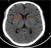
    Figure 1: CT axial image of the brain showing the length of the frontal horn of
lateral ventricles (captured at radiology department).
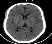
    Figure 2: Axial CT image of the brain shows the maximum distance between
the tip of frontal horns and maximum frontal horn width (captured at radiology
department 2019).
The computed tomographic measurements of frontal horn size were measured by the experienced radiologist. Cross checking numerical values were done at least three times while recording and transferring to the statistical Software Package for Social Sciences (SPSS). The collected data were checked for completeness, accuracy and clarity before analysis. The data were entered into a spreadsheet and analyzed using the IBM SPSS Statistics, version 20. The means (± standard deviation), ranges, minimum, maximum, and the 95% confidence intervals for the mean (in order to include the true population, mean in 95% of the cases) were all calculated. Data were grouped into six age groups 20-29 years, 30-39 years, 40-49, 50-59, 60-69 and 70-79 years. One-way analysis of variance was used to check for differences in frontal horn size across age groups. P-Value less than 0.05 is considered as statistically significant. Differences of continuous variables between two independent groups were assessed with the 2-tailed t test.
Results
A total of 169 adults encompassing 72.78% males and 27.22% females were enlisted. The age range of the study populations was between 20 and 79 years, with the mean age of 40.80 years (±18.06). As it is presented in Table 1, the frontal horn size indicated gradual increment in size from the age group of 30-39 years onwards with the greatest value in the age group 70-79 years in both sides. The minimum mean of frontal horn length, width and tips diameter were obtained in the age group of 30-39 years, whereas the maximum mean was obtained in the age group of 70-79 (Table 1).
![]()
    Age (years)
    Frequency
    Mean of frontal horn size and SD    (mm)
    Tips diameter


    Length
    Width


    Right
    Left
    Right
    Left


    20-29
    64
    25.21±2.22
    26.06±2.33
    4.08±1.10
    4.23±1.17
    29.17±2.50


    30-39
    25
    23.79±2.07
    24.66±1.94
    3.74±0.87
    3.92±0.83
    28.65±1.53


    40-49
    30
    28.56±1.69
    29.53±1.85
    4.79±0.79
    4.99±0.75
    30.47±1.81


    50-59
    13
    30.71±1.25
    31.53±1.13
    5.46±0.68
    5.63±0.67
    29.42±2.48


    60-69
    12
    31.55±2.16
    32.73±1.65
    6.37±0.79
    6.52±0.77
    31.83±1.63


    70-79
    25
    32.64±1.55
    33.51±1.56
    7.28±1.07
    7.53±1.10
    34.76±2.92
Table 1: Mean and standard deviations of frontal horn length, width and tips diameter of the adult age groups of 169 subjects, computerized tomographic study of
frontal horn of lateral ventricles.
Table 2 shows that various measurements taken on the frontal horn of the lateral ventricles. On analyzing these it was observed that the LFHL (males= 28.66 ± 3.67, 95% CI 27.98 – 29.32 mm and females= 27.95 ± 4.01, 95% CI 26.74 – 29.11 mm) was greater than that of the RFHL (males= 27.79 ± 3.62, 95% CI 27.13 – 28.44 mm and females= 26.96 ± 3.97, 95% CI 25.67 – 28.09 mm). Same thing also observed in the LFHW (males= 5.22 ± 1.61, 95% CI 4.91- 5.53 mm and females= 4.68 ± 1.42, 95% CI 4.30 – 5.06 mm) was greater than the RFHW (males= 5.04 ± 1.56, 95% CI 4.74 – 5.33 mm and females= 4.51 ± 1.45, 95% CI 4.13 – 4.90 mm).
![]()
    Statistics
    RFHL    (mm)
    LFHL    (mm)
    RFHW    (mm)
    LFHW    (mm)
    FHTD    (mm)


    Male
    Female
    Male
    Female
    Male
    Female
    Male
    Female
    Male
    Female


    Mean
    27.79
    26.96
    28.66
    27.95
    5.04
    4.51
    5.22
    4.68
    30.94
    28.82


    SD
    3.62
    3.97
    3.67
    4.01
    1.56
    1.45
    1.61
    1.42
    3.05
    2.4


    95%CI(L)
    27.13
    25.67
    27.98
    26.74
    4.74
    4.13
    4.91
    4.3
    30.38
    28.1


    95%CI(U)
    28.44
    28.09
    29.32
    29.11
    5.33
    4.9
    5.53
    5.06
    31.51
    29.45


    Minimum
    21.5
    19.1
    22.3
    19.4
    2.9
    2.5
    3
    3
    25
    24.4


    Maximum
    36.32
    33.6
    37.51
    35
    10
    9.5
    10.2
    9.6
    37.7
    34


    frequency
    123
    46
    123
    46
    123
    46
    123
    46
    123
    46
Table 2: Gender wise distribution of mean and standard deviation of frontal horn length, width and tips diameter. Computerized tomographic study of frontal horn of
lateral ventricles.
The mean length of frontal horn in male found to be 27.79 mm and 28.66 mm on right and left side respectively, while in the female it was 26.96 mm and 27.95 mm on right and left side respectively. The differences were statistically significant at p-value less than 0.001 (Table 3).
![]()
    Measurements    (mm)
    Male
    Female


    Mean
    SD
    P value
    Mean
    SD
    P value


    Frontal horn length
    Right side
    27.79
    3.62
    <0.001
    26.96
    3.97
    <0.001


    Left side
    28.66
    3.67
    27.95
    4.01


    Frontal horn width
    Right side
    5.04
    1.56
    <0.001
    4.51
    1.45
    <0.001


    Left side
    5.22
    1.61
    4.68
    1.42
Table 3: Paired sample t-test for the mean difference of measurements by side, CT study of the frontal horn of lateral ventricles, Northwest Ethiopia, 2019.
Table 4 shows the independent two-tailed t-test analysis indicated that a statistically significant difference (p<0.05) between mean frontal horn measurements (right and left frontal horn width and frontal horn tips diameter) of male and female subjects. Nevertheless, the right and left frontal horn length was not statistically significant (p>0.05) (Table 4).
![]()
Variable
Male
Female
Independent
sample t-testp- value
95% CI
LBUB
Mean
SD
Mean
SD
RFHL
27.79
3.62
26.96
3.97
1.292
0.198
-0.438
2.1
LFHL
28.66
3.67
27.95
4.01
1.096
0.275
-0.572
2
RFHW
5.04
1.56
4.51
1.45
2.005
0.047
0.008
1.053
LFHW
5.23
1.61
4.68
1.42
2.014
0.046
0.011
1.079
FHTD
30.94
3.05
28.82
2.4
4.236
0.001
1.131
3.105
Table 4: Independent sample t-test for the mean difference of frontal horn measurements by gender in CT study of the frontal horn of lateral ventricles, Northwest
Ethiopia, 2019.
Pearson`s correlation finding indicated that a strong positive statistically significant correlation (P<0.001) between right frontal horn length, left frontal horn length, right frontal horn width, left frontal horn width and frontal horn tips diameter and age (r= 0.786, 0.784, 0.748, 0.747 and 0.592, respectively) (Table 5).
![]()
    Age (in    years)
    RFHL
    LFHL
    RFHW
    LFHW
    FHTD


    Pearson Correlation
    0.786
    0.784
    0.748
    0.747
    0.592


    Sig.(2-tailed)
    <0.001
    <0.001
    <0.001
    <0.001
    <0.001


    N
    169
    169
    169
    169
    169
Table 5: Pearson`s correlation (r) of frontal horn size with age of the study
subjects, CT of the frontal horn of lateral ventricles, Northwest Ethiopia, 2019.
It was observed that as the age advances size of the ventricles also enlarges and this difference was statistically significant by ANOVA test for length of right Frontal horn f=88.796 p<0.001, length of left frontal horn f=87.902 p<0.001, right frontal horn width of f=53.196 p<0.001, for the Width of left frontal horn value f=52.888 p<0.001 and frontal horn tips diameter f=25.977 p<0.001(Table 6).
![]()
Sum of Squares
df
Mean Square
F
Sig
RFHL
Between Groups
Within Groups
Total1703.725
625.496
2329.2215
163
168340.745
3.83788.796
LFHL
Between Groups
Within Groups
Total2329.221
646.169
2388.495
163
168348.464
3.96487.902
<0.001
RFHW
Between Groups
Within Groups
Total248.504
152.292
400.7965
163
16849.701
0.93453.196
<0.001
LFHW
Between Groups
Within Groups
Total259.23
159.788
419.0185
163
16851.846
0.9852.888
<0.001
FHTD
Between Groups
Within Groups
Total686.583
861.64
1548.2235
163
168137.317
5.28625.977
<0.001
Table 6: Analysis of variance of means for the measurements of the frontal horn
size across age groups, Northwest Ethiopia 2019.
Discussion
The cerebral ventricular system is an important part of the human brain. The frontal horn size may give information about the diagnosis and course of ventriculomegaly. The ventricular size of the brain likely increased in a number of circumstances of several neurological disorders such as hydrocephalus, cerebral atrophy, Alzheimer’s disease, Parkinson’s disease. Measurements of the size of the frontal horns of lateral ventricle provide useful indicators of cerebral asymmetry and brain atrophy [7]. To the best of our knowledge, this is the first reported study in Ethiopia.
Moawia Gameraddin, Abdulrahim, Amir Ali & Mosleh in their study conducted in 2015 found the mean length of frontal horn in males to be 28.5mm on right and left sides, while in females it was 26.16mm and 26.17mm on right and left sides respectively. The finding of the present study was correlated well with the findings of the study conducted by Moawia et al. [8]. However, the readings obtained in Meerut on western Uttar Pradesh population of 100 male and 100 female study subjects were larger as compared to the present study. This may be due to the differences in the age group that was larger in a study at Uttar Pradesh populations, which were between 10 and 80 years [1].
A study conducted in an Indian population reported that the right frontal horn length was longer than the left in females, but the length of the left frontal horn was equal to the right one in the males. Nevertheless, it is inconsistent with the present study. This could be due to the geographical and racial distribution of the population [8].
The paired t-test showed frontal horn length by age has a statistically significant difference (p<0.001). This finding was consistent with a study done in Meerut on western Uttar Pradesh population [1]. However, it is inconsistent with the observations made in India that the left frontal horn length was equal to the right one in the males but slightly shorter than the right one in the female [7]. This might be due to the differences in the age group that was larger in India, which were between 12 and 80 years.
Regarding the age effect on the size of the frontal horn of lateral ventricle, many of the studies have noted increments in frontal horn of lateral ventricle size associated with advancing age. Moreover, in our study population, the length of the right and the left frontal horn of lateral ventricle increased with age. This may be due to generalized atrophy of the brain with aging [1,9,10,11].
Results of this study were in agreement with studies conducted by Yadav A, et al, which found that the values of the frontal horn length in the second and third decades of life were much different from those for all other age groups. It is therefore suggested that future studies should focus on a separate set of normal values for adolescents and young adults [12-15]. We confirm that the normal values in adults up to the age of 60 were relatively consistent with findings from previous studies. However, as observed in this study the values after the age of 60 were sharply increased.
On the application of ANOVA, followed by post hoc test found that statistically significant differences were found between age groups. This result was not in line with a study done in India, which reported as no statistically significant difference was found between any of the age groups (p>0.05) [3]. This might be due to the sample size different in each age group in the present study. Study subjects are only patients who come to the hospital, sample choice may not include the whole region of the northwest, Ethiopia, and this may limit the ability to generalize the result for the community. In addition, the study also shares the limitations of cross-sectional study designs.
Conclusion
In the present study, it was observed that length of frontal horn on the right side was 27.79 mm, 26.96 mm in males and females and on the left side 28.66 mm, 27.95 mm in males and females respectively. The frontal horn of left lateral ventricle shown to be larger than right in either sex while frontal horns of both lateral ventricles were larger in males. The size of the frontal horn of lateral ventricle increased with age. Differences observed between right and left sides, that the size was greater on the left side.
Acknowledgement
We would like to express our deep appreciation to the staff members of Department of Radiology at University of Gondar Comprehensive Specialized Hospital. We wish to acknowledge the medical director/ manager of the University of Gondar Comprehensive hospital for his support and permission. We would like to thank the University of Gondar and Debre tabor university for their support.
Funding
This study received money from the University of Gondar for data collection only. The authors declare that they have received no funds for the publication of this manuscript and that they have no external source of fund for both data collection and publication.
Availability of Data and Materials
The data set supporting this study are available in the manuscript.
Authors’ Contributions
AA initiated the research, wrote the research proposal, conducted the research, did data entry and analysis, and wrote the research and manuscript. BT, YG and MJ, involved in the write up of the proposal, data analysis, interpretation, and manuscript writing. All authors read and approved the final manuscript.
Ethical Approval and Consent to Participate
This study obtained ethical approval from College of Medicine and Health Sciences Institutional Review Board of the University of Gondar. The participants gave written informed consent prior to data collection.
References
- Yadav A, Sharma A, Nigam G, Yadav A, Chauhan K, Pandey V. Morphometric study of frontal horns of lateral ventricles of the brain by computed tomoraphy in western uttar pradesh population. Journal of Anatomy. 2015; 23: 22-27.
- Suliman ASM, Alawad AO, Ayad CE, AbdAlrahem AAH. The Effects of Gender and Age on the Cerebral Ventricles/Brain Ratio Morphometric Study. American Scientific Research Journal for Engineering, Technology, and Sciences. 2017; 28: 99-116.
- Singh BR, Gajbe U, Agrawal A, Reddy A, Bhartiya S. Ventricles of brain & 58; A morphometric study by computerized tomography. International Journal of Medical Research and Health Sciences. 2014; 3: 381-387.
- Igbaseimokumo U. Brain CT scans in clinical practice: Springer Science & Business Media. 2009.
- Gaser C, Nenadic I, Buchsbaum BR, Hazlett EA, Buchsbaum MS. Ventricular enlargement in schizophrenia related to volume reduction of the thalamus, striatum, and superior temporal cortex. American Journal of Psychiatry. 2004; 161: 154-156.
- Goel A, Shah A, Ramdasi R, Patni N. Orbital cortical approach to lesions around the frontal horn of the lateral ventricle: indication and surgical parameters. Acta neurochirurgica. 2014; 156: 825-830.
- Honnegowda T, Nautiyal A, Deepanjan M. A Morphometric Study of Ventricular System of Human Brain by Computerised Tomography in an Indian Population and its Clinical Significance. Austin Journal of Anatomy. 2017; 4: 1075.
- Gameraddin M, Alsayed A, Ali A, Al-Raddadi M. Morphometric analysis of the brain ventricles in normal subjects using computerized tomography. Open Journal of Radiology. 2015; 5: 13-19.
- Mallika B, Menasinkai SB, Brahmendra M. Morphometric study of lateral ventricles of brain by computed tomoraphy. International Journal of Anatomical Research. 2016; 4: 3294-3297.
- Jacob V, Kumar AK. Computerized tomography assessment of brain ventricular size based on age and sex: A study of 112 cases. Journal of Evolution of Medical and Dental Sciences. 2013; 2: 9842-9856.
- Parija B, Sahu N, Rath S, Padhy R. Age-related Changes in Ventricular System of Brain in Normal Individuals Assessed by Computed Tomography Scans. Siriraj Medical Journal. 2017; 66: 225-230.
- D'Souza e Dias Medora C, D’Souza e Dias Medora C. Morphometric study of the ventricular system of brain by computerised tomography. Journal of Anatomical Socity in India. 2007; 56: 19-24.
- Blakemore SJ, Choudhury S. Development of the adolescent brain: implications for executive function and social cognition. Journal of child psychology and psychiatry. 2006; 47: 296-312.
- Haug G. Age and sex dependence of the size of normal ventricles on computed tomography. Neuroradiology. 1977; 14: 201-204.
- Suliman ASM, Alawad AO, Ayad CE, AbdAlrahem AAH. The Effects of Gender and Age on the Cerebral Ventricles/Brain Ratio Morphometric Study. American Scientific Research Journal for Engineering, Technology, and Sciences (ASRJETS). 2017; 28: 99-116.
Citation: Agegnehu A, Tenaw B, Gebrewold Y and Jemberie M. Morphometric Study of Frontal Horn of Lateral Ventricle of Brain and Its Correlation with Age, Gender and Side among Adults in the University of Gondar Comprehensive Specialized Hospital, Gondar, Northwest Ethiopia, 2019. Austin J Anat. 2021; 8(1): 1096.