Research Article
Austin J Anesthesia and Analgesia.2014;1(1): 1001.
Non-Invasive Bioimpedence Monitor Use in Obstetric Patients Undergoing Spinal Anesthesia for Cesearean Section
Natesan Manimekalai, Izabela Wasiluk, Joana K. Panni, Igor Ianov, Moeen K. Panni*
Department of Anesthesiology, University of Florida, College of Medicine, Jacksonville, USA
*Corresponding author: Moeen K. Panni, Department of Anesthesiology, University of Mississippi Medical Center, 2500 N. State Street, Jackson, MS 39216, USA.
Received: January 08, 2014; Accepted: January 10, 2014; Published: January 12, 2014
Abstract
Background: Hypotension during spinal anesthesia remains a common clinical issue, particularly during Cesarean sections and can lead to adverse maternal or neonatal outcomes. ASA standard monitoring of heart rate (HR) and blood pressure (BP) are common maternal variables monitored throughout C-sections as surrogate markers for maternal cardiac output (CO), which more directly reflects uteroplacental perfusion
Methods: Here we present the first description of bioreactancenon invasive cardiac monitor (NICOMTM) use in a series of healthy parturients undergoing elective Cesarean section under spinal anesthesia, a monitor that is now routinely used at our institution in the obstetric operating room.
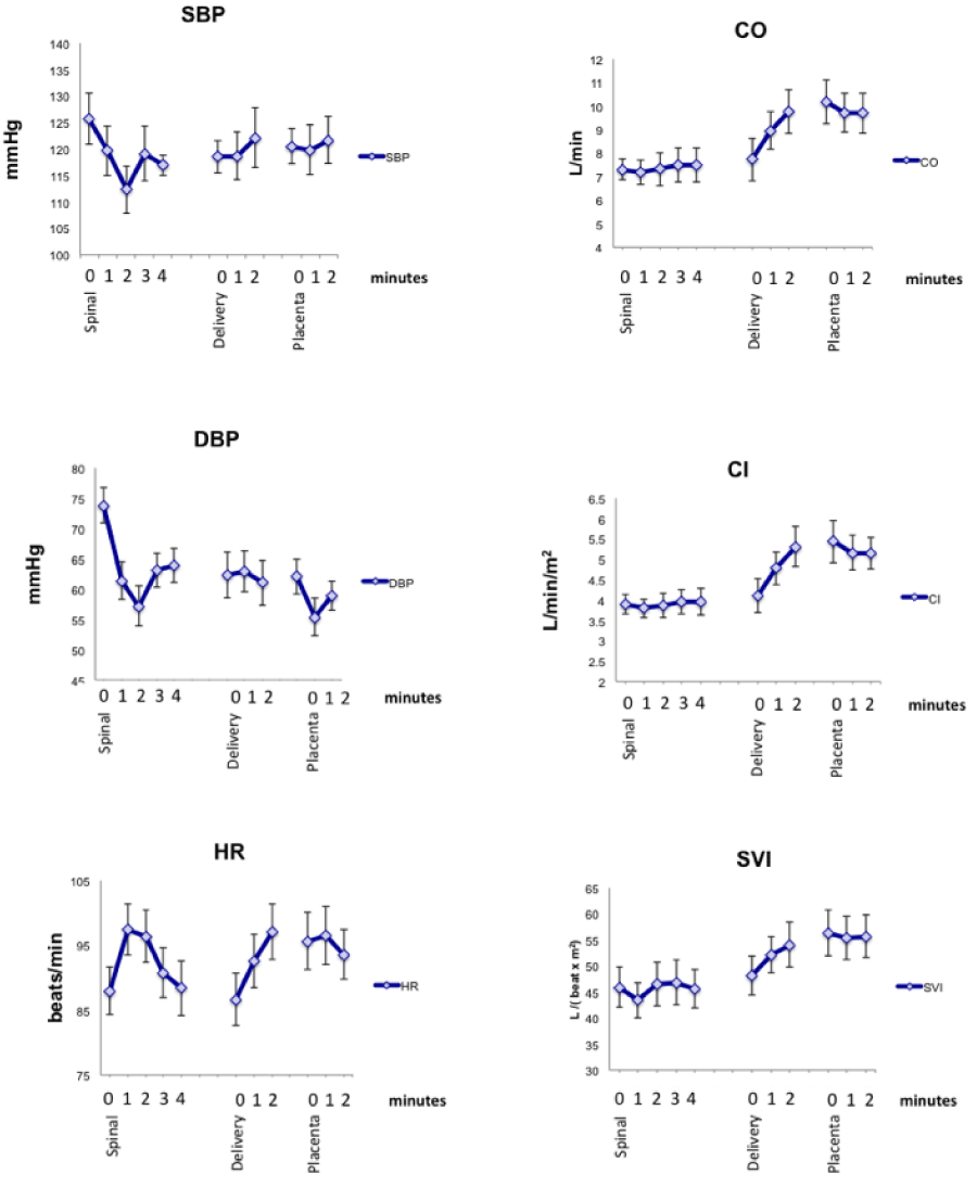
Results: There was a very significant decrease in both SBP and DBP and increase in HR (p<0.001) after spinal anesthesia placement, however there was no significant change in the CO, CI or SVI (p>0.05) during that same period. This change was maximal at 1 and 2 minutes after spinal anesthesia respectively for HR and blood pressure. In contrast after delivery of the fetus there were no significant changes in SBP, DBP and HR (p>0.05), but a dramatic increase in CO (22.5%), CI (16.3%) as well as SVI (13.6%). There were no further changes to any of these parameters at delivery of the placenta; however there was maintenance of the elevated cardiac output and stroke volume index compared to the base line at the time of the spinal placement.
Conclusion: While transient maternal hypotension does not seem to result in adverse short term neonatal outcome, it would be ideal to maintain maternal cardiac output for both maternal and fetal reasons. Routine use of NICOM in all C-section patients should be considered, particularly in high risk obstetric patients where early intervention for developing hypotension and more importantly reduced cardiac output, is critical.
Keywords: NICOM, spinal anesthesia, cardiac output
Introduction
Hypotension during spinal anesthesia remains a common clinical issue, particularly during Cesarean sections (C-sections) [1]. If hypotension does not resolve promptly, maternal complications of nausea and vomiting are likely occur [2], and if this is persistent; more serious consequences may result; such as decreased consciousness, pulmonary aspiration and in extreme but rare instances maternal cardiac arrest can occur [3]. Prolonged periods of hypotension can also lead to reduced placental blood flow compromising the fetus [4].
Early signs of hypotensive episodes are important to recognize, so that adequate and prompt treatment with vasopressorsand/or intravenous fluid resuscitation occurs, whichever is appropriate [5]. ASA standard monitoring of heart rate (HR) and blood pressure (BP) are common maternal variables monitored throughout the operative procedure for C-sectionsand are used as surrogate markers for maternal cardiac output (CO), which more directly reflectsuteroplacental perfusion [1]. Direct measurement and monitoring of maternal cardiac output would be ideal in the management of parturients undergoing Cesarean section; however these monitoring choices involve invasive techniques. Recently new non-invasive methods of monitoring hemodynamic status have been developed and clinically validated [6, 7],whichalsocan provide other useful hemodynamic indicatorsin addition to cardiac output (CO), such as cardiac index (CI) and stroke volume index (SVI). To date NICOM has not been extensively used in the obstetric operating room, likely due in part to cost and equipment availability.
Here we present the first description of bioreactancenon invasive cardiac monitor (NICOMTM, Cheetah Medical Inc., Wilmington, Delaware, USA) use in a series of healthy parturientsundergoing elective Cesarean section under spinal anesthesia, a monitor that is now routinely used at our institution in the obstetric operating room.
Methods
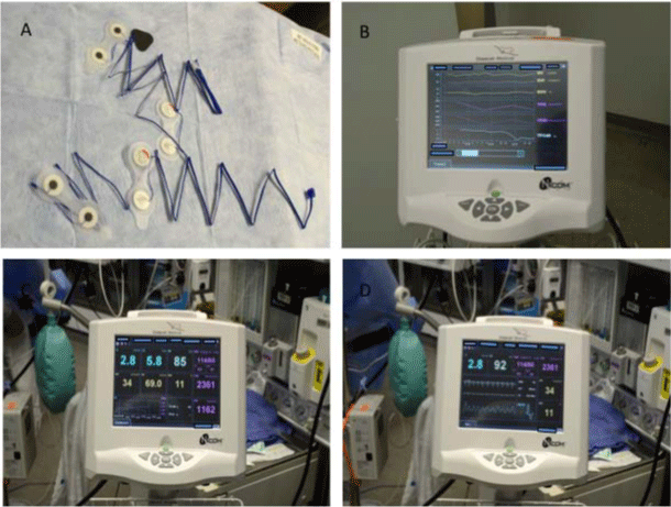
Following Institutional Review Board (IRB) approval, a retrospective review of 13 randomly selected healthy parturients who had received spinal anesthesia for elective C- section was performed; patients that had also had a NICOMTM monitor placed (Figure 1) as well as other standard hemodynamic monitoring was maintained (NIBP, Pulse Ox and ECG) in addition to NICOMTM. Inclusion criteria for analysis; were pregnant patients undergoing elective C-section under spinal anesthesia that had full hemodynamic monitoring during the case, including use of NICOMTM. Exclusion criteria were patients that either did not undergo elective C-section with spinal anesthesia or did not have all the hemodynamic monitor information recorded during the C-section. Standard demographic data, such as age, height, weight, gestational age, IV fluid bolus, thespinal anesthesia regimen, and vasopressor use, such as epinephrine/ phenylepherinewas also collected from these cases.
Figure 1: NICOMTM monitor used (Cheetah Medical Inc., Wilmington, Delaware, USA) including lead sets and examples of data and trace recorded.
Hemodynamic variables collected by NICOMTM were:Systolic blood pressure (SBP), diastolic blood pressure (DBP), heart rate (HR), cardiac output (CO), cardiac index (CI) and stroke volume index (SVI). This data was collected and then analyzed at pre spinal (baseline) value and following 1 minute intervals post spinal up to 10 minutes or until delivery of the fetus, 1 minute intervals after fetus delivery until delivery of the placenta, and finally 1 minute intervals after the delivery of the placenta, also up to 20 minutes or if placenta was removed.
Results
The changes in each of the variables; systolic/diastolic blood pressure (SBP/DBP), heart rate (HR), cardiac output/index (CO/CI) and stroke volume index (SVI) are summarized in Table 1 and Figure 2 displays the most significant changes in the first couple of minutes, for each significant event during the C-section (e.g. before/after spinal dosing, before/after fetus delivery and before/after placental delivery). Patient demographic data is summarized in Table 2 and reflects a typical obstetric patient population in our obstetric operating room.
    NICOM
      HemodynamicVariable
    Immediatelyfollowspinal
    Immediatelyfollowspinal:
      lowest/highestvaluecfbaseline
    Immediatelyfollowingfetusdelivery
      cfpriortodeliveryoffetus
    Immediatelyfollowingfetusdelivery:
      lowest/highestvaluecfbaseline


    SBP
    4.8%DECREASE***
    2MINUTESAFTERSPINALDOSE10.6%
      BELOWBASELINE***
    NOSIGNIFICANTCHANGE
    5.7%DECREASEcfBASELINE*


    DBP
    16.8%DECREASE***
    2MINUTESAFTERSPINALDOSE22.5%
      BELOWBASELINE*
    NOSIGNIFICANTCHANGE
    15.5%DECREASEcfBASELINE***


    HR
    10.8%INCREASE***
    1MINUTEAFTERSPINALDOSE10.6%
      ABOVEBASELINE***
    NOSIGNIFICANTCHANGE
    PEAKAT2MINUTESAFTERDELIVERY
      (10.4%ABOVEBASELINE)**


    CO
    NOSIGNIFICANTCHANGE
    NOSIGNIFICANTCHANGE
    22.5%INCREASE***
    PEAKAT2MINUTESAFTERSPINAL(33.3%
      ABOVEBASELINE)***


    CI
    NOSIGNIFICANTCHANGE
    NOSIGNIFICANTCHANGE
    16.3%INCREASE***
    PEAKAT2MINUTESAFTERSPINAL(36.7%
      ABOVEBASELINE)***


    SVI
    NOSIGNIFICANTCHANGE
    NOSIGNIFICANTCHANGE
    13.6%INCREASE*
    PEAKAT2MINUTESAFTERDELIVERY
      (12.3%ABOVEBASELINE)*


    *
    p<0.05


    **
    p<0.01


    ***
    p<0.01
Table 1: Estimated number of new cancer cases and deaths in 2013 (SEER stat fact sheet).
Figure 2: Graphs showing the means (+/- SD) of each variable; Systolic blood pressure (SBP), Diastolic blood pressure (DBP), Heart rate (HR), Cardiac output (CO), Cardiac index (CI) and Stroke volume index (SVI); minutes after spinal placement, fetus and then placenta delivery.
Table 2: Mean (SD).
Discussion
While most anesthesia practitioners utilize the standard ASA hemodynamic parameters such as SBP, DBP and HR, as can be seen from our data these may not always reflect cardiac output; which is a more important determinant of uterine fetal perfusion.In addition to fetal heart rate monitoring, which will drop as uterine blood flow is reduced [8]there have been different monitoring techniques utilized such as fetal brain Doppler monitoring, to asses adequate fetal perfusion [9]; however it would be better to prevent this from occurring by keeping maternal cardiac output optimal before these adverse downstream changes develop.
More commonly used non-invasive cardiac monitor's usebioimpedence technology; however this methodology did not apply as well in the obstetric setting, for a number of reasons; notwithstanding the susceptibility to patient movement and other artifacts. Bioreactance monitors analyze the variations in voltage in each beat as a response to the application of a high-frequency, trans-thoracic electrode and have a number of advantages over the previously used bioimpedence based monitors, inaccurate concerns resulting from signal-noise ratio, variations seen with different patient body habitus and electrode / skin conductivity . NICOM monitoring has been successfully used in cardiac patients, ICU and in pregnant patients.Validation studies comparing NICOM to more invasive cardiac monitoring, such as semi-continuously monitoring by thermodilution using a pulmonary artery catheter (PAC-CCO) have been utilized in cardiac patients and have shown NICOM to be comparable[10].
A recent study investigated potential predictive risk factors for hypotensive events following spinal anesthesia, and found 50% of spinal anesthetic C-section cases resulted in hypotensionwith independent risk factors for this being age, body mass index and peak level of sensory block. Treatment options for hypotension include intravenous prophylaxis such as colloid or crystalloid pre-loading and interventional treatment with fluids or vasopressor medications such as ephedrine or phenylephrine [11]. Past studies have investigated the effectiveness of these treatments and analyzed outcomes for both mother and fetus. However these treatments though effective for restoring blood pressure readings to normal, may have varying effects on other hemodynamic variables such as CO, which are more relevant to fetal perfusion.The other measurements used to assess cardiac function and hemodynamics in addition to heart rate (HR) and mean arterial blood pressure (MAP), that are assessed with non invasive (NICOM) monitoring, may be more relevant to fetus placental perfusion. These include a variety of cardiac parameters such as beat to beat cardiac output (CO), stroke volume index (SVI) and cardiac index (CI).
The overall incidence rate of Cesarean section has increased as has the co-morbidities and complicated them [12].NICOM has been used mostly in ICU settings [13] but has been applied during pregnancy [14,15].NICOM has also very recently been applied in pediatric hemorrhagic shock model [16], but there are few reports of NICOM use during surgical procedures.We report the successful use of bioreactance NICOM monitoring of key events such as post spinal anesthesia, fetal and placental delivery in a case series of parturients during C-section.Electrical velocimetry has been used to measure cardiac output to best position a pregnant patient in the ICU to prevent aorta caval compression [17]. Similarly our use of bioreactance NICOM was shown to be an easy to use monitor throughout the C-sections, and could provide reliable and valuable additional hemodynamic information during the surgery. The profile of each cardiac variable added to the picture of the hemodynamic status of the patient, and provided information that could be used in clinical management decisions, as needed.
In conclusion while transient maternal hypotension does not seem to result in adverse short term neonatal outcome [18,19], it would be ideal to maintain maternal cardiac output for both maternal and fetal reasons. Routine use of NICOM in all C-section patients should be considered, particularly in high risk obstetric patients where early intervention for developing hypotension and more importantly reduced cardiac output, is critical.
References
- Ngan Kee WD. Prevention of maternal hypotension after regional anaesthesia for caesarean section. Curr Opin Anaesthesiol.2010; 23:304-309.
- Balki M, Carvalho JC. Intraoperative nausea and vomiting during cesarean section under regional anesthesia. Int J Obstet Anesth.2005; 14:230-241.
- Charuluxananan S, Thienthong S, Rungreungvanich M, Chanchayanon T, Chinachoti T, et al. Cardiac arrest after spinal anesthesia in Thailand: a prospective multicenter registry of 40,271 anesthetics. Anesth Analg.2008; 107:1735-1741.
- Erkinaro T, Kavasmaa T, Päkkilä M, Acharya G, Mäkikallio K, et al. Ephedrine and phenylephrine for the treatment of maternal hypotension in a chronic sheep model of increased placental vascular resistance. Br J Anaesth.2006; 96:231-237.
- Cyna AM, Andrew M, Emmett RS, Middleton P, and Simmons SW. Techniques for preventing hypotension during spinal anaesthesia for caesarean section. Cochrane Database of Systematic Reviews.2006; 4:CD002251.
- Marqué S, Cariou A, Chiche JD, Squara P. Comparison between Flotrac-Vigileo and Bioreactance, a totally noninvasive method for cardiac output monitoring. Crit Care. 2009; 13:R73.
- Raval NY, Squara P, Cleman M, Yalamanchili K, Winklmaier M, et al. Multicenter evaluation of noninvasive cardiac output measurement by bioreactance technique. J Clin Monit Comput.2008; 22:113-119.
- Skillman CA, Plessinger MA, Woods JR, Clark KE. Effect of graded reductions in uteroplacental blood flow on the fetal lamb. Am J Physiol.1985; 249:H1098-H1105.
- Cruz-Martínez R, Figueras F, Hernandez-Andrade E, Oros D, Gratacos E. Fetal brain Doppler to predict cesarean delivery for nonreassuring fetal status in term small-for-gestational-age fetuses. Obstetrics and Gynecology. 2011; 117: 618-626.
- Squara P, Denjean D, Estagnasie P, Brusset A, Dib JC, et al. Noninvasive cardiac output monitoring (NICOM): a clinical validation. Intensive Care Med.2007; 33:1191-1194.
- Brenck F, Hartmann B, Katzer C, Obaid R, Brüggmann D, et al. Hypotension after spinal anesthesia for cesarean section: identification of risk factors using an anesthesia information management system. J ClinMonitComput.2009; 23:85-92.
- Coleman VH, Lawrence H, Schulkin J. Rising cesarean delivery rates: the impact of cesarean delivery on maternal request. Obstet Gynecol Surv.2009; 64:115-119.
- Marqué S, Cariou A, Chiche JD, Squara P. Comparison between Flotrac-Vigileo and Bioreactance, a totally noninvasive method for cardiac output monitoring. Crit Care.2009; 13:R73.
- San-Frutos LM, Fernández R, Almagro J, Barbancho C, Salazar F, et al. Measure of hemodynamic patterns by thoracic electrical bioimpedance in normal pregnancy and in preeclampsia. European Journal of Obstetrics Gynecology and Reproductive Biology.2005; 121: 149-153.
- Ohashi Y, Ibrahim H, Furtado L, Kingdom J, Carvalho JC. Non-invasive hemodynamic assessment of non-pregnant, healthy pregnant and preeclamptic women using bioreactance. [Corrected]. Rev Bras Anestesiol.2010; 60:603-613, 335-340.
- Ballestero Y, Urbano J, López-Herce J, Solana MJ, Botrán M, et al. Pulmonary arterial thermodilution, femoral arterial thermodilution and bioreactance cardiac output monitoring in a pediatric hemorrhagic hypovolemic shock model. Resuscitation. 2012; 83:125-129.
- Archer TL, Suresh P, Shapiro AE. Cardiac output measurement, by means of electrical velocimetry, may be able to determine optimum maternal position during gestation, labour and caesarean delivery, by preventing vena caval compression and maximising cardiac output and placental perfusion pressure. Anaesthesia Intensive Care.2011; 39:308-311.
- Corke BC, Datta S, Ostheimer GW, Weiss JB, Alper MH. Spinal anaesthesia for Caesarean section. The influence of hypotension on neonatal outcome. Anaesthesia.1982; 37:658-662.
- Maayan-Metzger A, Schushan-Eisen I, Todris L, Etchin A, Kuint J. Maternal hypotension during elective cesarean section and short-term neonatal outcome. American Journal of Obstetrics and Gynecology.2010; 202:e1-e5.

