
Case Report
Austin J Anesthesia and Analgesia. 2025; 13(1): 1126.
Double-Lumen Tube for One-Lung Ventilation in a Tracheostomized Patient: A Case Report
Jidal M*, Amahroq R, Halhoul Y, Rachqui M, Elwali A and Bensghir M
Department of Anesthesiology, Anesthesia and Intensive Care Unit, Mohammed V Military Hospital in Rabat, Faculty of Medicine and Pharmacy of Rabat, Morocco
*Corresponding author: Jidal Marouane, Department of Anesthesiology, Anesthesia and Intensive Care Unit, Mohammed V Military Hospital in Rabat, Faculty of Medicine and Pharmacy of Rabat, Morocco Email: marouane.j96@gmail.com
Received: July 30, 2025 Accepted: August 15, 2025 Published: August 19, 2025
Abstract
Background: One-lung ventilation (OLV) is essential for many thoracic surgical procedures, yet it poses a unique challenge in tracheostomized patients due to altered anatomy and limited airway access. While bronchial blockers are commonly preferred in such cases, the use of a double-lumen tube (DLT) is rarely reported but may offer certain advantages.
Case Presentation: We describe the case of a 69-year-old male with a history of total laryngectomy and permanent tracheostomy following treatment for laryngeal squamous cell carcinoma. The patient was scheduled for videoassisted thoracoscopic surgery (VATS) for left upper lobectomy due to a newly diagnosed pulmonary carcinoma. One-lung ventilation was successfully achieved via a Carlens-type double-lumen tube inserted through the mature tracheostomy stoma. Lung isolation and surgical exposure were satisfactory, and no perioperative complications were noted.
Discussion: This case demonstrates the feasibility and safety of using a DLT for OLV in a patient with a long-term, well-formed tracheostomy. Although less commonly used than bronchial blockers in such settings, DLTs may offer faster lung isolation, lower malposition risk, and easier suctioning when anatomical and technical conditions allow.
Conclusion: One-lung ventilation using a double-lumen tube through a tracheostomy is a viable alternative in selected patients. Proper preoperative planning, airway assessment, and bronchoscopic guidance are essential to ensure success and minimize complications.
Keywords: One lung ventilation; Tracheotomized patient; Double lumen tube; Case report
Abreviations
OLV: One-Lung Ventilation; DLT : Double-lumen Tuve; VATS : Video-assisted Thoracoscopic Surgery; BMI : Body Mass Index.
Introduction
One-lung ventilation (OLV) during thoracic surgery in tracheostomized patients represents a true anesthetic challenge in terms of airway management and successful lung isolation— particularly as such situations are becoming increasingly common. Indeed, patients with laryngeal cancer, who often require total laryngectomy with permanent tracheostomy, are at particularly high risk of developing lung cancer, with an estimated incidence ranging from 2.8% to 11.2% [1]. In these patients, OLV is typically achieved using bronchial blockers. Our case highlights the feasibility and safety of performing one-lung ventilation using a Carlens-type doublelumen tube, provided that certain conditions are met.
Case Presentation
We report the case of a 69-year-old man with a history of chronic smoking, abstinent for the past five years, and previously diagnosed with squamous cell carcinoma of the larynx, for which he underwent total laryngectomy followed by adjuvant chemotherapy in 2020. He was recently diagnosed with a squamous cell carcinoma of the left upper lung lobe, for which a video-assisted thoracoscopic left upper lobectomy was planned.
The preanesthetic assessment revealed a WHO performance status of 1. His body mass index (BMI) was 27.2 kg/m², and there was no reported weight loss. Cardiovascular evaluation showed NYHA class II dyspnea, good exercise tolerance with a functional capacity > 4 METs, and a 6-minute walk test of 640 meters without desaturation. From a respiratory standpoint, the patient had a well-established tracheostomy with a size 8 Krishaber tracheostomy tube in place. The stoma was clean, sufficiently wide to accommodate a doublelumen tube (DLT), and there were no secretions. He maintained an oxygen saturation of 98% on room air, and pulmonary auscultation was normal. Cervico-thoracic CT revealed a 32 × 20 mm nodule in the left upper lobe, associated with apical and paraseptal emphysema, without significant mediastinal lymphadenopathy. Pulmonary function testing could not be performed due to the lack of equipment adapted to tracheostomized patients. Perfusion lung scintigraphy estimated the predicted postoperative FEV1 at 47%. In the operating room, the Krishaber tracheostomy tube was replaced with a size 8 standard tracheostomy cannula.
After induction of anesthesia, flexible bronchoscopy was performed to evaluate the airway, followed by careful placement of a left-sided 37 Fr Carlens double-lumen tube without carinal hook through the tracheostomy stoma under endoscopic guidance. Through the bronchial lumen, the carina was visualized (Figure 1), and the bronchial lumen was gently advanced into the left mainstem bronchus (Figure 2). The tube was then securely fixed to prevent displacement (Photo 1), and correct positioning was confirmed by auscultation after placing the patient in the right lateral decubitus position. The surgical procedure was uneventful, and the patient was discharged on postoperative day six. At two-month follow-up, no complications related to the insertion of the DLT through the tracheostomy stoma were observed.
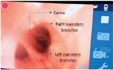
Figure 1: Endoscopic view showing the carina and both mainstem bronchi.
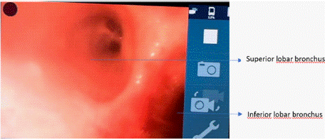
Figure 2: Endoscopic view showing the division of the left main bronchus.
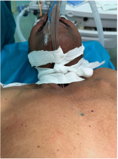
Photo 1: Fixation of the double-lumen tube through the tracheostomy.
Discussion
One-lung ventilation (OLV) in tracheostomized patients presents a significant anesthetic challenge due to the inability to use conventional oral devices. This clinical situation is increasingly encountered, particularly among patients previously treated for cancers of the upper aerodigestive tract—a population commonly associated with the development of a second primary pulmonary malignancy [1].
The present case is in line with previous reports [2-4] demonstrating the feasibility and safety of using a double-lumen tube (DLT) in patients with a long-standing tracheostomy. Although, such use has been reported in the literature(4), DLT insertion is contraindicated in cases of recent tracheostomy (< 7 days) [5] due to the risk of false passage creation or loss of airway control during tube placement.
In our case, a Carlens left-sided DLT was gently introduced through the tracheostomy stoma under bronchoscopic guidance, which is essential to ensure a safe and accurate approach in this setting. According to the retrospective study by Campos et al. [5], which included 70 tracheostomized patients who underwent OLV, DLTs were used in only 6% of cases, whereas bronchial blockers were employed in 84%. The limited use of DLTs is explained by their large external diameter, rigidity, and risk of tracheal injury, particularly when the tracheostomy stoma is narrow or not fully matured. Nevertheless, in selected cases [2-4], as illustrated in our observation, DLT use may be considered when the stoma is well-healed, of adequate size, and bronchoscopic expertise is available.
The main advantage of the DLT is its reliability in achieving lung isolation and its ability to allow rapid differential lung recruitment, making it the standard technique for thoracic surgery in nontracheostomized patients [6]. In more complex cases, the preferred alternative is the use of independent bronchial blockers (e.g., Arndt, Cohen, EZ-Blocker), introduced either through a Shiley tracheostomy tube or a single-lumen tube (SLT), as was the case in most patients in the Campos series [5].
In our patient, several factors favored the use of a DLT: a mature and stable tracheostomy, absence of stenosis, wide stoma, lack of secretions, anatomical compatibility with a 37 Fr DLT, and most importantly, the team’s experience in bronchoscopic techniques. No intraoperative or postoperative complications were observed.
This case thus contributes to the limited but growing body of evidence supporting the successful use of DLTs via a tracheostomy stoma. It highlights that, in well-selected patients, with a healed stoma, appropriate equipment, and an experienced team, the DLT remains a viable option, even if not currently considered first-line in existing recommendations.
Finally, it is worth noting the existence of specialized DLTs designed specifically for tracheostomized patients, such as the Naruke DLT or the Rüsch Tracheoport [7]. These devices are shorter and better adapted to altered airway anatomy, allowing safer insertion through the tracheostomy stoma, although they are not widely available across centers.
Conclusion
This case highlights that, although the use of double-lumen tubes (DLTs) in tracheostomized patients is uncommon and technically challenging, it remains a feasible and safe option in carefully selected individuals. A mature and wide tracheostomy stoma, absence of local complications, and the availability of bronchoscopic expertise are essential prerequisites for successful DLT placement via the tracheostomy route. While bronchial blockers remain the first-line devices in most cases, DLTs may be considered when optimal lung isolation and rapid differential ventilation are required. Further studies and broader access to specifically designed DLTs for tracheostomized patients could help refine airway management strategies in this growing population.
Author Statements
Authors Contributions
All the authors participated in this work, read and approved the final version of the manuscript.
References
- Rispoli M, Nespoli MR, Salvi R, Corcione A, Buono S. One-lung ventilation in tracheostomized patients: our experience with EZ-Blocker. J Clin Anesth. 2016; 31: 288-290.
- Yaney LL. Double-lumen endotracheal tube for one-lung ventilation through a fresh tracheostomy stoma: a case report. AANA J. 2007; 75: 411-415.
- Renton MC, Conacher ID. Single-lung ventilation via a double lumen tube in a patient with a tracheostomy. Anaesthesia. Févr. 2002; 57: 197-198.
- Shih CK, Kuo YW, Lu IC, Hsu HT, Chu KS, Wang FY. Application of a doublelumen tube for one-lung ventilation in patients with anticipated difficult airway. Acta Anaesthesiol Taiwanica Off J Taiwan Soc Anesthesiol. Mars. 2010; 48: 41-44.
- Campos JH, Musselman ED, Hanada S, Ueda K. Lung Isolation Techniques in Patients With Early-Stage or Long-Term Tracheostomy: A Case Series Report of 70 Cases and Recommendations. J Cardiothorac Vasc Anesth. Févr. 2019; 33: 433-439.
- Lohser J, Ishikawa S. Clinical Management of One-Lung Ventilation. In: Slinger M FRCPC, Peter, éditeur. Principles and Practice of Anesthesia for Thoracic Surgery [Internet]. New York, NY: Springer; 2011: 83-101.
- Dincq AS, Lessire S, Mayné A, Putz L. Double-lumen tubes for tracheostomized patients. J Cardiothorac Vasc Anesth. 2015; 29: e35-36.