Research Article
Austin J Anesthesia and Aalgesia. 2014;2(5): 1031.
Patient Variability in Sciatic Nerve Branch Point Distance Using Ultrasound Guided Localization
Lollo L*, Stogicza A
Department of Anesthesiology and Pain Medicine, University of Washington, USA
*Corresponding author: Lollo L, Department of Anesthesiology and Pain Medicine, University of Washington, Box 356540, 1959 NE Pacific St, BB-1469,Seattle, WA 98195-6540, USA.
Received: May 20, 2014; Accepted: June 30, 2014; Published: July 01, 2014
Abstract
Background and Objectives: Improved ultrasound and needle technology make popliteal sciatic nerve blockade a popular anesthetic technique and imaging to localize the branch point of the common peroneal and posterior tibial components is important because successful blockade techniques vary with respect to injection of the common trunk proximally or separate injections distally. Nerve stimulation, ultrasound, cadaveric and magnetic resonance studies demonstrate variability in distance and discordance between imaging and anatomic examination of the branch point. The popliteal crease and imprecise, inaccessible landmarks render measurement of the branch point variable and inaccurate. The purpose of this study was to use the tibial tuberosity, a fixed bony reference, to measure the distance of the branch point.
Method: During popliteal sciatic nerve blockade in the supine position the branch point was identified by ultrasound and the block needle was inserted. The vertical distance from the tibial tuberosity prominence and needle insertion point was measured.
Results: In 92 patients the branch point is a mean distance of 12.91 cm proximal to the tibial tuberosity and more proximal in male (13.74 cm) than female patients (12.08 cm). Body height is related to the branch point distance and is more proximal in taller patients. Separation into two nerve branches during local anesthetic injection supports notions of more proximal neural anatomic division.
Limitations: Imaging of the sciatic nerve division may not equal its true anatomic separation.
Conclusion: Refinements in identification and resolution of the anatomic division of the nerve branch point will determine if more accurate localization is of any clinical significance for successful nerve blockade.
Introduction
Sciatic nerve blockade (SNB) at the popliteal fossa is a frequently used regional anesthetic technique for foot and ankle procedures and advances in ultrasound imaging, echogenic needle coatings and practitioner training and proficiency have contributed to this increase [1]. Successful placement of SNB is assessed by the level of intraoperative anesthesia, postoperative analgesia and satisfaction with the anesthetic course by both patients and practitioners [2]. The point of injection with respect to the branch point of the sciatic nerve (SN) for the most effective neural blockade varies among authors [3]. Some report that injection near the common nerve trunk proximal to its division results in superior anesthesia whereas others observe that distal blockade of one or both divisions, usually the posterior tibial (PT), provides a faster and more profound level of analgesia [4]. The choice of injection techniques have compared speed of onset time, volume of local anesthetic used, duration of time for nerve block performance and patient satisfaction for each and have been found to be relatively comparable [5]. The high success rate and rapid onset time of a sub-sheath proximal SNB technique are based on evaluation for the para-neural and sub-epineural spaces and require accurate localization of the SN branch point in order to achieve these results [6,7].
The classic nerve stimulator guided SNB technique at the popliteal fossa recommends needle insertion at a distance of 5 to 7 cm proximal to the popliteal crease and to observe for toe plantar flexion at the lowest possible current strength as an endpoint for SNB [8]. This is a point where the SN is considered undivided but the success rate for adequate anesthesia is 82% compared to the near 100% rate reported for USG SNB [9]. In cadavers the SN branch point was observed in the lower posterior compartment of the thigh in 40%, the popliteal fossa in 35% and in the gluteal region in 15% [10]. Magnetic resonance imaging studies reported the SN branch point at a mean distance of 7.5 cm from the lateral joint line and 93% of patients had the branch point within 10 cm of the joint line and the authors recommend needle placement for SNB at 10 cm proximal to the popliteal fossa [11]. Placing a needle 7 cm proximal to the knee in cadavers caused dye to spread 5 to 10 cm along the SN [12]. This observation was hypothesized to be due to the presence of a continuous neural sheath extending proximally around both branches of the SN [13]. The mean SN branch point distance in cadavers was 60 mm proximal to the popliteal crease and a common binding epineural sheath for the two branches of the SN was also observed in this group [14]. The sub-gluteal approach for sciatic nerve blockade would avoid the risk of inadequate analgesia but patient body habitus, degenerative joint disease of the hip, added discomfort in maintaining the hip fully extended, greater time interval for onset of action and trainee difficulty in performing this technique prolong procedural time.
These studies provide guidance for needle placement for SNB but are not feasible for use in everyday practice. The use of the popliteal crease as a landmark is subjectively variable and difficult to ascertain in morbidly obese patients or those with occlusive dressings over the area and knee flexibility causes the popliteal crease to be a mobile reference point. The purpose of this study was to use ultrasound guidance (USG) to measure the distance of the SN branch point from the tibial tuberosity, a fixed bony landmark that avoids the subjective use of the popliteal crease.
Method
After receiving IRB approval from the University of Washington Human Subjects Division, patients provided written informed consent prior to undergoing foot and ankle surgery and were enrolled for participation in a sciatic nerve block study. The preoperative data collected were age, ASA physical status, height, weight, and calculated BMI. Ultrasound imaging was performed using a Sonosite M machine with a 10 MHz 38 mm linear array probe in short axis view and interpretation and measurements were performed at the time of nerve blockade by concordance between two supervising faculty experienced in ultrasound guided regional anesthesia. During the nerve block procedure the SN was localized in the popliteal fossa and then tracked proximally to the point where the two branches ("Figure of eight") coalesced into a single trunk and this was used as the needle insertion point. The vertical distance of the SN branch point at the site of needle insertion under USG from the tibial tuberosity prominence was then measured and recorded.
Results
104 patients were enrolled over a six month period. Complete data were available for 92 patients. 41 (44.56%) patients were female and 51 were male (55.43%). The mean ASA physical status for both groups was 2 and the range was 1 to 3. The mean age for females was 53.41 years and for males was 52.1 years. The mean overall BMI was 28.53 kg/m2. The mean female BMI was 28.08 kg/m2 and the male BMI was 28.97 kg/m2. The mean overall height was 170.21 cm. Mean height for females was 163.83 cm and for males was 176.59 cm. The overall mean SN branch point distance from the tibial tuberosity was 12.91 cm. The SN branch point distance measured 12.08 cm for females and 13.74 cm for males.
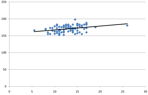
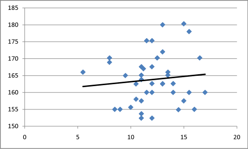
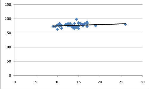
Table 1 summarizes the patient demographics and Table 2 the measured data. Graph 1 is a plot of patient height versus the SN branch point for all patients. Graphs 2 and 3 are plots of patient height versus the SN branch point with relation to female and male gender respectively. Trendlines have been calculated and plotted for each of the graphs.
Patient
ASA Status(Range)
Age (yrs)
Age Range (yrs)
BMI (kg/m2)
BMI Range (SD)
Total (n=92)
2 (1 - 3)
52.76
22 -83
28.53
20.1 48.8 (5.94)
Males (n=51)
2 (1 - 3)
52.1
22 - 81
28.97
20.5 48.8 (6.4)
Females (n=41)
2 (1 - 3)
53.41
26 - 83
28.08
20.1 40.7 (5.47)
Table 1: In vitro models used to study opioid/induced hyperalgesia.
Patients
Mean Height (cm)
Height Range (SD)
Mean Branch Point (cm)
Branch Point Range (SD)
Branch Point 95% CI
Total (n=92)
170.21
152.4 198.1 (7.18)
12.91
7 26 (4.21)
0.86
Males (n=51)
176.59
165 -198.1 (6.94)
13.74
7 26 (2.95)
0.81
Females (n=41)
163.83
152.4 180.3 (7.42)
12.08
8 17 (2.47)
0.76
Table 2: In vivo models used to study opioid/ induced hyperalgesia.
Figure 1: Relation of Body Height and Sciatic Nerve Branch Point, Total Patients (n=92). X-axis: Distance of Sciatic nerve branch point from the tibial tuberosity (cm) Y-axis: Body Height (cm)
Figure 2: Relation of Body Height and Sciatic Nerve Branch Point, Female Patients (n=41). X-axis: Distance of Sciatic nerve branch point from the tibial tuberosity (cm) Y-axis: Body Height (cm)
Figure 3: Relation of Body Height (cm and Sciatic Nerve Branch Point (cm), Male Patients (n=51). X-axis: Distance of Sciatic nerve branch point from tibial tuberosity (cm) Y-axis: Body Height (cm)
Discussion
The purpose of this study was to measure the distance of the SN branch point from the tibial tuberosity as the bony reference landmark to the needle insertion point proximal to the separation of the common peroneal (CP) and PT nerves as determined by USG. The tibial tuberosity was readily identified and palpated and vertical measurement was easily obtained in all patients. The 92 patients were uniform for ASA status, age and BMI. The mean height for females was lower than for males but there was overlap between the two groups.
The SN branch point was more proximal for males than for females but there was overlap in the height and SN branch point distance between the two gender groups. The data suggest that taller patients have more proximal SN branch points with respect to the tibial tuberosity. This has some clinical significance because needle placement in taller patients will in general need to be directed more proximally in order to avoid missing the common SN trunk resulting in partial or inadequate SNB.
Although the ultrasound interpretation of the image was assessed by the operators to be a point proximal to the SN branch point, this did not always correlate with the true anatomic division of the nerve as demonstrated by the two branches separating and appearing adjacent to each other within a common sheath during local anesthetic injection. This finding implies that the true anatomic division of the SN is more proximal than the ultrasonographic image suggests in some patients. This observation supports prior reports that demonstrated the presence of two nerve branches in a common para-neural and sub-epineural sheath and that the anatomic point of separation in some cases is more proximal than the distance assessed by USG [15].
In summary the SN branch point could be readily measured from the tibial tuberosity and there is a relation to body height in the sense that taller patients have a more proximal point of nerve division by USG. This study had limitations related to the observed fact that although the sciatic nerve may appear to be fused at the branch point by ultrasound visualization that the true anatomic division of the nerve may be at a more proximal point and the clinical significance of this with respect to successful nerve blockade needs to be assessed.
Conclusion
The SN branch point was measured using the tibial tuberosity as the bony landmark. This provided consistency in the measurement by using a fixed bony prominence as opposed to a subjective mobile and difficult to precisely localize region such as the popliteal crease. The SN branch point was observed to be more proximal in taller patients than others. The SN appears to be undivided proximal to the branch point by USG but it was observed that the two branches would separate as local anesthetic was injected in some patients. Further studies and improvement in ultrasonographic resolution of SN anatomy are suggested to provide more accurate localization of the anatomic division of the SN branch point and determine if there is any clinical significance of this discordance with respect to the efficacy of SNB.
References
- Perlas A, Brull R, Chan VW, McCartney CJ, Nuica A, Abbas S, et al. Ultrasound guidance improves the success of sciatic nerve block at the popliteal fossa. Reg Anesth Pain Med. 2008; 33: 259-265.
- Choquet O, Capdevila X. Ultrasound-guided nerve blocks: the real position of the needle should be defined. Anesth Analg. 2012; 114: 929-930.
- Morau D, Levy F, Bringuier S, Biboulet P, Choquet O, Kassim M, et al. Ultrasound guided evaluation of the local anesthetic spread parameters required for a rapid surgical popliteal sciatic nerve block. Reg Anesth Pain Med. 2010; 35: 559-564.
- Tran de QH, González AP, Bernucci F, Pham K, Finlayson RJ. A randomized comparison between bifurcation and prebifurcation subparaneural popliteal sciatic nerve blocks. Anesth Analg. 2013; 116: 1170-1175.
- Buys MJ, Arndt CD, Vagh F, Hoard A, Gerstein N. Ultrasound-guided sciatic nerve block in the popliteal fossa using a lateral approach: onset time comparing separate tibial and common peroneal nerve injections versus injecting proximal to the bifurcation. Anesth Analg. 2010; 110: 635-637.
- Lopez AM, Sala-Blanch X, Castillo R, Hadzic A. Ultrasound guided injection inside the common sheath of the sciatic nerve at division level has a higher success rate than an injection outside the sheath. Rev Esp Anestesiol Reanim. 2014; 61: 304-310.
- Techasuk W, Bernucci F, Cupido T, González AP, Correa JA, Finlayson RJ, et al. Minimum effective volume of combined lidocaine-bupivacaine for analgesia subparaneural popliteal sciatic nerve block. Reg Anesth Pain Med. 2014; 39: 108-111.
- Hara K, Sakura S, Yokokawa N. The role of electrical stimulation in ultrasound-guided subgluteal sciatic nerve block: a retrospective study on how response pattern and minimal evoked current affect the resultant blockade. J Anesth. 2013.
- Sala-Blanch X, de Riva N, Carrera A, López AM, Prats A, Hadzic A. Ultrasound-guided popliteal sciatic block with a single injection at the sciatic division results in faster block onset than the classical nerve stimulator technique. Anesth Analg. 2012; 114: 1121-1127.
- Prakash, Bhardwaj AK, Devi MN, Sridevi NS, Rao PK, Singh G, et al. Sciatic nerve division: a cadaver study in the Indian population and review of the literature. Singapore Med J. 2010; 51: 721-723.
- Grasu RM, Costelloe CM, Boddu K. Revisiting anatomic landmarks: lateral popliteal approach for sciatic nerve block based on magnetic resonance imaging. Reg Anesth Pain Med. 2010; 35: 227-230.
- Vloka JD, HadziÄ A, Kitain E, Lesser JB, Kuroda M, April EW, et al. Anatomic considerations for sciatic nerve block in the popliteal fossa through the lateral approach. Reg Anesth. 1996; 21: 414-418.
- Saleh HA, El-fark MM, Abdel-Hamid GA. Anatomical variation of sciatic nerve division in the popliteal fossa and its implication in popliteal nerve blockade. Folia Morphol (Warsz). 2009; 68: 256-259.
- Andersen HL, Andersen SL, Tranum-Jensen J. Injection inside the paraneural sheath of the sciatic nerve: direct comparison among ultrasound imaging, macroscopic anatomy and histologic analysis. Reg Anesth Pain Med. 2012; 37: 410-414.
- Karmakar MK, Shariat AN, Pangthipampai P, Chen J. High-definition ultrasound imaging defines the paraneural sheath and the fascial compartments surrounding the sciatic nerve at the popliteal fossa. Reg Anesth Pain Med. 2013; 38: 447-451.


