Special Article - Regional Anesthesia
Austin J Anesthesia and Analgesia. 2017; 5(3): 1062.
Regional Anesthetic Technique for a Patient with Neurofibromatosis Type 1 and an Unusual Appearance of the Lower Extremity Nerves
Suvar T*and Mehaffey
Department of Anesthesiology, University of Arkansas for Medical Sciences, USA
*Corresponding author: Suvar T, University of Arkansas for Medical Sciences, USA
Received: November 01, 2017; Accepted: November 21; 2017; Published: November 28, 2017
Abstract
Neurofibromatosis type 1 (NF1) is predominantly a neurocutaneous disorder, but can affect different organ systems; numerous considerations must be made in a NF1 patient who will undergo anesthesia. We report a case of a soft tissue biopsy in a 27-year-old Caucasian male with a history of NF1 complicated by multiple cervical spinal fusion surgeries with significant airway concerns. The decision was made to proceed with a regional anesthetic technique with moderate sedation.
Objective: This case demonstrates challenges associated with regional anesthesia and demonstrates safe anesthetic techniques for a surgical procedure in an NF1 patient.
Case Report: A 27-year-old patient with neurofibromatosis 1 had developed a rapidly growing mass in his right foot that required biopsy. History of multiple cranio-cervical-thoracic fusions, scoliosis, and limited neck extension raised concerns for a difficult airway.
Conclusion: The procedure was successfully managed with regional anesthesia and moderate sedation, with no subsequent complications.
Introduction
Neurofibromatosis type 1 (NF1) is predominantly a neurocutaneous disorder, but can affect different organ systems; numerous considerations must be made in a NF1 patient who will undergo anesthesia. We report a case of a soft tissue biopsy in a 27-year-old Caucasian male with a history of NF1 complicated by multiple cervical spinal fusion surgeries with significant airway concerns. The decision was made to proceed with a regional anesthetic technique with moderate sedation.
Case Presentation
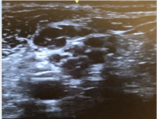
A 27-year-old male with a past medical history of neurofibromatosis type 1 presented to the clinic for a 3-month history of a rapidly growing mass located on the dorsum of his right foot. MRI was performed and revealed a 3.8 x 4 x 3.8 mass, appearing to involve the 2nd and 3rd metatarsals. Surgical biopsy of the mass was planned. On pre-operative assessment, patient had a Mallampati score of 2, but has had numerous cranio-cervical thoracic spine fusions due to neuromuscular scoliosis, with significantly limited neck extension rendering him a difficult endotracheal intubation. The decision was made to proceed with regional anesthesia combined with intravenous sedation. The patient was pre-medicated with 2mg of midazolam. We attempted a sciaticpopliteal and adductor canal nerve blocks under ultrasound guidance when we noticed several neurofibromas in his popliteal nerve (Figure 1). Given this aberrant anatomy, we decided to take a more distal approach and proceed with an ankle block using a 22Gshort-bevel needle. The right superficial and deep peroneal and tibial nerves were visualized and blocked with bupivacaine, 0.5 % (3mL in each nerve) under ultrasound guidance. A field block was also performed on the right saphenous nerve using bupivacaine, 0.5%, 5mL. Given the location of the surgery, a sural nerve block was not performed. Once in the operating room, a propofol infusion was started and the surgical procedure lasted less than two hours. Biopsy of the mass revealed spindle cell proliferation within a fibrous background and areas of myxoid change, with no significant mitotic activity and negative for S100 protein, suggestive of low grade malignant peripheral nerve sheath tumor.
Discussion
Neurofibromatosis type 1 (NF1) is an autosomal dominant neurocutaneous condition caused by an NF1 gene mutation, resulting in decreased activity or nonfunctional neurofibromin protein. It affects 1 in 2600 to 3000 individuals. Clinical manifestations include café-au lait spots, axillary freckling, Lisch nodules, optic gliomas, and neurofibromas. Neurofibromas are the most common tumor found in NF1 patients. They can be cutaneous, plexiform, or nodular, and they can undergo malignant transformation with a risk of 5-13% [1].
NF1 may involve multiple organ systems, and therefore raise anesthetic concerns during surgical procedures. Neurofibromas may be present in the airway which can make intubation difficult [2].
Lovell et al. reports a case of cervical vertebrae dislocation that was discovered post surgery in an asymptomatic NF1 patient with multiple cervical neurofibromas. There must be consideration for a radiologic examination prior to anesthesia for NF1 patients with a history of cervical surgery to assess for any unknown cervical dislocations [3]. Vertebral deformities, like scoliosis present in our patient, or neurofibromas in the spinal cord may also complicate spinal or extradural blocks. Other considerations include undiagnosed neurofibromas in the lungs and GI tract, hypertension, renal artery stenosis, and pheochromocytoma [2].
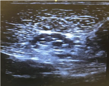
Shahid and Sebastian [4], report a case of lower limb wound debridement in a66-year-old NF1 patient successfully managed with femoral and sciatic nerve block. Another case was reported by Bagam et al [5], of a 60-year-old female with NF1 who needed dynamic hip screw fixation of left femur and intramedullary interlocking nail for the humerus. The surgical procedures were managed with a subarachnoid block and a brachial plexus block, respectively. Given this patient’s extensive history of multiple cranio-cervical-thoracic fusions surgeries, NF1 complicated by scoliosis, and neurofibromas extensively found throughout his body, a decision was made to proceed with a regional block as demonstrated by various case reports to be the safest anesthetic option. Our ultrasound scan of the popliteal fossa demonstrated multiple hypoechoic structures within the popliteal nerve. The common peroneal and tibial nerve branching was not easily identified as they should have been seen converging from the popliteal nerve. As seen in Figure 1, there were concerns for having adequate local anesthetic infiltration of the endoneurium as well as considerations for nerve damage and insult to the neuromas with an inadvertent puncture of the nerve by the 22G short-bevel needle. A more caudal approach was utilized to block the right superficial and deep peroneal nerves, and tibial nerve as seen in (Figure 2).

    Figure 1:  Hypoechoic structures within the popliteal nerve, superficial to the
popliteal artery.

Figure 2: Tibial nerve.
Numerous challenges are faced when administering anesthetics to patients with NF1; significant airway concerns in our patient lead to a regional technique with mild sedation as a safer alternative to general anesthesia for lower extremity surgery. While there were concerns for performing regional blocks at the saphenous and popliteal level, a more distal approach can be used in the presence of neurofibromas in major peripheral nerves.
Written Consent Statement
The authors of this case report here by state that the involved patient provided written permission for the report to be published.
References
- Korf BR. Neurofibromatosis type 1 (NF1): Pathogenesis, clinical features, and diagnosis. Up To Date. Waltham. 2017.
- Hirsch NP, Murphy A, Radcliffe JJ. Neurofibromatosis: Clinical presentations and anaesthetic implications. Br J Anaesth. 2001; 86: 555-564.
- Lovell AT, Alexander R, Grundy EM. Silent, unstable, cervical spine injury in multiple neurofibromatosis. Anaesthesia.1994; 49: 453-454.
- Shahid M, Sebastian BA. Safe Alternative in Neurofibromatosis for Lower Limb Surgeries: Combined Femoral and Sciatic Nerve Block. Journal of Clinical and Diagnostic Research: JCDR. 2015; 9: 03-04.
- Bagam KR, Vijaya DS, Mohan K, Swapna T, Maneendra S, Murthy S. Anaesthetic Considerations in a Patient with Von Recklinghausen Neurofibromatosis. Journal of Anesthesiology. Clinical Pharmacology. 2010; 26: 553-554.