
Special Article - Abdominal Surgery
Ann Surg Perioper Care. 2016; 1(3): 1015.
Rupture of a Simple Renal Cyst into the Pyelocalyceal System Induced by Laparoscopic Surgery of the Prostate
Garcia-Segui A*, Ortiz-Gorraiz M, Soler-López C and Costa MA
Department of Urology, Hospital General Universitario de Elche, Alicante, Spain
*Corresponding author: Alejandro Garcia-Segui, Department of Urology, Hospital General Universitario de Elche, Camí de l’Almazara, Elche, Alicante, Spain
Received: November 13, 2016; Accepted: December 09, 2016; Published: December 12, 2016
Abstract
The spontaneous rupture of renal cysts with communication to the renal cavities has been reported very rarely. We report a patient with a asymptomatic renal cyst and prostate cancer undergoing extraperitoneal laparoscopic radical prostatectomy in Trendelenburg position complicated with a rectal injury repaired laparoscopically and the passing CO2 into peritoneal cavity by a peritoneal gap. He had a torpid post-surgery evolution due to a rupture of renal cyst into the pyelocalyceal system, that it was successfully treated by a percutaneous nephrostomy.
We report the case for its rarity and the possible relationship with laparoscopic surgery.
Keywords: Spontaneous rupture of renal cyst; Rupture of renal cyst into pyelocaly-ceal system; Renal cyst
Introduction
The spontaneous rupture of renal cysts with communication to the renal cavities has been reported very rarely [1-11]. The rupture of a simple cyst is an event that may occur into the peri-renal space, the peritoneal cavity or the pyelocalyceal system [2,3]. There are few reports of spontaneous rupture of the renal cyst into the collector system [1,4-11]. We report a patient undergoing laparoscopic radical prostatectomy (LRP) with a torpid post-surgery evolution due to a rupture of renal cyst into the pyelocalyceal system on possible relationship with laparoscopic surgery.
Case Presentation
A 63-year-old male patient with prostate cancer Gleason 7(4 + 3)- T2a- PSA 5.8ng/ml. He underwent transurethral resection of prostate (TURP) in 2012. He had a CT scan taken 2 months earlier that showed a renal cyst in the lower pole of the left kidney measuring 59mm. The patient underwent LRP by extraperitoneal approach and he was placed in 300-Trendelenburg position. Unfortunately, the total abdominal wall was distended by passing CO2 into peritoneal cavity (pneumoperitoneum) caused by a potential peritoneal gap. We employed a gas pressure of 12mmHg. An intraoperative rectal injury occurred, that it was repaired laparoscopically with two lines of interrupted suture lines plus placement of interposition tissue. The total operative time was 180 minutes. At 2nd day of postoperative, the patient had a acute flank pain on left side with vomiting and elevated serum creatinine, without evidence of infection or peritonitis or urinary infection. A computed tomography (CT) scan of the abdomen with a contrast enema revealed the rectal wall indemnity and presence of liquid in left paracolic gutter, thickening of the left periconal fascia without evidence of contrast extravasation or dilation of the urinary tract, or urolithiasis. After the restoration of renal function with fluid therapy, a new multiphase CT scan was requested for perpetuating pain at 5th day of postoperative. The images revealed a diminution of the previously recognized simple renal cyst with irregular contours, partially filled with contrast material. These finding demonstrate a spontaneous rupture of the cyst with communication to the pyelocalyceal system (Figure 1 and 2). A percutaneous nephrostomy positioned toward the collecting system was done. The antegrade pyelography revealed retrograde filling of the renal cyst by radiopaque contrast material, confirming the connection with the urinary tract (Figure 3). The patient had immediate pain relief after urinary diversion and he evolved favorably and was discharged a few days later. The nephrostomy tube remained in place for 2 weeks and it had been removed after a control pyelogram. At 6 months follow-up the patient remains asymptomatic and without radiologic evidence of the renal cyst communication with the collecting system (Figure 4).
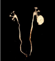
    Figure 1:  Reconstruction from excretory phase of CT scan showed simple
renal cyst filled with contrast material.
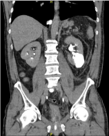
    Figure 2:  Coronal projection reconstruction from excretory phase of CT scan
showed diminution of the previously recognized simple renal cyst filled with
contrast material.
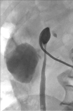
    Figure 3:  Antegrade pyelography through percutaneous nephrostomy
revealed backflow contrast material from the pyelocalyceal system into the
renal cyst.
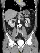
    Figure 4:  Follow-up CT scan obtained at 4 months showed that the
communication of the renal cyst to collecting system has obliterated.
Discussion
The spontaneous rupture of simple renal cysts into the renal cavities is a very rare complication, which pathogenesis is unclear, that it may be caused by trauma, by urinary infection, by acute rise in intracystic pressure following internal hemorrhage, or rise in intra pyelocalyceal pressure following ipsilateral obstructive ureteropathy like stones or alterations in fluid dynamics [1,2]. Tuna M et al presented a report about renal cyst rupture possibly after a laparoscopic intervention in a patient suffering flank pain with massive hemorrhage that may be caused by rise pressure of abdominal cavity due to the pneumoperitoneum of the laparoscopic procedure. They concluded that increased intra-abdominal pressure may also lead to renal cyst rupture, because the peritoneal pressure is usually increased during laparoscopic procedures for the sake of pneumoperitoneum maintenance, and flank or Trendelenburg patient positioning during the surgeries may augment the pressure and affect the renal tissue [12]. In our understanding, spontaneous rupture of renal cyst into the renal cavities provoked by laparoscopic surgery of prostate has not been reported previously. Our patient was underwent to a laparoscopic surgery with prolonged operative time due the intraoperative complications, requiring that we keep in the forced Trendelenburg position and with an intra abdominal pressure inadequately controlled due to extraperitoneal approach with a peritoneal gap (Figure 5). The sum of these factors may have caused an increase in the abdominal pressure lead to an acute tear in the cyst wall.
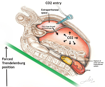
    Figure 5:  Possible pathogenesis of renal cyst rupture in our patient caused by
in-crease in the abdominal pressure due to: laparoscopic surgery in a forced
Trendelenburg position with prolonged operative time and inadequately
control of the pneumoperitoum pressure due to extraperitoneal approach with
a peritoneal gap.
Radiological examination with contrast medium is essential to ensure an accurate diagnosis to demonstrate backflow of contrast medium from the pyelocalyceal system into the ruptured cystic cavity [1-4]. In these patients, the conservative medical management must be the first option; and the percutaneous renal puncture, percutaneous nephrostomy or the ureteral stenting could be reserved for special cases with refractary pain, urinary obstruction or poor evolution.
References
- McLaughlin AP 3rd, Pfister RC. Spontaneous rupture of renal cysts into the pyelocaliceal system. J Urol. 1975; 113: 2-7.
- Papanicolaou N, Pfister RC, Yoder IC. Spontaneous and traumatic rupture of renal cysts: diagnosis and outcome. Radiology. 1986; 160: 99-103.
- Nussbaum A, Hunter TB, Stables DP. Spontaneous cyst rupture on renal CT. AJR Am J Roentgenol. 1984; 142: 751-752.
- Marques DT, Bezerra RO, de Brito Siqueira LT, Menezes MR, de Souza Rocha M, Cerri GG. Spontaneous combined rupture of a pelvicalyceal cyst into the collector system and retroperitoneal space during the acquisition of computed tomography scan images: a case report. J Med Case Rep. 2012; 6: 386.
- Hernández Castrillo A, de Diego Rodríguez E, Rado Velázquez MA, Lanzas Prieto JM. Spontaneous rupture of a simple renal cyst to the pyelocalyceal system. Evolution from Bosniak I to IIF. Arch Esp Urol. 2008; 61: 439-441.
- Jiménez López-Lucendo N, García Cobo E, García Rodríguez P. Spontaneous rupture of multiple cysts into the excretory tract in a case of cysts of thehilar sinus. Arch Esp Urol. 1989; 42: 916-919.
- Arrufat JM, Cervelló E, Pastor JM, Isusquiza JI, Casas V, Albella F. Rupture of a single renal cyst in the excretory ducts. Arch Esp Urol. 1979; 32: 61-70.
- Solsona-Narbon E, Celma-Marín J, Alcalá-Santaella F. [Spontaneous rupture of a renal serous cyst in a pathologic calix]. Actas Urol Esp. 1977; 14: 219- 222.
- López-López JA. [2 cases of renal cyst rupture into the urinary system: single cyst and ilicystosis]. Actas Urol Esp. 1977; 1: 333-336.
- Okubo Y, Ogawa M, Tochimoto M, Tsuchiya A. Spontaneous communication between a simple renal cyst and the pyelocaliceal system with a gasproducing infection. Urol Int. 2003; 70: 335-336.
- Mayayo T, Maganto E, Mateos J, Tallada M, Romero-Maroto J, Perales L, et al. Hemorrhage and caliceal communication of renal serous cyst. Diagnostic difficulties. Actas Urol Esp. 1978; 2: 85-88.
- Tuna Mut, Mehmet Çetinkaya, Rüstü Türkay, Nesat Çullu, Leyla Tekin. Life Threatening Retroperitoneal Hematoma: Renal Cyst Rupture induced by Laparoscopy? Acta Medica Mediterranea. 2013. 29: 447-449.