Case Presentation
Ann Surg Perioper Care.2016; 1(3): 1016.
Unusual Presentation of Cutaneous Metastasis from Bladder Tumour: Two Case Reports of Rare Implantation
Pastore AL¹*, Palleschi G¹, Fuschi A¹, Velotti G¹, Leto A¹, Yazan A¹ Salhi Y¹, Porta N², Petrozza V² and Carbone A¹
¹Urology Unit, Department of Medico-Surgical Sciences and Biotechnologies, Faculty of Pharmacy and Medicine, Sapienza University of Rome, Latina, Italy
²Pathology Unit, ICOT, Department of Medico-Surgical Sciences and Biotechnologies, Faculty of Pharmacy and Medicine, Sapienza University of Rome, Latina, Italy
*Corresponding author: Antonio Luigi Pastore, Department of Medico-Surgical Sciences and Biotechnologies, Urology Unit, ICOT, Faculty of Pharmacy and Medicine, Sapienza University of Rome, Via Franco Faggiana 1668, Latina 04100, Italy
Received: November 17, 2016; Accepted: December 21, 2016; Published: December 23, 2016
Abstract
Cutaneous metastasis from the transitional cell carcinoma (TCC) of the bladder is extremely rare. This paper describes two uncommon cases of patients presented with a single fast-growing skin lesion accompanied by pain. The first case involved a 55-year-old male patient with a lesion in the anterior abdominal wall who underwent radical cystectomy five months earlier. The second case involved a 70-year-old female patient with a single, nodular bleeding skin lesion raised in the left submammary region that underwent radical mastectomy two years earlier. CT-scan showed a 3cm nodular solid, subcutaneous lesion with contrast enhancement involving the left rectus abdominis muscle in the male patient and a 2cm nodular solid lesion to the left hypochondria suspicious for metastatic disease in the female patient. The histopathologic examination subsequently confirmed TCC metastasis in both the cases. This report highlights the importance of a high-index of suspicion of skin urothelial metastases to avoid misdiagnosis and also supports the use of immunohistochemical staining for the determination of the primary origin of the tumour.
Keywords: Cutaneous metastasis; Urothelial cancer; Skin lesion; Bladder cancer; Immunohistochemical staining
Introduction
Cutaneous metastases from genitourinary cancers are rare entities. Hence, the practising urologist plays an essential role in the management (treatment and follow-up) of patients with malignancies arising from the genitourinary tract.
Consequently, the differential diagnosis of a skin lesion should include cutaneous metastasis to avoid misdiagnosis.
The most common sites of metastatic disease from urologic tumours include the regional lymph nodes, liver, lungs, and bones. Some rare cases may also involve the axial and peripheral skeletal muscle, central nervous system, pancreas, stomach and testes [1].
The current study describes two cases of metastasis of transitional cell carcinoma (TCC) of the bladder: a 55-year-old man with a lesion in the abdominal wall involving the left rectus abdominis muscle, and a 70-year-old woman with a “zosteriform”-like lesion to the left submammary region mimicking herpes zoster or a likely metastasis from the breast tumour. To the best of our knowledge, both are rare cases of early metastatic disease of bladder cancer in the abdominal wall.
Case Presentation
Case 1
On January 2015, a 55-year-old Caucasian male presented to the outpatient clinic with a three-week history of gross haematuria and a month history of a single fast-growing skin lesion accompanied by pain in the abdominal wall. Five months prior to this, in August 2014, the patient had been diagnosed with muscle-invasive bladder cancer for which he underwent intracorporeal laparoscopic radical cystectomy along with orthotopic ileal neobladder [2] for stage pT3b N1 TCC.
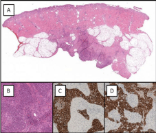
The histopathological examination revealed an epithelial malignant proliferation, superficially organised into papillary structures, which is further organised in solid nests and cords that infiltrate the subepithelial corium and the muscularis. The cells showed immunoreactivity for CK AE1/AE3 and CK7 (Figure 1).

    Figure 1:  (A) Low-power photomicrograph showing malignant bladder
epithelial proliferation superficially organised in papillary structures, deeply
organised in solid nests and cords that infiltrate subepithelial corium and
diffusely the muscularis (magnification 4x); (B) High-power photomicrograph
showing epithelial cells of medium size, with altered nucleus-cytoplasm
ratio, and leptocromatinic nucleus, nucleolus often prominent, and large
eosinophilic cytoplasm (magnification 40x); the cell showed immunoreactivity
for CK AE1/AE3 (C) and CK7 (D) (magnification 40x).
After the cystectomy, the patient received adjuvant chemotherapy with cisplatin and gemcitabine. On admission, the patient’s physical examination showed a rubbery, subcutaneous, and painful welldemarcated nodule of 3cm, overlying the left periumbilical area. Further cystoscopy revealed a bleeding neoplasm on neovesicalurethral anastomosis, which was consistent with local recurrence of bladder tumour. The nodule was surrounded by erythematous skin. The laboratory data were within normal limits. Further clinical staging through CT-scan demonstrated a 3.3cm nodular solid lesion with contrast enhancement involving the left side of the neovesical-urethral anastomosis. In addition, a 3cm nodular solid, irregular shaped, subcutaneous lesion with contrast enhancement involving the left rectus abdominis muscle suspicious for metastatic disease was also observed. The histopathological examination of the transurethral resection samples of the neovesical-urethral anastomosis lesion and excisional biopsy of the cutaneous lesion revealed an epithelial malignant proliferation with morphological and immunohistochemical features, consistent with the recurrent TCC of bladder (Figure 2).

    Figure 2:  (A) Low-power photomicrograph showing skin with infiltration in
deep dermis and subcutis of a malignant epithelial proliferation organised in
solid nests and cords (magnification 1x); (B) High-power photomicrograph
showing morphological features of atypical epithelial cells (magnification 40x)
that showed immunoreactivity for CK AE1/AE3; (C) and CK7 (D), consistent
with recurrent TCC of bladder (magnification 40x).
Case 2
A 70-year-old Caucasian woman presented to our outpatient clinic with a four-week history of occasional gross haematuria and a five-week history of a 2cm painful and itchy nodular solid lesion, which occasionally exuded blood to the left hypochondria. The patient reported a surgical history of left radical mastectomy with axillary lymph node dissection and postoperative irradiation for breast cancer. On admission, the laboratory data were within normal limits and except for breast cancer, her past medical history was unremarkable. The physical examination showed a “zosteriform”-like, erythematous eruption to the left submammary region surrounding a 2cm solid nodule. The first clinical suspicion was for metastatic breast cancer or its direct extension to the abdominal wall. Consequently, she underwent a wide excision of the cutaneous lesion that revealed a pattern of atypical cells in nests and cords with abundant cytoplasm and hyperchromatic atypical nuclei that was positive for CK AE1/AE3 and CK7 immunohistochemical stains, confirming the diagnosis of metastatic TCC. The CT-scan imaging and bone scan demonstrated multiple papillary lesions in the bladder with muscle-layer invasion and pelvic and para-aortic lymph node enlargement. The cystoscopy showed multiple large papillary tumours on the left abdominal wall and trigone area of the bladder. The biopsy as well as the pathological report revealed a poorly differentiated muscle-invasive TCC. The patient received one cycle of chemotherapy with cisplatin and gemcitabine, along with palliative treatment. The condition of the patient got worse, and she died of the metastatic disease within seven months after the diagnosis of cutaneous metastasis.
Discussion
The incidence of cutaneous metastasis from the primary internal malignancies was reported to range from 0.2% to 10.4% [3], with an incidence of cutaneous involvement by all urological malignancies that ranged from 1.1% to 2.5% [4]. Cutaneous metastases from bladder TCC are uncommon [5] and are mainly due to iatrogenic implantation after open or laparoscopic radical cystectomy [6]. Following the radical cystectomy, bladder TCC recurred in about 22% of patients; the pelvis, liver, lungs, adrenals, bowel, and bones were the most commonly affected sites, having a mean recurrence time of 15.1 months [7]. TCC is the most common cancer of the bladder, representing 90% of such cases [8]. The incidence of metastatic TCC of the bladder is directly related to the depth of penetration of the bladder wall, tumour grade and tumour size, with depth being the single most important prognostic factor in TCC [9,10]. Although rarely found, cutaneous metastases have also been described in the non-muscle-invasive bladder cancer [1,11,12]. Cutaneous metastases of bladder TCC occur with an incidence of 0.84—3.6 % [1,5], and are frequently misdiagnosed as they mimic benign dermatological conditions such as furuncles or sebaceous cysts [4], erysipeloid [13], and herpes zoster [14]. Mueller et al. [1] categorised the clinical appearance of skin metastases into three groups: (a) nodular type (as in our cases), which is found in most cases, (b) inflammatory type, and (c) fibrotic (cicatricial) or sclerodermoid type. Multiple cutaneous lesions are frequently found in patients, but these lesions may also present as solitary metastasis [15]. The lymphatic spread represents the most frequent mechanism by which a metastatic visceral tumour may reach the skin [1]. Studies have also reported other cases of cutaneous metastases such as by iatrogenic implantation at the time of surgery [6], by haematogenous spread [1,11], and by direct invasion [1].
In both the cases, the most likely mechanism of metastatic spread was haematogenous in nature as the nodular lesions were at a distance from the former port-site, and they had developed in the periumbilical area of the abdominal wall. Histologically, the cutaneous metastasis from visceral carcinoma often showed infiltrative growth of atypical epithelial cells among the collagen bundles, with a pattern of mixed single cells, narrow strands, and clusters of cells [1]. Metastatic lesions can be poorly differentiated, and this characteristic could make it difficult to identify primary tumours. Therefore, immunostaining could be a valuable aid in the diagnosis of cutaneous metastasis.
Patients with urologic tumours and cutaneous metastases showed universally poor prognosis. However, cutaneous metastases of bladder TCC are considered a late manifestation of systemic spread. In addition, most of the studies on cancer-specific survival reported that more than 98% of patients lived for less than 12 months after the diagnosis of a cutaneous metastasis from a urologic tumour [1,5,11].
Conclusion
In conclusion, both the cases show the importance of having a high index of suspicion during the evaluation of a newly formed skin lesion in patients with a history of genitourinary tumours. Therefore, in this pattern of patients, the urologists should increase attention to the physical examination of the skin and carefully evaluate patients for any new cutaneous lesions.
References
- Mueller TJ, Wu H, Greenberg RE, et al. Cutaneous metastases from genitourinary malignancies. Urology 2004; 63: 1021-1026.
- Pastore AL, Palleschi G, Silvestri L, et al. Pure intracorporeal laparoscopic radical cystectomy with orthotopic “U” shaped ileal neobladder. BMC Urol. 2014; 14: 89.
- Nashan D, Meiss F, Braun-Falco M, et al. Cutaneous metastases from internal malignancies. Dermatol Ther 2010; 23: 567-580.
- Block CA, Dahmoush L, Konety BR. Cutaneous metastases from transitional cell carcinoma of the bladder. Urology. 2006; 67: 846.
- Krathen RA, Orengo IF, Rosen T. Cutaneous metastasis: a meta-analysis of data. South Med J. 2003; 96: 164-167.
- Miyamoto T, Ikehara A, Araki M, et al. Cutaneous metastatic carcinoma of the penis: suspected metastasis implantation from a bladder tumor. J Urol. 2000; 163: 1519.
- Hassan JM, Cookson MS, Smith JA, et al. Patterns of initial transitional cell recurrence in patients after cystectomy. J Urol 2006; 175: 2054-2057.
- Oosterlinck W, Lobel B, Jakse G, et al. Guidelines on bladder cancer. Eur Urol. 2002; 41: 105-112.
- Beautyman EJ, Garcia CJ, Sibulkin D, et al. Transitional cell bladder carcinoma metastatic to the skin. Arch Dermatol. 1983; 119: 705-707.
- Hinshaw M, Stratman E, Warner T, et al. Metastatic transitional cell carcinoma of the bladder presenting as genital edema. J Am Acad Dermatol. 2004; 51: 143-145.
- Akman Y, Cam K, Kavak A, et al. Extensive cutaneous metastasis of transitional cell carcinoma of the bladder. Int J Urol. 2003; 10: 103-104.
- Swick BL, Gordon JR. Superficially invasive transitional cell carcinoma of the bladder associated with distant cutaneous metastases. J Cutan Pathol. 2010; 37: 1245-1250.
- Aloi F, Solaroli C, Paradiso M, et al. Inflammatory type cutaneous metastasis of bladder neoplasm: erysipeloid carcinoma. Minerva Urol Nefrol. 1998; 50: 205-208.
- Somani BK, Prita D, Grant S, et al. Herpetiform cutaneous metastases from transitional cell carcinoma of the urinary bladder: immunohistochemical analysis. J Clinic Pathol. 2006; 59: 1331-1333.
- Reingold IM. Cutaneous metastases from internal carcinoma. Cancer. 1966; 19: 162-168.