
Case Presentation
Ann Surg Perioper Care. 2016; 1(3): 1017.
Giant Syphilitic Ascending Aortic Anuerysm
Natraj Setty HS*, kumar V, Raghu TR, Kharge J, Geetha BK, Patil SS, Jadhav SK and Manjunath CN
Sri Jayadeva Institute of Cardiovascular Sciences and Research, Bengaluru, Karnataka, India
*Corresponding author: Natraj Setty HS, Assistant Professor of Cardiology, Sri Jayadeva Institute of Cardiovascular Sciences and Research, Bangalore, Karnataka, India
Received: November 17, 2016; Accepted: December 26, 2016; Published: December 28, 2016
Abstract
Aneurysms are usually defined as a localized dilation of an arterial segment greater that 50% its normal diameter. The occurrence and expansion of an aneurysm in a given segment of the artery involves local hemodynamic factors. Aneurysmal degeneration occurs more commonly in the aging population. Ascending aortic aneurysm is the second most common aortic aneurysm after abdominal aortic aneursyms. Most ascending aortic aneurysm is diagnosed in sixth or seventh decade of life. Patient with connective tissue disorder and bicuspid aortic valve are diagnosed earlier in life. Ascending aortic aneurysms can involve proximally from the aortic annulus and can extend distally till the innominate artery. They may compress or erode into the sternum and ribs, superior vena cava or airway. They can rupture or dissect involving the pericardium, aortic valve, or coronary arteries. We report a case of gross ascending aortic anuersym who presented with Dyspnoea NYHA class III and chest pain since six months. Options of surgery were discussed with patient’s relatives. But, the patient and her relatives were reluctant to give consent for surgery. Hence, Patient is on conservative management.
Keywords: Ascending aortic aneurysm; CT aortogram; 2D Echocardiography; Syphilis
Background
Aneurysms are usually defined as a localized dilation of an arterial segment greater that 50% its normal diameter. The occurrence and expansion of an aneurysm in a given segment of the artery involves local hemodynamic factors. Aneurysmal degeneration occurs more commonly in the aging population. The incidence of late manifestations of syphilis have declined almost to a rare entity since the era of antibiotics. Before the discovery of penicillin [1]. The primary lesion of cardiovascular syphilis is aortitis, an inflammatory response to the invasion of the aortic wall by the Treponema pallidum that evolves to obliterative endarteritis of the vasa vasorum and results in necrosis of the elastic fibers and connective tissue in the aortic media. The resulting weakening of the aortic wall will progress into the late vascular manifestations of syphilis. The ascending aorta is the segment most commonly affected (50%), followed by the arch (35%) and the descending aorta (15%). The rich lymphatic arrangement in the ascending aorta that may predispose greater mesoaortitis is believed to be the cause for larger involvement of this segment [2].
Indications for surgical treatment of ascending aortic aneurysms are based on size or growth rate and symptoms. Because the risk of rupture is proportional to the diameter of the aneurysm, aneurysmal size is the criterion for elective surgical repair. To repair an extensive aortic aneursym is a challenging task because it poses problems of bleeding, end organ ischemia, underlying disease and staged repairs.
We report a case of gross ascending aortic aneursym who is on medical management.
Case Presentation
We report a case of gross ascending aortic anuersym who presented with Dyspnoea NYHA class III and chest pain since six months. On examination, early diastolic murmur of grade 2 in Erbs area. Routine investigation was normal. VDRL and TPHA were positive. HIV and connective tissue profile was normal. Chest XRAY revealed Cardiomegaly and Ascending aorta aneursym (Figure 1). 2D Echocardiography apical four chamber view shows ascending aortic aneurysm. Parasternal long axis shows gross ascending aortic aneurysm (Figure 2A and 2B). CT Aortogram revealed proximal ascending aorta – 82 X 83 mm (AP x RL) and distal ascending aorta – 85x77 mm (AP x RL). Arch and descending thoracic aorta was normal (Figure 3A and 3B). Patient was advised surgical management but patient and their relatives did not give consent for surgery. Patient was managed conservatively with anti failure medications. Patient improved symptomatically.
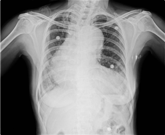
Figure 1: Chest X-Ray shows ascending aortic aneurysm and cardiomegaly.
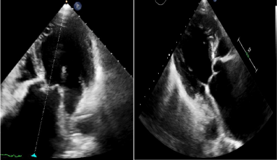
    Figure 2:  A: 2D Echocardiography apical four chamber view shows dilated
LV and ascending aortic aneurysm. B: Parasternal long axis shows gross
ascending aortic aneurysm.
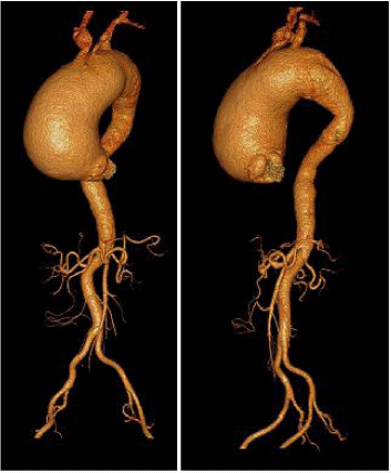
    Figure 3:  A: CT Aortogram revealed proximal ascending aorta – 82 X 83 mm
(AP x RL). Descending thoracic aorta at this level - 22x23 mm (AP x RL). B:
Distal ascending aorta - 85x77 mm (AP x RL).
Discussion
The basic lesion of cardiovascular syphilis is aortitis and myocarditis. Syphilis is a bacterial STD caused by the spirochete Treponema pallidum, subspecies pallidum, which had developed as a new world disease out of evolving treponemal species [3]. The disease progresses through primary, secondary, and tertiary phases. Involvement of the cardiovascular system is the most dangerous sequela of the tertiary phase (late syphilis). This usually manifests as syphilitic aortitis and infrequently as gummatous myocarditis. In untreated syphilis, aortitis may manifest 10 to 40 years after the initial sexual contact [4]. Syphilitic Aneurysms arise during tertiary syphilis due to chronic inflammation in the tunica adventitia of large elastic arteries, particularly the aorta. The inflammatory response to the spirochetes causes an obliterative endarteritis of the vaso vasorum, leading to a loss of blood supply to the elastic tunica media. Ischemia of the tunica media, combined with further syphilitic invasion into the tunica media itself, results in medial destruction and weakening, ultimately causing dilation and aneurysm-formation. The ascending aorta is most often affected (in 50% of cases), followed by (in decreasing order of incidence) the aortic arch, the descending aorta, and the abdominal aorta. Impairment of coronary ostia and aortic valve is also possible. The main cause of death in approximately 80% of cases is rupture of saccular aneurysms, which requires surgical intervention [5]. CT angiogram is the best imaging study to define the size and anatomy of the aneurysm, but in the setting of an aneurysm, the echocardiogram and coronary angiogram are mandatory to exclude aortic regurgitation and coronary flow-limiting lesions [6]. Surgical intervention may be required for luetic aneurysm and aortic regurgitation. Coronary ostial stenosis may be treated by endarterectomy or bypass grafting [7]. The definitive treatment of aortic aneurysm is surgical repair, which involves resection of the dilated portion of the aorta and replacing it with a synthetic vascular graft. Surgery and specific antibiotic treatment does not exclude future manifestations of the disease, even after erradication of the Treponema pallidum, which makes permanent follow-up needed [8].
Conclusion
In the era of decreasing incidence of syphilis, high index of suspicion with timely diagnosis and prompt medical management with surgical interventions might definitely improve the outcome. The cardiovascular complications of syphilis predominantly involve the aorta, leading to the formation of aneurysms and aortic-valve incompetence. Treatment of cardiovascular syphilis with penicillin prevents further progression of the disease. Open surgical repair remains the standard approach to treating most large aortic aneurysm and results are believed to be more predictable and satisfactory.
References
- Duncan JM, Cooley DA. Surgical considerations in aortitis. Part III: syphilitic and other forms of aortitis. Tex Heart Inst J. 1983; 10: 337-341.
- Heggtveit HA. Syphilitic aortitis: a clinicopathologic autopsy study of 100 cases, 1950 to 1960. Circulation. 1964; 24: 346-355.
- Harper KN, Ocampo PS, Steiner BM, et al. “On the origin of the treponematoses: a phylogenetic approach”. PLoS Neglected Tropical Diseases. 2008; 2: e148.
- Sacks R, Tipple C, and Goldmeier D. “Syphilitic aorta: exploring the bigger picture”. International Journal of STD and AIDS. 2010; 21: 608.
- Saleem MA, McNeeley M, Zias E, Pucillo A, Ro JH and Weiss MB. “Penetrating ulcer of ascending thoracic aorta in syphilis”. Catheterization and Cardiovascular Interventions. 2004; 61: 16–19.
- Jackman JD, Radolf JD. Cardiovascular syphilis. Am J Med. 1989; 87: 425- 433.
- Komaranchath AS, Abhilash A, Kareem MMA. Heller-Dohle syndrome: a rare case of syphilitic aortitis with calcified ascending aorta aneurysm in an elderly lady. Am J Med Case Report. 2013; 1.
- Salas Millán J, Martínez Calzón JL, González deVega N, Castillo Castro JL. Cardiovascular syphilis: a case report. Rev Esp Cardiol. 2000; 53: 1656-1658.