Case Report
Ann Surg Perioper Care. 2017; 2(3): 1034.
Kimura Disease in Infraorbital Region of Face Manifesting with a Huge Mass Lesion: A Rare Case Report
Sun B1*, Zhao B2, Yu Y1, Liu K1 and Zhang W1
¹Department of Oral and Maxillofacial Surgery, School and Hospital of Stomatology, Jilin University, Changchun, P.R.China
²Department of Periodontics, School and Hospital of Stomatology, Jilin University, Changchun, P.R.China
*Corresponding author: Sun B, Department of Oral and Maxillofacial Surgery, School and Hospital of Stomatology, Jilin University, Qinghua road 1500, Changchun, Jilin province, P.R.China
Received: November 19, 2017; Accepted: December 12, 2017; Published: December 19, 2017
Abstract
Kimura disease (KD) is a chronic inflammatory disorder that presents as painless or pruritic subcutaneous lesions with in the head and neck, especially in the parotid and submandibular regions. Peripheral eosinophilia and elevated serum IgE levels are the characteristics of the disease. Here, we present a case of Kimura Disease in Infraorbital region of face manifesting with a huge mass lesion.
Keywords: Kimura disease; Infraorbital region; Huge mass
Introduction
Kimura’s disease is a benign chronic condition of unknown cause, which usually presents as a painless swelling of subcutaneous tissue that develops into either single or multiple locations in the head and neck region, especially in the major salivary glands and regional lymph nodes. Other sites such as the oral cavity, axilla, extremity, trunk and groin have also been involved. Peripheral eosinophilia and elevated serum IgE levels are the characteristics of the disease [1]. Surgical resection and low dose radiotherapy are the main treatment methods. Here, we present a case of Kimura Disease in infraorbital region of face manifesting with a huge mass lesion.
Case Presentation
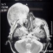
A 65-year-old Chinese man presented with a huge subcutaneous tissue mass lesion in right infraorbital region of face, which had been repeated growth and contraction for 20 years. Examination revealed that the mass was rubbery, painless, and surface skin of the mass was similar to the orange peel (Supplemental Figure 1). Magnetic resonance imaging (MRI) showing a huge mass with partial low signal intensity accompanied with high signal intensity septum in the right infraorbital and buccal area (T1 Short T2 inversion Recovery) (Figure 1, Supplemental Figure 2). Peripheral blood test showed that the leukocyte count was 14.2×10⁹/L with 58% eosinophils.

Figure 1: Magnetic resonance imaging (MRI) of the mass lesion.
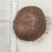
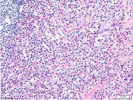
The patient was operated on under general anaesthesia. First, the huge mass was resected, which was orbicular and about 11cm in diameter (Figure 2). And then an adjacent soft tissue flap of right buccal was used to repair the defect of the infraorbital region. Histological examination of the specimen showed inflammatory cell infiltration consisting of a large number of eosinophils (Figure 3, Supplemental Figure 3).

Figure 2: The huge mass was orbicular and about 11cm in diameter.

Figure 3: Histopathological examination of the mass.
Kimura’s disease is a rare disease entity and the disease manifesting with a giant mass that occur in the infraorbital region is extremely rare. The pathogenesis of Kimura’s disease remains uncertain. Many scholars believe it to be an allergic reaction or Parasitic to an elevated serum IgE level and the deposition of IgE in the germinal centre, as well as peripheral blood eosinophilia [2].
Surgical resection and radiotherapy are the main treatment methods [3]. Surgery plays a pivotal role in providing the diagnosis and the remove of giant massed or unsightly lesions. However, recurrence of the disease is high after surgery alone, especially for incompletely excised lesions. A moderate dose of radiation (25 to 30 Gy) achieves excellent local control, especially to recurrent, large or inoperable disease. In this case, the lesion invaded the right eyeball surrounding tissue and could not been completely removed, so radiotherapy was used after the operation and good treatment results was achieved.
References
- Kittipong D, Pornchai J, Prasan T, et al. Kimura’s Disease. Asian J Oral Maxill -ofac Surg. 2006; 18: 151-156.
- Obata Y, Furusu A, Nishino T, et al. Membranous nephropathy and Kimura’s disease manifesting a hip mass. A case report with literature review. Intern Med. 2010; 49: 1405-1409.
- Kuroda K, Kashiwagi S, Teraoka H, et al. Kimura’s disease affecting the axillarylymph nodes: a case report. BMC Surgery. 2017; 17: 63.